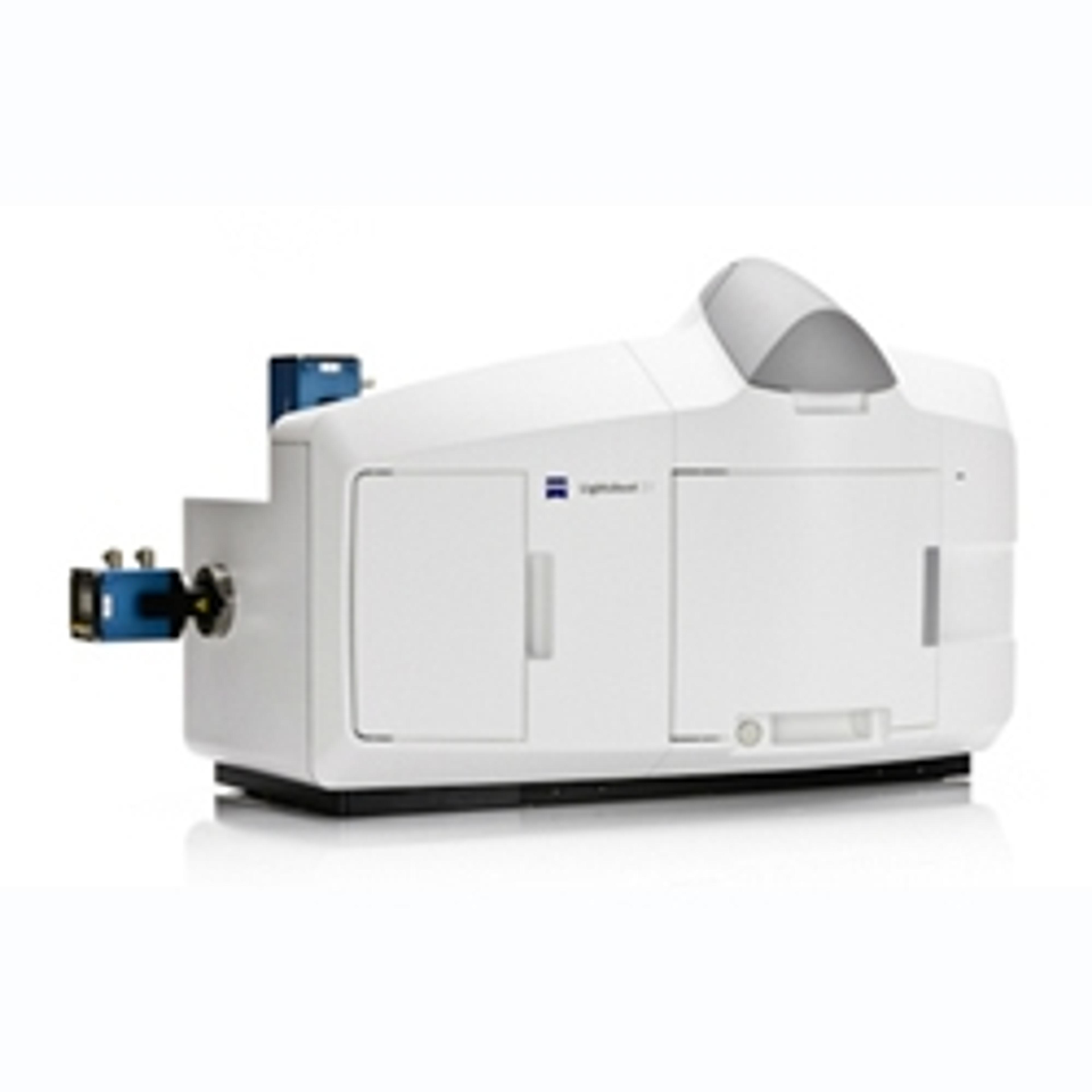
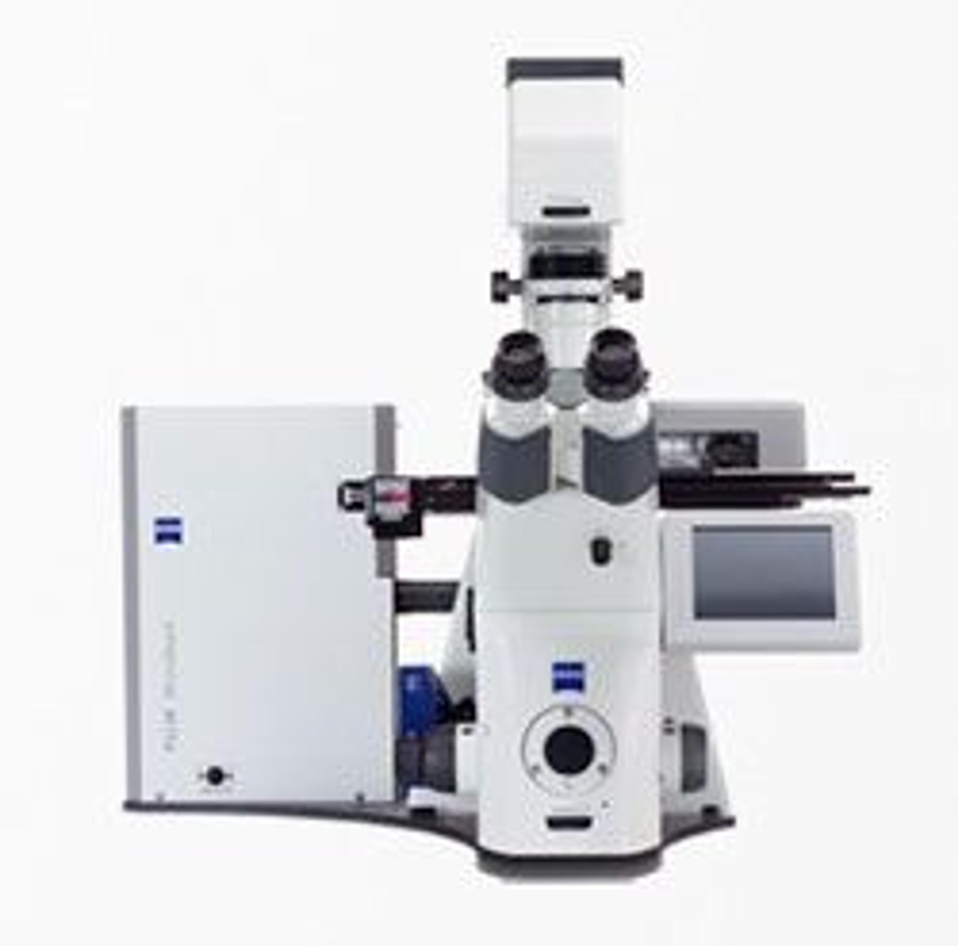
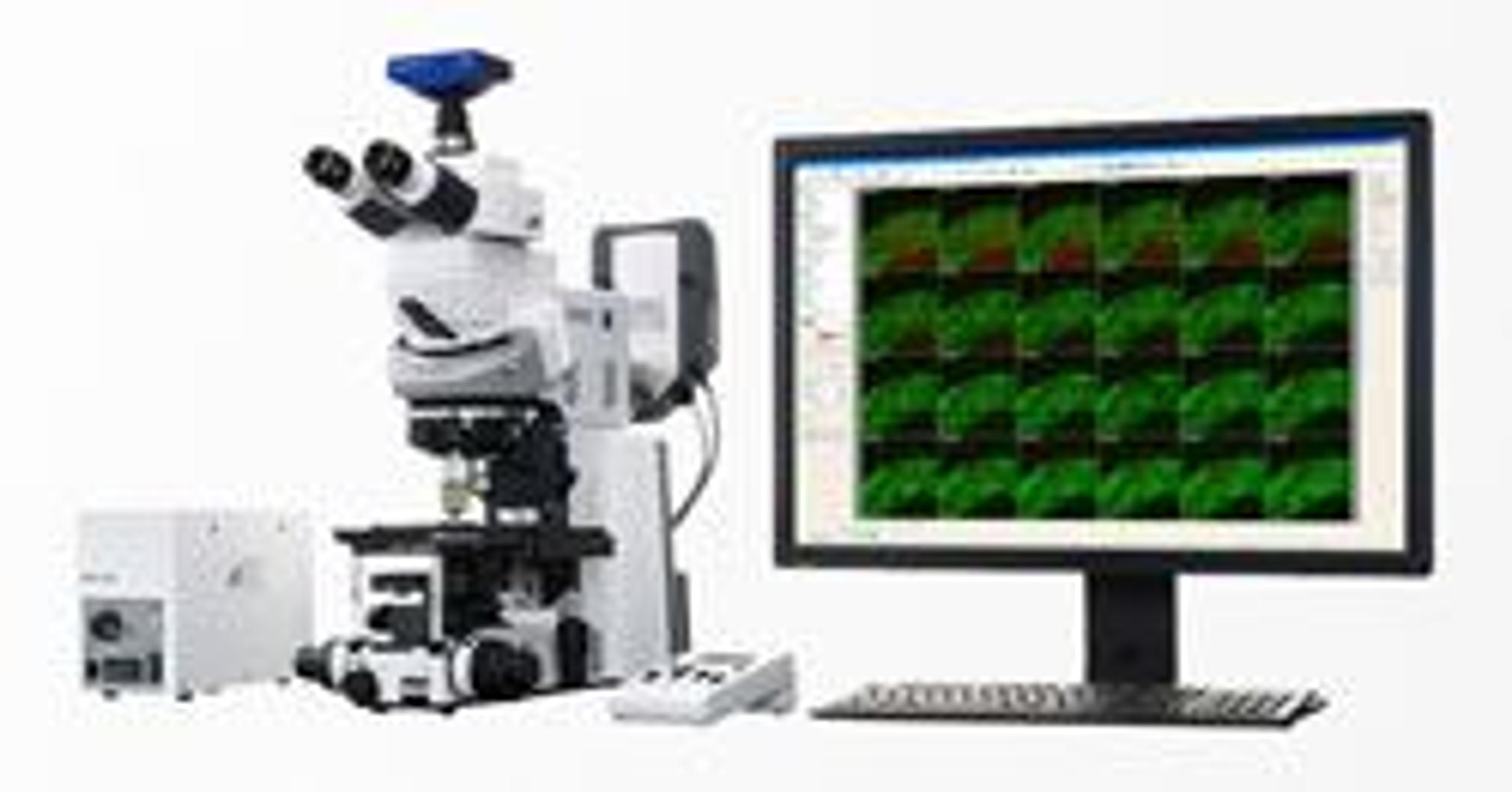
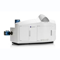
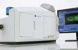
ZEISS Lightsheet Z.1 Microscope
Light Sheet Fluorescence Microscopy for Multiview Imaging of Large Specimens.


Receive your quote directly from the manufacturer.
Good product if maintained well
To analyze Fungal smears
The Microscope is easy to use and and it serve its expectations to the value.
Review Date: 9 Jun 2023 | ZEISS Research Microscopy Solutions
Great result
Histopathology
The Zeiss microscope is one the best instrument that show images clearly and high quality that released beside Zeiss company
Review Date: 14 Apr 2023 | ZEISS Research Microscopy Solutions
Amazing, Must buy.
Blood microscopy.
Used it in university labs for hemocytometery and DLC. Amazing product, writing this to claim the T shirt. Amazing product, A must have for a lab.
Review Date: 29 Jul 2022 | ZEISS Research Microscopy Solutions
Easy to use and gives a clear image.
Research
The beauty of the Zeiss microscope is the ease of use and clarity of the image. Thank you!
Review Date: 12 Mar 2020 | ZEISS Research Microscopy Solutions
Very helpful for the analysis of steel microstructure.
Analysis for steel
This Zeiss microscope has been very helpful for the analysis for steel and its microstructure. It is very fast for defect analysis in the steel.
Review Date: 12 Mar 2020 | ZEISS Research Microscopy Solutions
Compact, user-friendly and versatile equipment. Clean design, good support form ZEISS.
Cell biology, developmental biology, cancer, plant biology
The product contains several accessories for sample preparation. Great support from ZEISS to train users to use the system and software. Fast scanning of big samples, great quality
Review Date: 8 May 2019 | ZEISS Research Microscopy Solutions
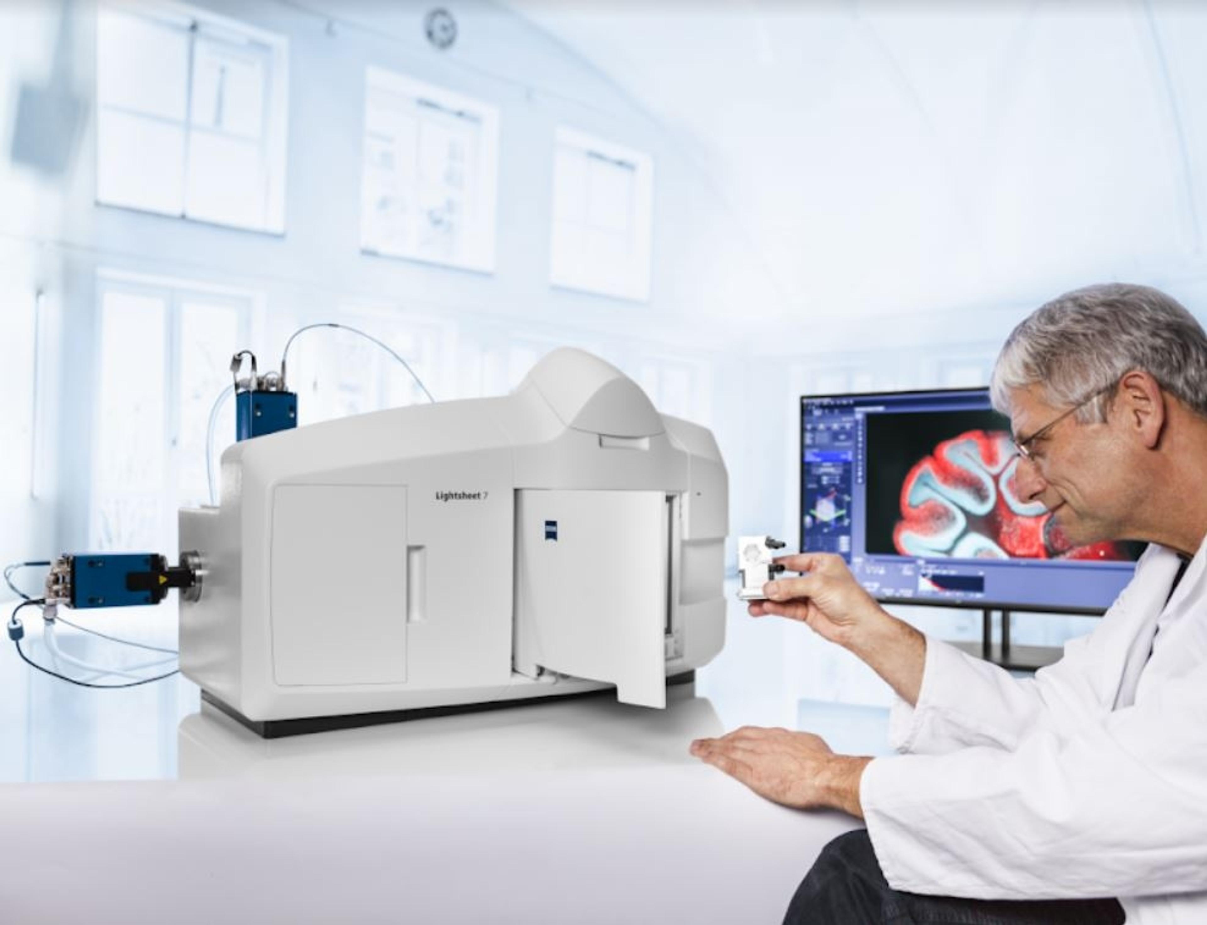
Discover the benefits of the first light sheet microscope designed to image fluorescently labeled living samples.
Now you can observe your model organisms, tissues and cells as they develop over days with virtually no phototoxicity or bleaching. Lightsheet Z.1 lets you acquire images of your whole sample volume at sub-cellular resolution in a fraction of the time it takes with other techniques.
Follow the development of your most valuable specimens in a gentle and stable environment – with up to a thousand times less light exposure.

ZEISS Lightsheet Z.1 Sample Preparation
This paper describes theoretical and practical aspects of sample preparation for light sheet fluorescence microscopy (LSFM). It presents general rules for sample handling and mounting, as well as guidelines with respect to the best preparative technique to use, taking into account sample type, structure and properties. Step-by-step protocols and recommended materials for ZEISS Lightsheet Z.1 samples are included. These protocols cover sample preparation ranging from micrometer-sized fluorescent beads to millimeter-sized insects, providing detailed information relating to preparation and observation techniques. Finally, this paper identifies the main artifacts and problems that can result from the preparation techniques.
Improved Imaging of Cleared Samples with ZEISS Lightsheet Z.1: Refractive Index on Demand
This application note demonstrates a novel technique to formulate the optimum refractive index (RI) without compromising image quality or damaging the solvent-vulnerable Light Sheet Fluorescence Microscopy (LSFM) system. LSFM uses a thin plane of light (light sheet) to optically section transparent tissues or whole organisms that have been fluorescently labeled. This “RI matching” solution is based on the published 3DISCO, an organic-solvent based protocol, which requires no special equipment to provide a faster imaging solution compared to laser scanning microscopes.
Adjusting Refractive Index for Clearing Applications using ZEISS’ Lightsheet Z.1
Scientists must consider specimen integrity, fluorescent labelling and refractive indices (R.I.) when working with clearing applications and adapting the clearing procedures to their specimens. It is crucial to check the R.I. of clearing solutions to minimize mismatches and resulting optical errors, such as spherical aberrations, and to increase the image quality and depth penetration, before and during the clearing procedure. This technology note describes a simple method to check the R.I. for clearing and imaging solution necessary for successful clearing imaging with Lightsheet Z.1.
ZEISS Lightsheet Z.1; Light Sheet Fluorescence Microscopy for Multiview Imaging of Large Specimens
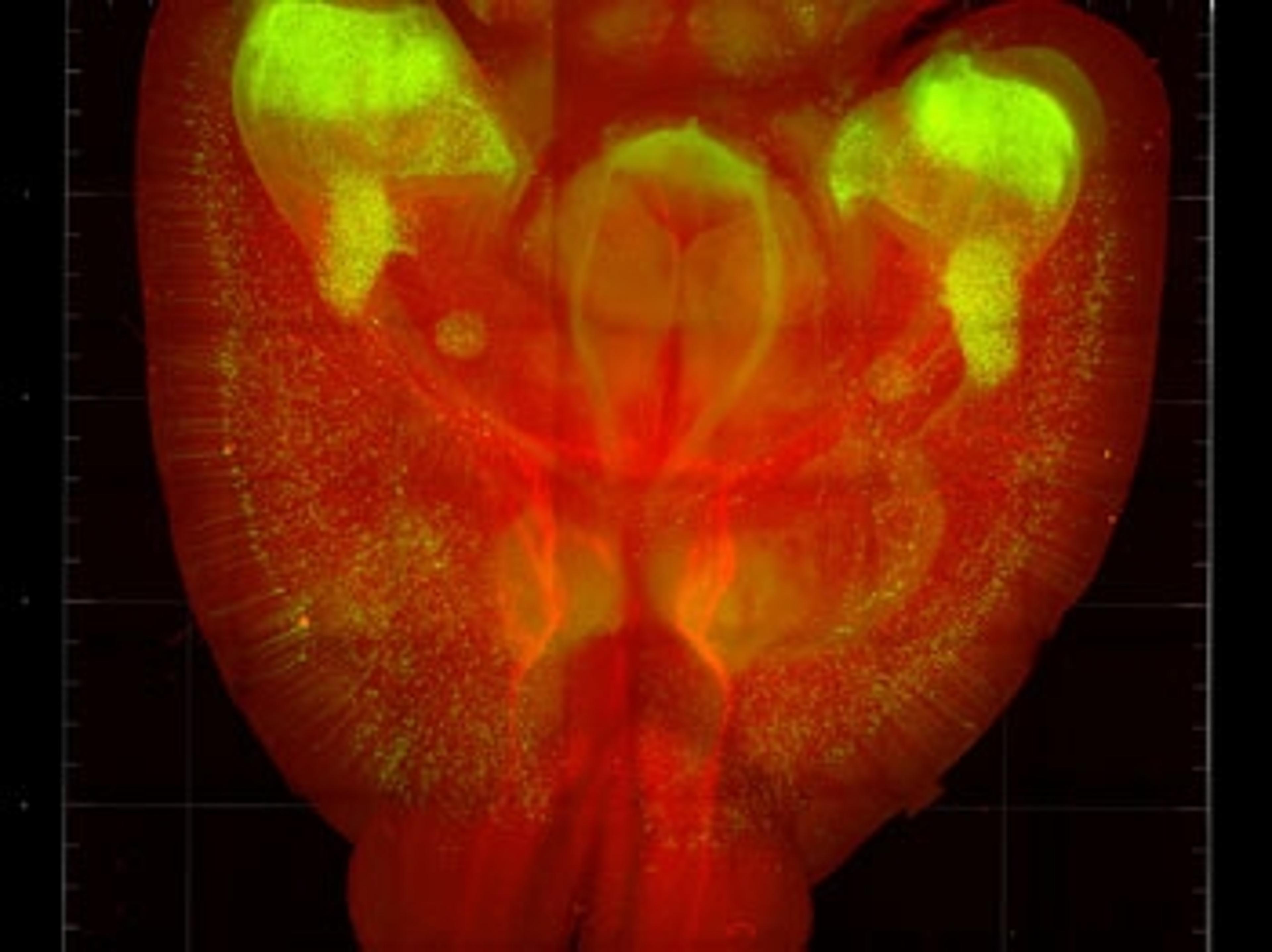
Light sheet fluorescence microscopy has a wide range of applications including use in developmental biology, systems biology, organogenesis and cell dynamics, 3D cell culture, plants, imaging of marine animals as well as structural imaging of large, fixed specimens and imaging of optically cleared specimens. The Zeiss Lightsheet Z.1 allows you to observe model organisms, tissues and cells as they develop over days with virtually no phototoxicity or bleaching. This product note describes the benefits of Zeiss’ Lightsheet Z.1; the first light sheet microscope designed to image fluorescently labeled living samples.
ZEISS Lightsheet Z.1 Quick Guide: Imaging with Water or Clearing Immersion
The ZEISS Lightsheet Z.1 imaging system is capable of delivering optical sections of large samples, with virtually no phototoxicity or bleaching and with high temporal resolution. Light Sheet Fluorescence Microscopy has advantages for imaging live as well as cleared samples, with the Lightsheet Z.1 as equally capable in clearing applications. This document describes the necessary steps to change between water immersion and clearing immersion imaging options.
Light Sheet Microscopy at the Harvard Center for Biological Imaging; Evaluation of the Lightsheet Z.1 Microscope from ZEISS
In the spring of 2013 the Harvard Center for Biological Imaging (HCBI) was able to demo, and eventually acquire, Lightsheet Z.1 microscope from ZEISS. The HCBI have now performed a number of long term embryonic development imaging experiments including imaging of zebrafish, fly, worm, cricket, and salamander embryos. Throughout these experiments, ZEISS Lightsheet Z.1 was employed to acquire both fast dynamics and long-term changes during development; this application note evaluates the use of ZEISS’ Lightsheet Z.1 in such experiments.
ZEISS Lightsheet Z.1: Sample Preparation
This white paper describes the theoretical and practical aspects of sample preparation for Light Sheet Fluorescence Microscopy. General rules for sample handling, as well as guidelines with respect to the best preparative technique to use are presented. Step-by-step protocols, covering sample preparation ranging from micrometer-sized fluorescent beads to millimeter-sized insects, are also included.
Control of External Devices during Time Series Acquisition with ZEISS Lightsheet Z.1
This technology note demonstrates the integration of daylight illumination into time lapse experiments.
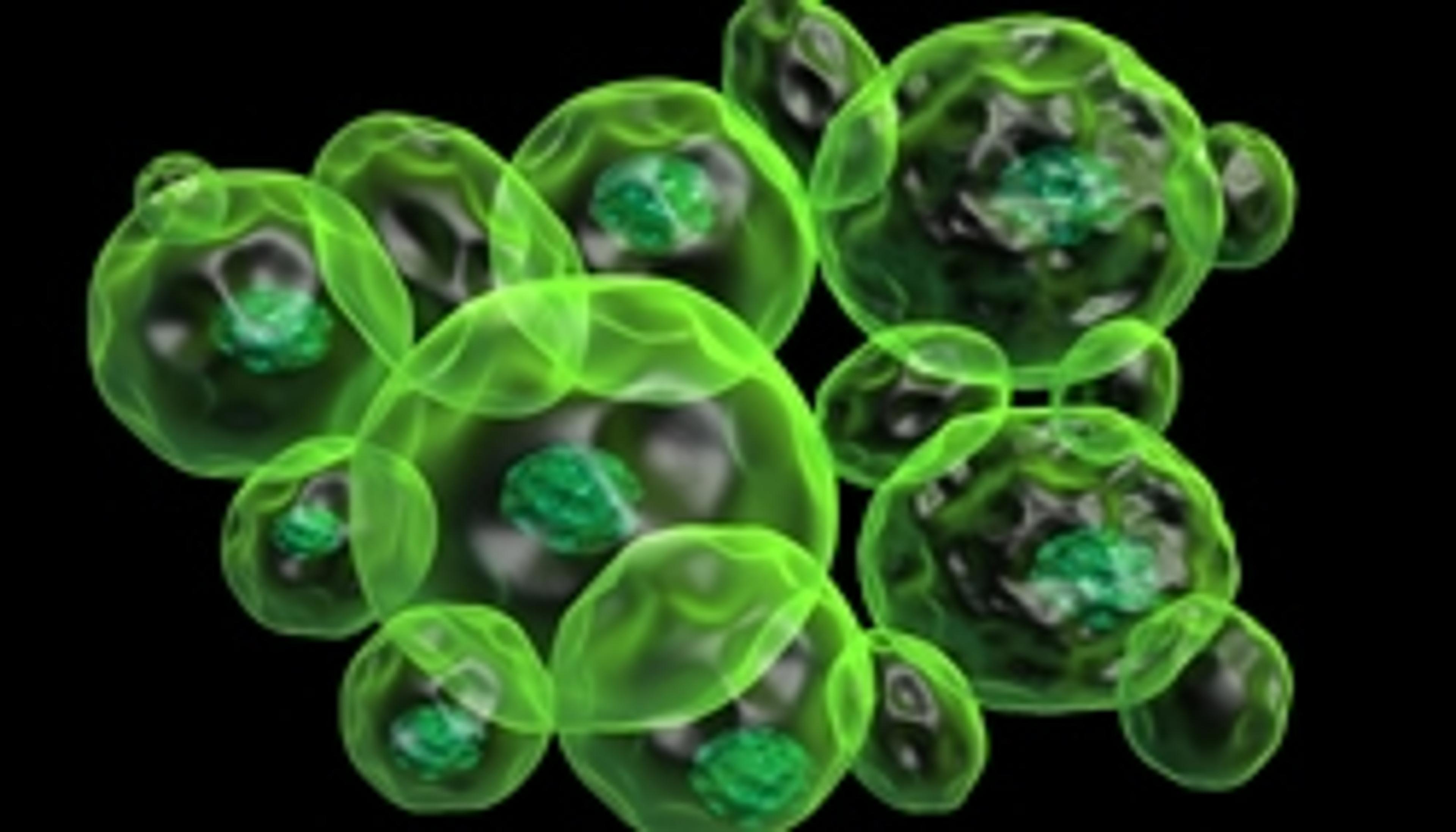
Fast Imaging of Cellular Spheroids with Light Sheet Fluorescence Microscopy
This application note presents an application of the commercially available high-resolution light sheet based microscope, Lightsheet Z.1 from ZEISS, to the investigation of large cultured organotypic tissues, so called cellular spheroids.
Sample Preparation for ZEISS Lightsheet Z.1
Lightsheet Z.1 allows you to record the development of large, living samples or 3D tissue cultures and gently image them to deliver exceptionally high information content. In this video see how easy it is to prepare samples for imaging with Lightsheet Z.1 - the MultiView Light Sheet Fluorescence Microscopy system by ZEISS.
Long-Term Imaging for Large Samples with Light Sheet Microscopy
Hear how the Lightsheet Z.1 Multiview light sheet fluorescence microscope from ZEISS enables you to image large living samples in physiologically relevant conditions for long periods of time, with virtually no phototoxicity or bleaching. The system also allows for imaging of large cleared specimens, with exceptional light efficiency and speed.
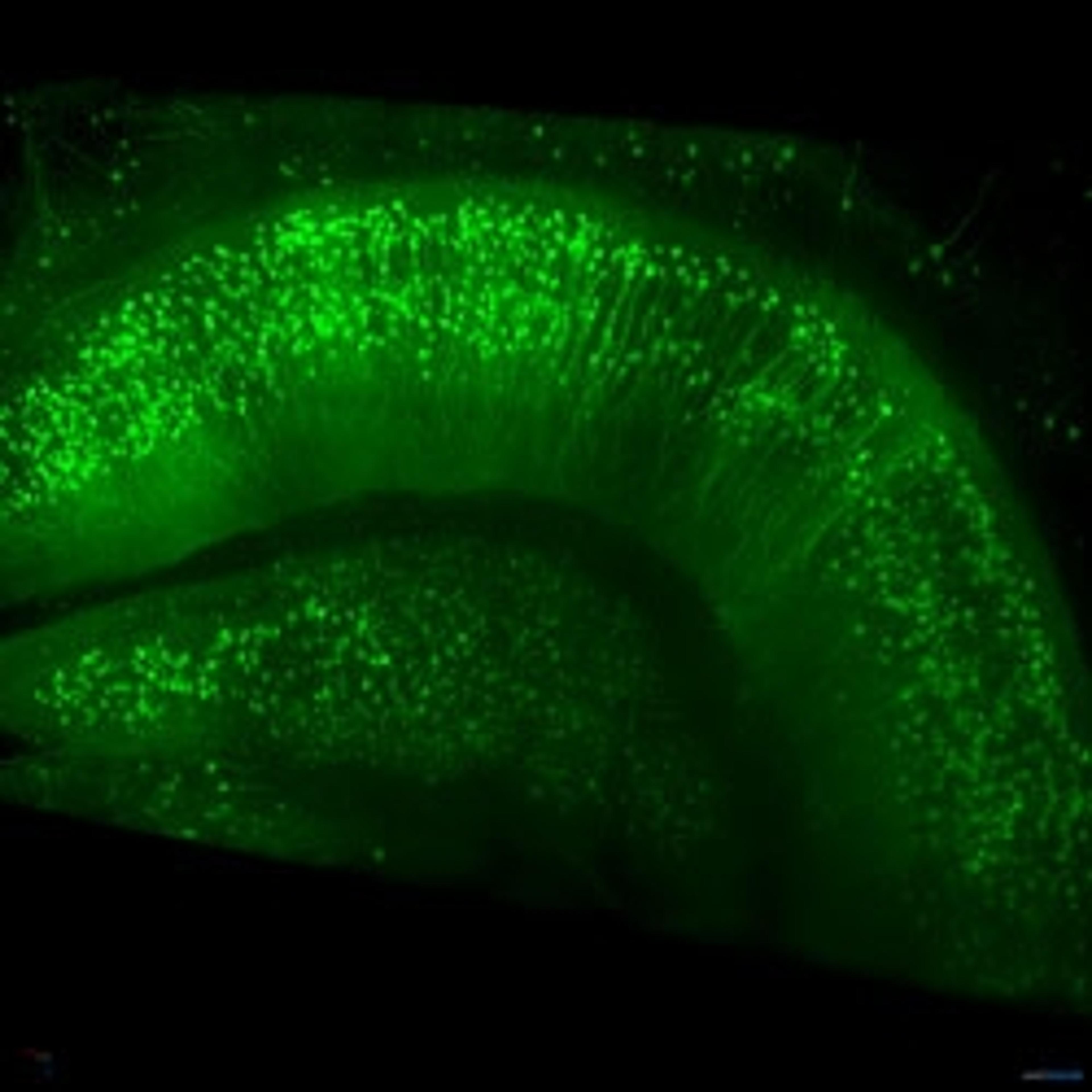
Software for Handling of Large Microscopy Datasets
Hear how ZEISS has worked together with arivis AG to provide a software platform for the Lightsheet Z.1, which can handle the large datasets produced by 3D imaging of large samples, enabling analysis of tiled stacks and large time dependent series. An example data set of a mouse hippocampus is presented.
Fast, Gentle Imaging for Living Samples with the Lightsheet Z.1
In this video Scott Oleynch, Marketing Manager for Zeiss Microscopy, tells SelectScience how the new Lightsheet Z.1 microscope system enables scientists to perform fluorescence imaging on large, living samples, with virtually no phototoxicity or bleaching and with high temporal resolution. Scott discusses some of the unique features of the system and some of the applications for imaging model organisms such as zebrafish, c. elegans and drosophila. Interview filmed by SelectScience at AACR 2013.
Fast, Gentle Imaging for Living Samples
Watch this video to learn how the innovative Lightsheet Z.1 from Carl Zeiss Microscopy enables you to perform 3D fluorescence imaging on large, living samples for prolonged periods of time, with virtually no phototoxicity or bleaching and with high temporal resolution. The unique Multiview Light Sheet Fluorescence Microscope allows you to record the development of large, living samples and gently image them to deliver exceptionally high information content. Watch this video to see examples of how the new Lightsheet Z.1 gives you the tools you need to work at unprecedented speed and acquire images of your whole sample volume at sub-cellular resolution in a fraction of the time it takes using other techniques.
ZEISS light sheet microscopy allows multiview imaging of cleared tissues
Lightsheet 7 platform is designed to reduce imaging artifacts in optical sections
Solutions for the Visualization and Analysis of Big Image Data in Life Sciences
ZEISS and arivis AG partner to provide leading 3D imaging systems

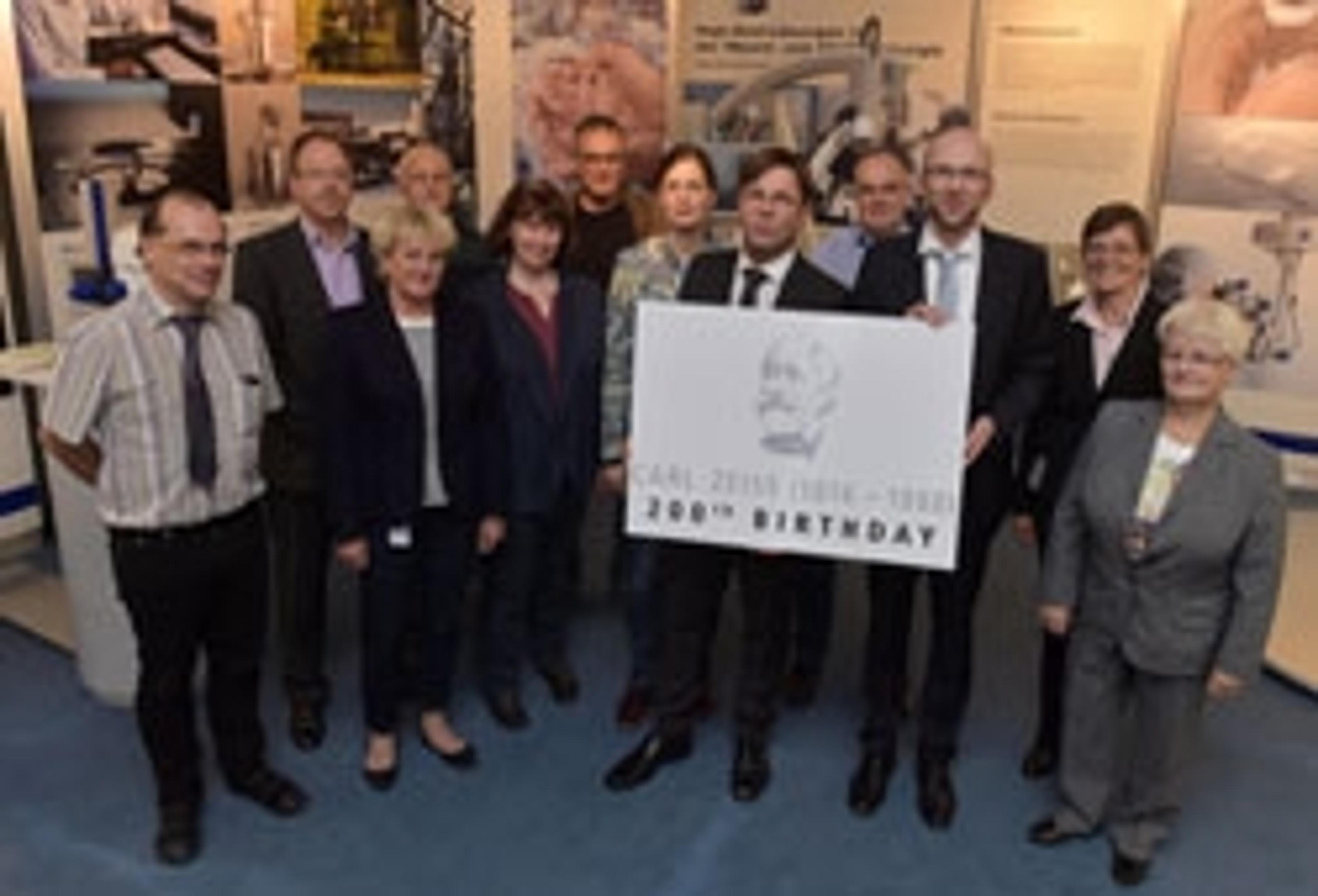
ZEISS Opens New Microscopy Customer Center
From Phenotype to Genotype: Unlocking the Power of Correlative Microscopy for Cell Biology
SelectScience® spoke to Dr Peter O'Toole, Head of Imaging and Cytometry, Department of Biology at the University of York, about the technology serving his department
8 Recent Advances in Bio Imaging and Microscopy
Discover the latest products and methods for the imaging of biological samples