RNAscope™ Multiplex Fluorescent Assays
The RNAscope Multiplex Fluorescent assays are ideal for simultaneous detection studies of any genes in nearly any tissue-type using fluorescent labels.

The supplier does not provide quotations for this product through SelectScience. You can search for similar products in our Product Directory.
Technical support team is nice and always there to help, Its good value for money
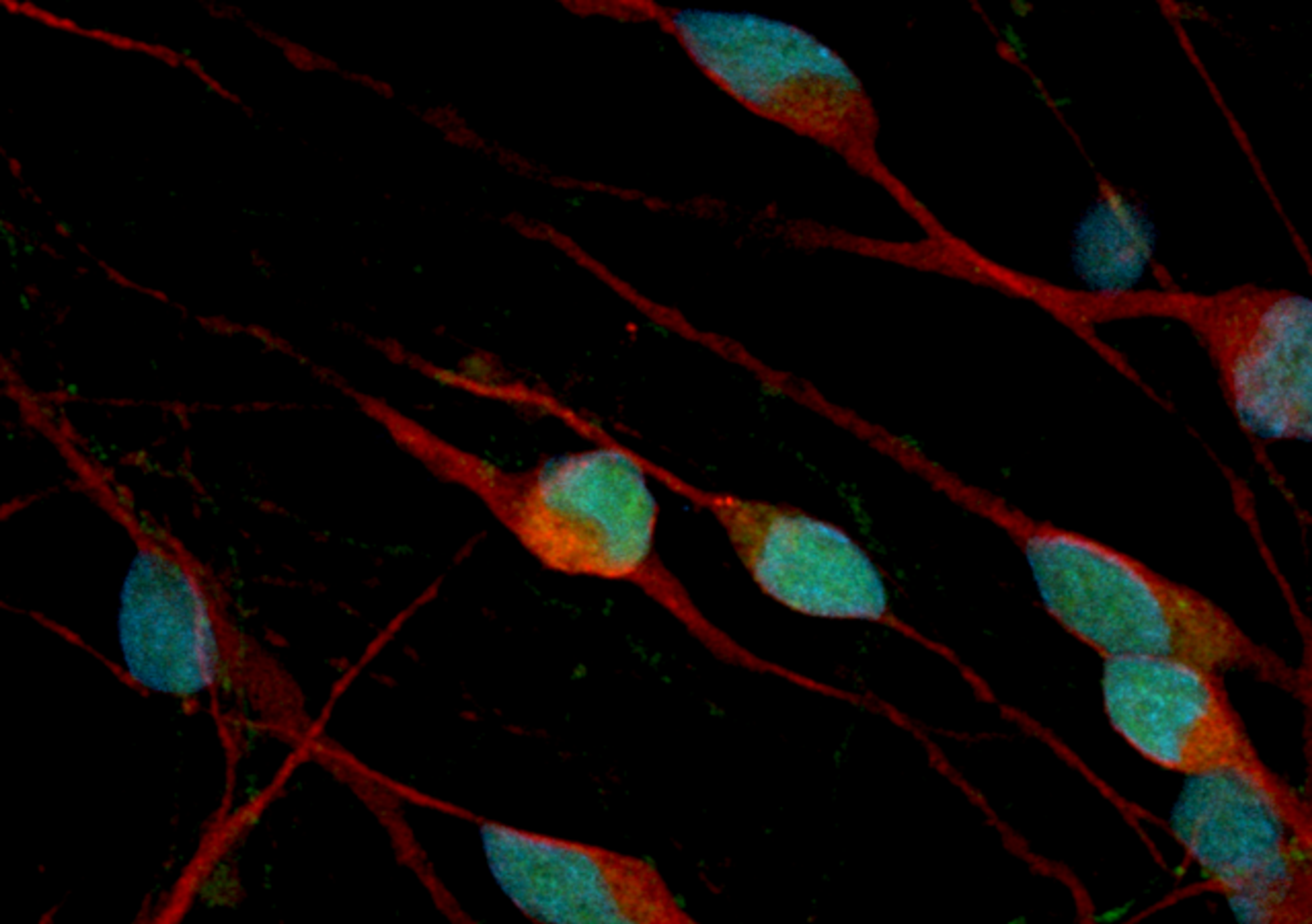
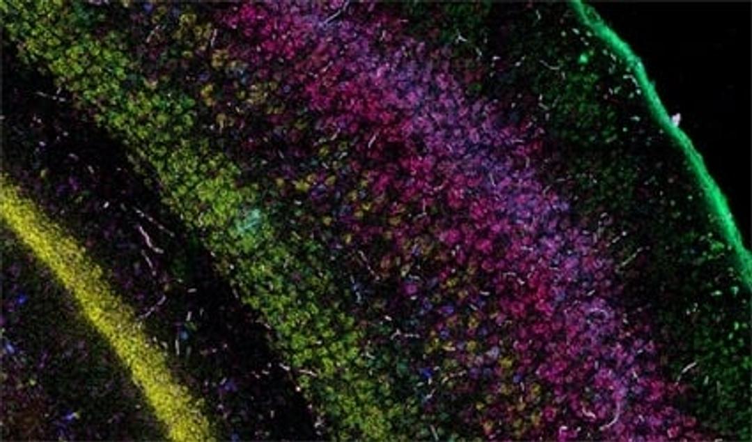
Detection of target RNA within intact cells.
Customer support is great. I had a tough time optimizing the assay due to the nature and type of my target tissues, but I got outstanding technical support from the ACD team during the standardization process. Results are reproducible if the quality of tissues is maintained. They have hundreds of ready-to-go probes so it saves a lot of time and effort to make your own probes. The detection method is very easy compared to the standard ISH. The protocol is easy to follow.
Review Date: 6 Oct 2021 | Advanced Cell Diagnostics
Great for brain tissue.
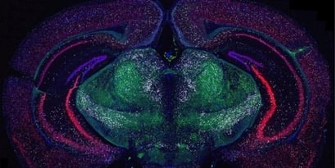
Analyze gene expression in brain samples
Easy to use and mostly works well. Found the best application in brain samples. It can be tricky to optimize for other tissues, tried adrenal and placenta as well and they neve work as well even after fiddling with many parameters.
Review Date: 24 Sept 2021 | Advanced Cell Diagnostics
So far good results. Go ahead if you are looking to buy it.
Detecting RNA using ISH
I have been using the RNAscope multiplex fluorescent V2 assay kit for few months now. The kit comes with a manual which guides you step by step through the protocol and it is very easy to use. Since I am new with this kit, I got the technical service package as well. The sales care and technical support team is very nice and always there to help, be it a phone conversation or a google team meet. Its good value for money except I feel that they should sell a few reagents like proteases and Amp buffers separately and not as a kit. Overall, I don't have any complaints.
Review Date: 6 Aug 2021 | Advanced Cell Diagnostics
Would recommend using RNAscope Multiplexing Fluorescent Assay for doing ISH.
Detecting RNA using ISH
The protocol is easy to follow. Customer support is great. The detection method is very easy with reduced background fluorescence compared to the standard ISH. They have hundreds of ready to go probes so it saves a lot of time and effort to make your own probes.
Review Date: 14 May 2021 | Advanced Cell Diagnostics
Great technology to detect gene targets where traditional IHC has been a challenge.
Gene expression in rodent brain samples
In-person technical support was available to guide the process from beginning to end. Many steps but protocol was clear and easy to follow.
Review Date: 18 Nov 2020 | Advanced Cell Diagnostics
One of the best techniques to visualize and quantify RNA molecules in a tissue section.
RNA in situ hybridization
I have used RNAscope V2 (fluorescent) and chromogenic in-situ hybridization assays for spatial detection of target RNA species in plants and animal tissue sections. This technique is amazingly WONDERFUL. Once optimized, this method is straightforward and very easy to use. After sales customer care is one of the best in industry. I had a tough time to optimize the assay due to the nature and type my target tissues, but I got outstanding technical support from ACD team during the standardization process. Results are reproducible if quality of tissues are maintained. Initial investment on RNAscope tools, kit and probe designing is a bit expensive but full value for money. Now RNscope is fully optimized in our lab and we are using it routinely.We published a modified protocol on it (doi: 10.1186/s13007-020-00614-4) and employ this technology for a few other projects too. We got beautiful pictures of multiple target RNAs as per our research objective and now many of our collaborators are interested in RNAscope technology.
Review Date: 25 Jun 2020 | Advanced Cell Diagnostics
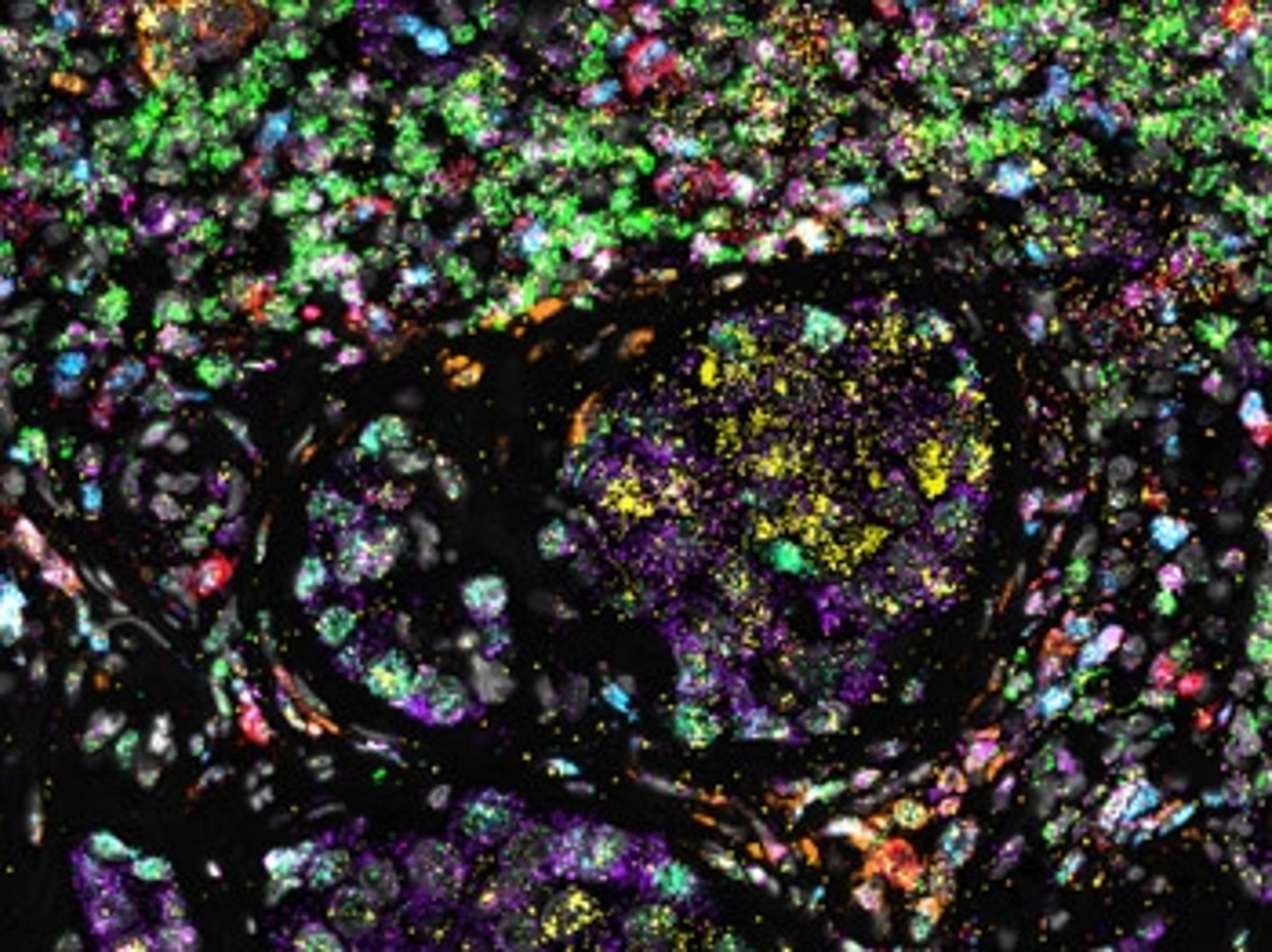
The RNAscope Multiplex Fluorescent assays are ideal for simultaneous detection studies of any genes in nearly any tissue-type using fluorescent labels. We offer with various levels of plexing to:
- FIND cells of interest by using cell specific markers
- CONFIRM cells by simultaneous detection of additional targets
- CHARACTERIZE cellular heterogeneity of complex tissues by mapping and identifying multiple cell subtypes simultaneously
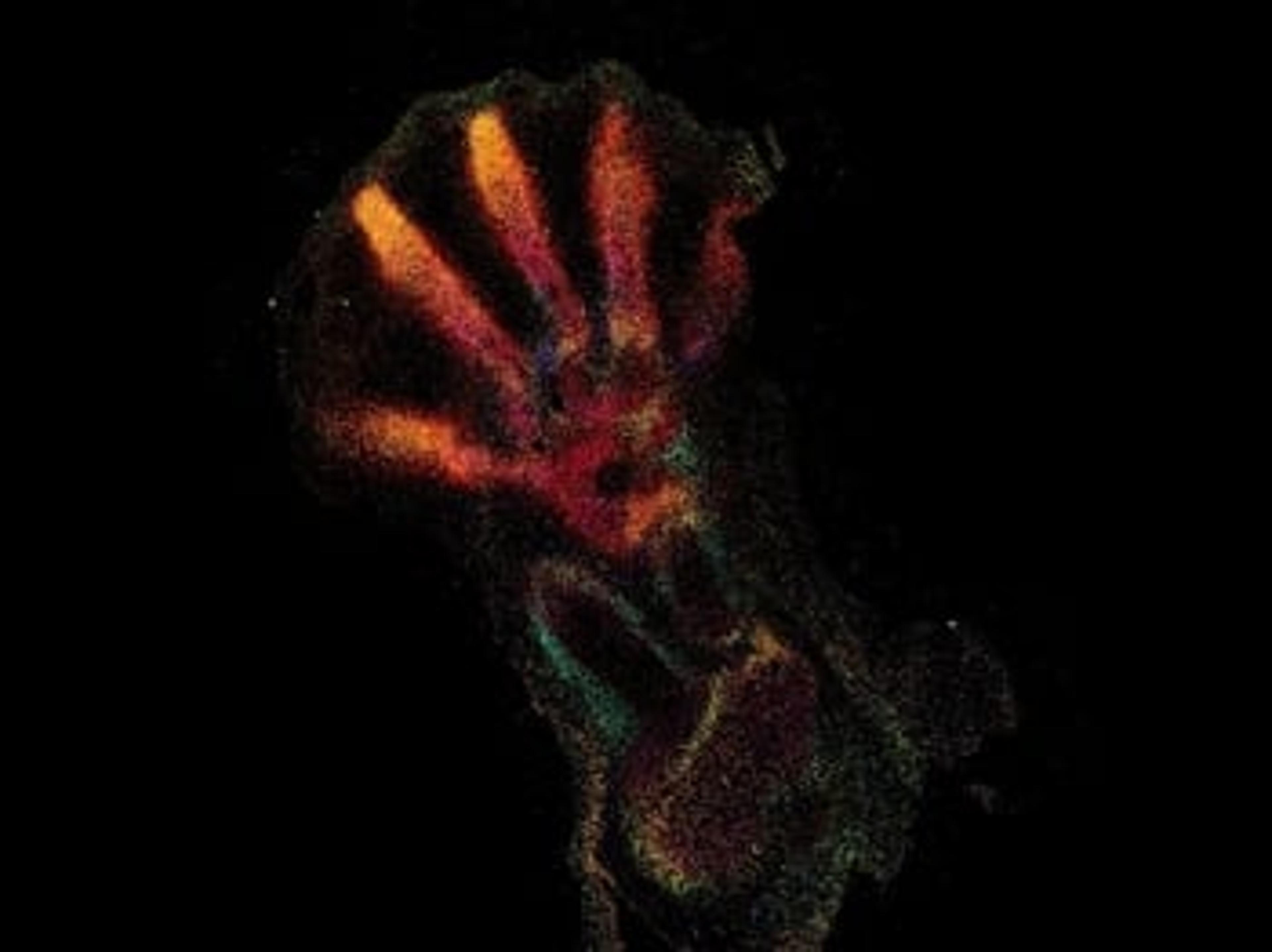
Spatial analysis of gene expression is an essential tool for comprehensive studies of complex, highly heterogeneous tissues, such as the brain, tumors, and developing organs. Traditional RNA-ISH can be challenging due to low sensitivity, inconsistent detection accompanied with limited plexing. RNAscope Fluorescent Multiplex assays provide exceptional sensitivity, allowing single-molecule detection of RNA targets at the single cell level.
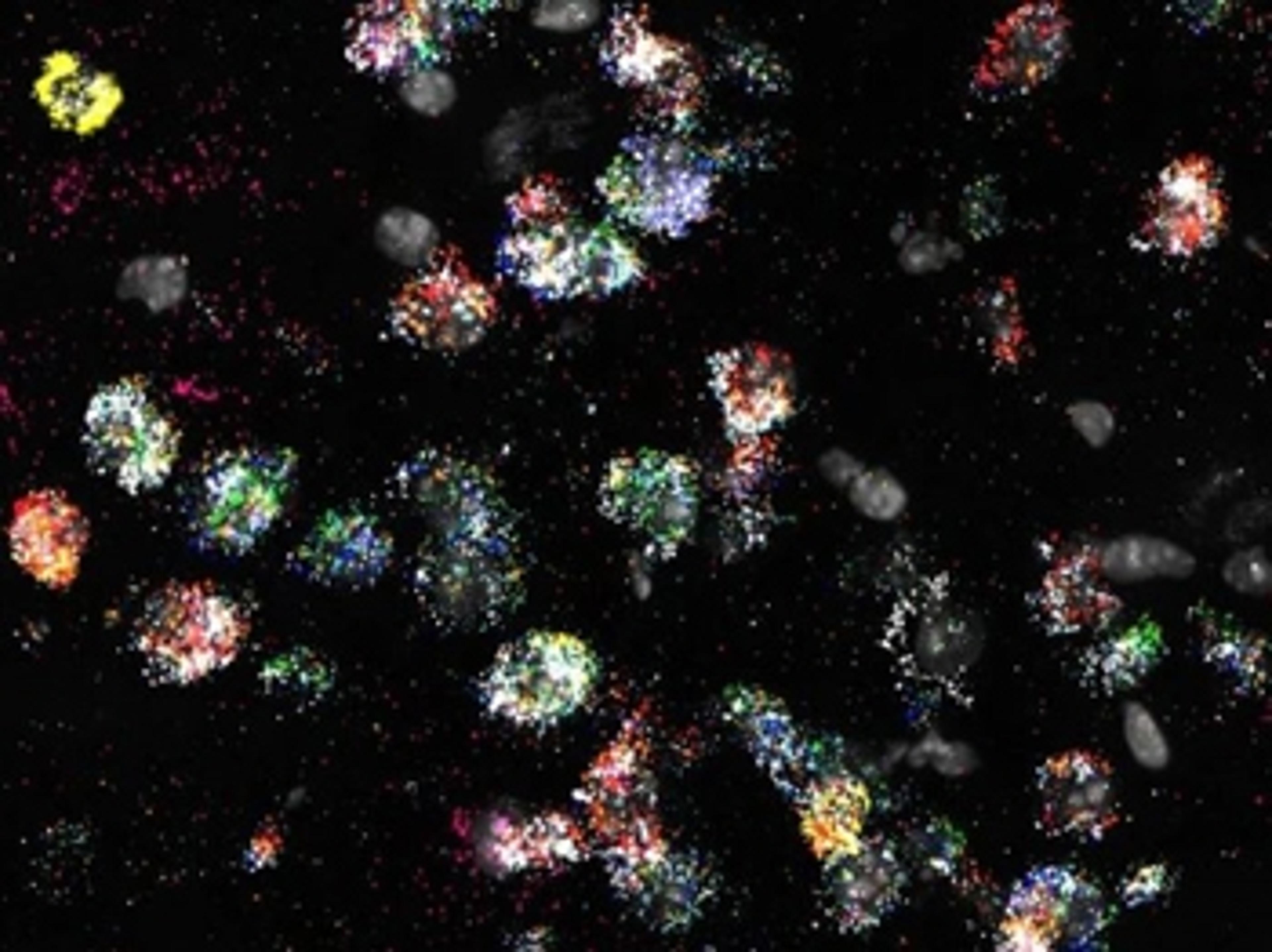
The multiplex assays offer exceptional sensitivity and allow single-molecule detection of up to 12 RNA targets simultaneously. These assays offer a novel and proprietary method of in situ hybridization (ISH) to achieve single cell resolution for RNA detection in a multitude of slide mounted samples. The assays are compatible with various sample types such as fresh frozen, fixed frozen and FFPE.
- Single molecule RNA detection.
- Single cell resolution
- Sensitive
- Specific
- Spatial Context
Taking a multi-omic multiplexing approach to spatial biology
Multiomics has begun to take the center stage in biomedical research as it combines two or more individual omics studies such as genomics, epigenomics, transcriptomics, proteomics, and metagenomics, allowing scientists to interpret and visualize the mechanisms of biological processes with spatial context.
In this free guide, we present a series of case studies illustrating how a multi-omic multiplexing approach can be leveraged to gain a comprehensive view of spatial biology, including a multiplexed in situ transcriptomic method for the spatial mapping of target genes in highly complex and heterogenous FFPE tumor tissues, and much more.

Combined workflow of GeoMx Cancer Transcriptome Atlas and RNAscope assays
This application note demonstrates the concordance between the RNAscopeTM and GeoMx® assays and highlights the detailed molecular analysis that can be performed with this type of high-plex spatial data. Also described are qualified RNAscopeTM probe combinations selected for cell type markers shown to be compatible with the GeoMx® CTA assay.
Quantitative RNAscope image analysis guide
In this application note, Advanced Cell Diagnostics (ACD) provides examples of exemplary assays, discusses experimental considerations, addresses common challenges, introduces HALO® image analysis solutions, and answers common quantitative image analysis questions. Plus, learn how RNAscopeTM technology is designed to enable RNA target expression analysis within intact cells and tissues with high sensitivity and specificity.
RNAscope technology enables the incorporation of spatial mapping and confirmation of gene signatures into single cell RNA sequencing workflows
In this application note, the diverse cell types in the mouse striatum that have been previously identified by scRNA-seq were confirmed and spatially mapped using the RNAscopeTM Multiplex Fluorescent assay and the RNAscopeTM HiPlex assay. The major and minor gene signatures identified by scRNA-seq, including discrete D1 and D2 medium spiny neuron (MSN) subtypes, were confirmed.
Further cellular heterogeneity within the MSN subpopulations was marked by a transcriptional gradient, which was spatially resolved with the RNAscopeTM technology.
Quantitative RNAscope image analysis guide
The RNAscope® Duplex and Multiplex assays enable simultaneous detection of more than one target RNA thus facilitating the evaluation of multiple cell phenotypes and spatial relationships within a complex tissue microenvironment. Download the guide below to find out how HALO® imaging software can create an easier solution with RNAscope® assays.
Upgrade your single-cell data with higher-plexing
RNA plays an important role in the regulation of cellular processes, making it an ideal biomarker for dynamic gene expression changes in both healthy cells and disease phenotypes. In situ hybridization (ISH) technology provides a powerful method to detect this gene expression, at single-cell resolution, within the spatial and morphological context of an intact tissue. Advanced Cell Diagnostics offer a comprehensive portfolio of ISH assays, with various levels of plexing, that can be applied to a wide range of applications, from neuroscience to immuno-oncology to cell therapy.
In this application eBook, we present:
- A series of case studies illustrating the research capabilities of high-plexing assays
- An outline of the benefits of the RNAscope™ technology
- Top tips on how to achieve accurate, reliable results for virtually any target species, tissue, or gene
Visualize gene expression and splice junctions at the single-cell level using the RNAscope and BaseScope in situ hybridization assays
This application note highlights how the BaseScope ISH assay allows for the visualization of splice junctions between adjacent exons and/or retained introns in order to characterize alternative splicing events in cells and tissues.
Simultaneous visualization of multiple immuno-oncology related markers in the tumor environment
In this application note, Advanced Cell Diagnostics discusses the use of the RNAscope® Multiplex Fluorescent assay to visualize the interactions and spatial relationships between immune and tumor cells in tumor microenvironments.
Detection of RNA in the central and peripheral nervous system using the RNAscope <i>in situ</i> hybridization assay
In this application note, Advanced Cell Diagnostics demonstrates the specificity and sensitivity of the RNAscope® in situ hybridization assay as a method to chromogenically and fluorescently examine gene expression with the morphological tissue environment.
Visualization of Lgr5+ stem cells and the immune response in the inflamed mouse colon
In this application note, Advanced Cell Diagnostics demonstrates the utility of the RNAscope® technology in elucidating direct effects of inflammatory cues on intestinal stem cells (ISCs), and their niche, during pathogenesis of intestinal bowel disease (IBD) and other inflammatory diseases.

Spatial organization of multicellular immune hubs in human colorectal cancer
Immune responses to cancer are highly variable, with mismatch repair-deficient (MMRd) tumors exhibiting more anti-tumor immunity than mismatch repair-proficient (MMRp) tumors. To understand the rules governing these varied responses, researchers at the Broad Institute of MIT and Harvard transcriptionally profiled 371,223 cells from human colorectal tumors. Analysis revealed extensive transcriptional and spatial remodeling across tumors. To discover hubs of interacting malignant and immune cells, expression programs were identified in different cell types that co-varied across tumors from affected individuals and spatial profiling was used to localize coordinated programs. By identifying interacting cellular programs, the logic underlying spatially organized immune-malignant cell networks was revealed.
In this webinar, Dr. Jon Chen, Massachusetts General Hospital Cancer Center, Harvard Medical School, will discuss this study in greater detail and demonstrate a path to discovering multicellular interaction networks that underlie immunologic and tumorigenic processes in human cancer..
Key learning objectives
- How scRNA-seq can be used to reveal shared and distinct features of human MMRd and MMRp colorectal cancer
- How multicellular immune hubs can be predicted based on co-variation of gene programs
- Why understanding the molecular mechanisms underlying immune hubs and their temporal and spatial regulation upon treatment is critical for advancing cancer therapy
Certificate of attendance
All webinar participants can request a certificate of attendance, including a learning outcomes summary, for continuing education purposes.

Combining RNAscope™ Technology with Protein Detection: A Multi Omics Approach Using RNA-Protein Integrated Co-Detection Workflow (ICW)
The RNAscope in situ hybridization (ISH) technology is a powerful method for detecting gene expression with spatial and morphological tissue context. In this webinar, we will focus on our Integrated Co-Detection Workflow (ICW), which combines RNAscope ISH technology with immunohistochemistry (IHC) or immunofluorescence (IF) procedures to detect RNA and protein in the same sample. The webinar will cover guidelines for sample preparation, pretreatment, and detection, including tips for troubleshooting.
Key learning objectives
- Overview of RNAscope technologies
- Introduction to the Integrated Co-Detection Workflow (ICW)
- Guidelines on sample preparation and pretreatment
- Guidance on assay setup and appropriate controls
- Troubleshooting tips
- Examples of RNAscope co-detection results
Certificate of attendance
All webinar participants can request a certificate of attendance, including a learning outcomes summary, for continuing education purposes.
‘Google Maps’ for the human body – How spatial genomics is revolutionizing our understanding of disease
Discover how the Wellcome Sanger Institute is helping to develop a physical atlas of all human organs

Leveraging multi-omic technology to better understand cognitive dysfunction
Learn how a group of MIT researchers are using novel assay technology to better understand anterior thalamic dysfunction

Bio-Techne has announced the commercial release of novel DNAscope in situ hybridization assays
Pushing the spatial genomics frontier one biomolecule at a time from RNA to DNA
New RNA in situ hybridization application guide for single-cell analysis
Check out the new eBook guide that covers a wide range of higher-plexing applications, from neuroscience to immuno-oncology to cell therapy

UCLA neuroscientist reveals how RNAscope HiPlex assays can help improve our understanding of vision
In this SelectScience interview, Dr. Arlene Hirano discusses the advantages of using RNAscope HiPlex assays to explore the mammalian retina