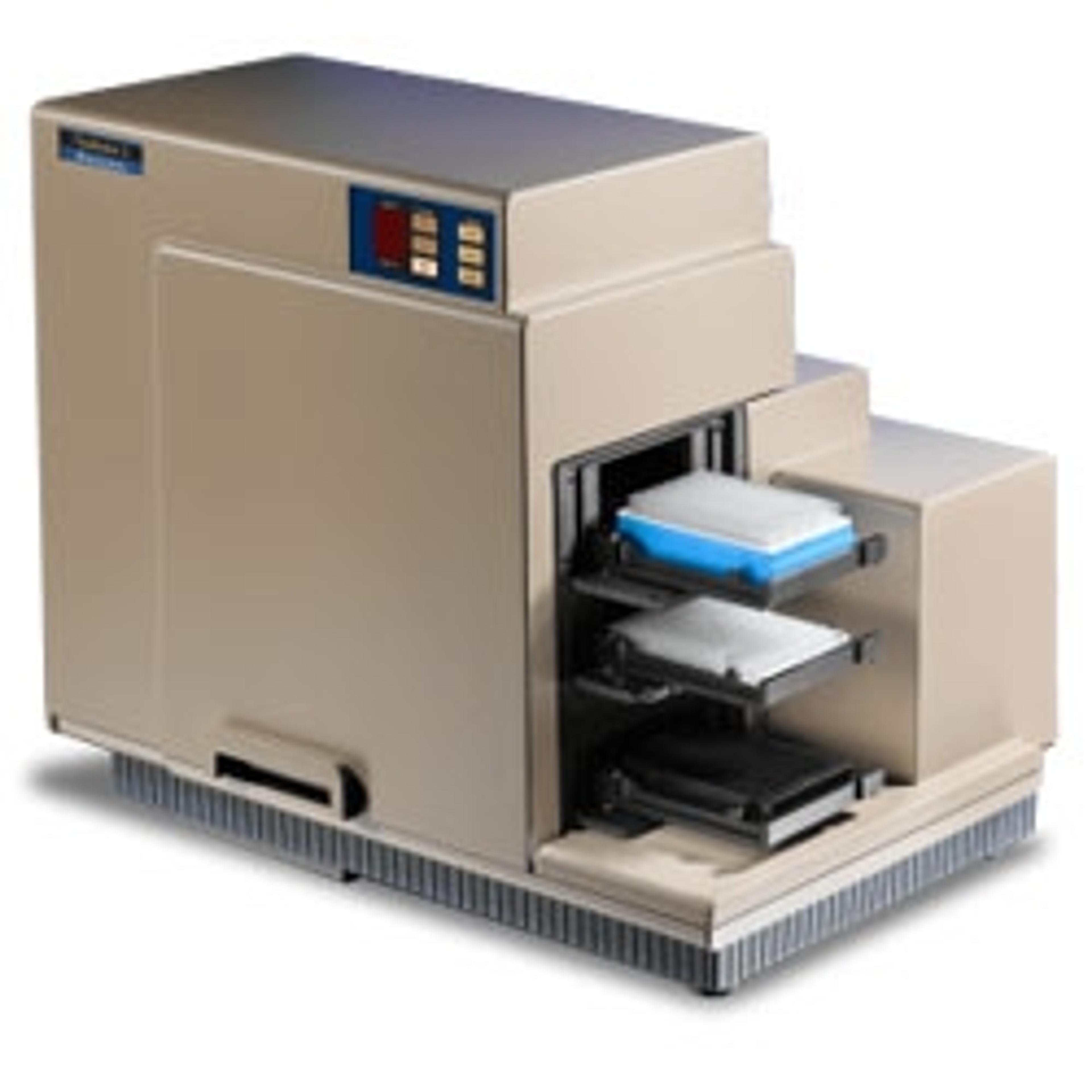
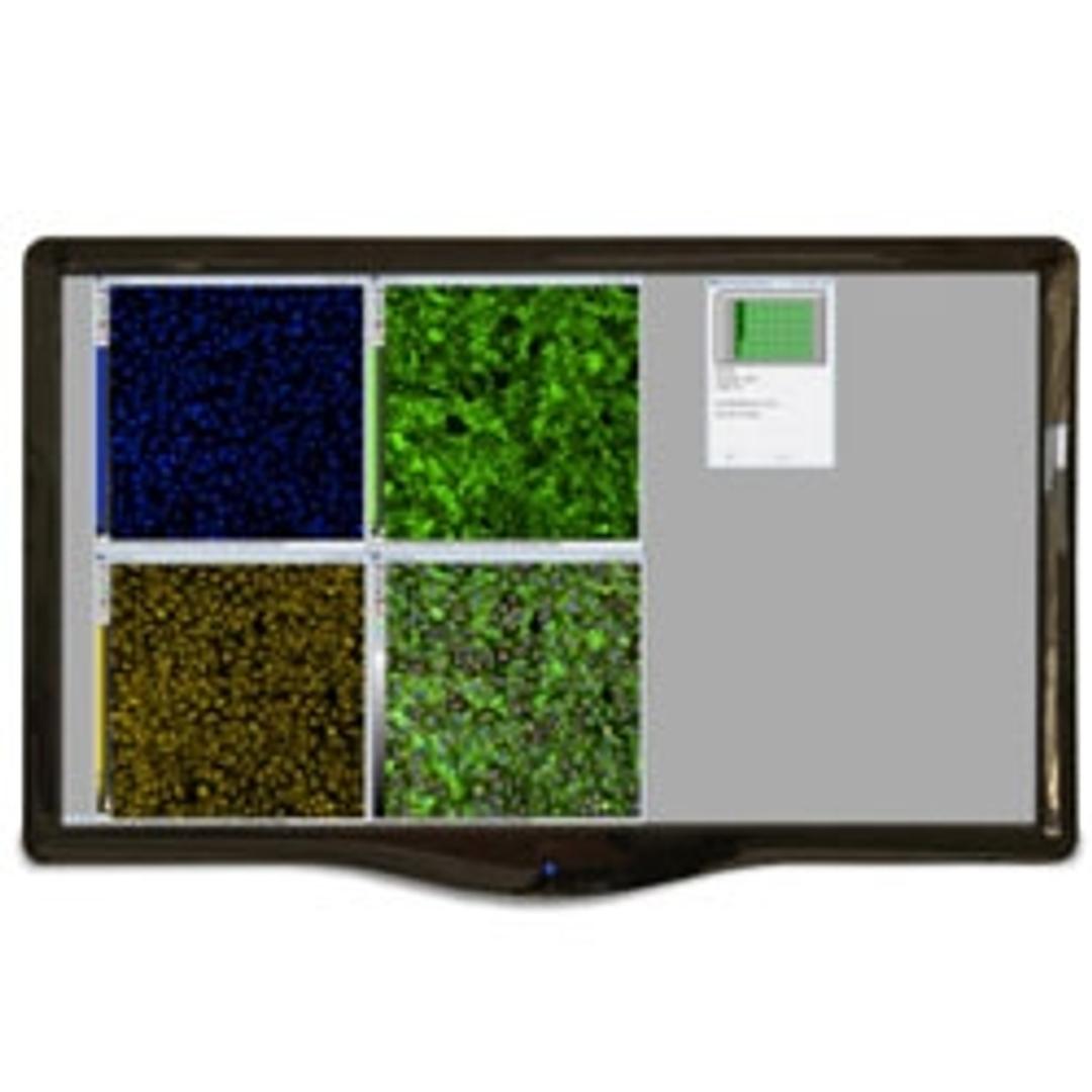
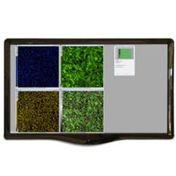
MetaXpress High-Content Image Acquisition and Analysis Software
High-content image analysis software featuring time lapse analysis

Receive your quote directly from the manufacturer.
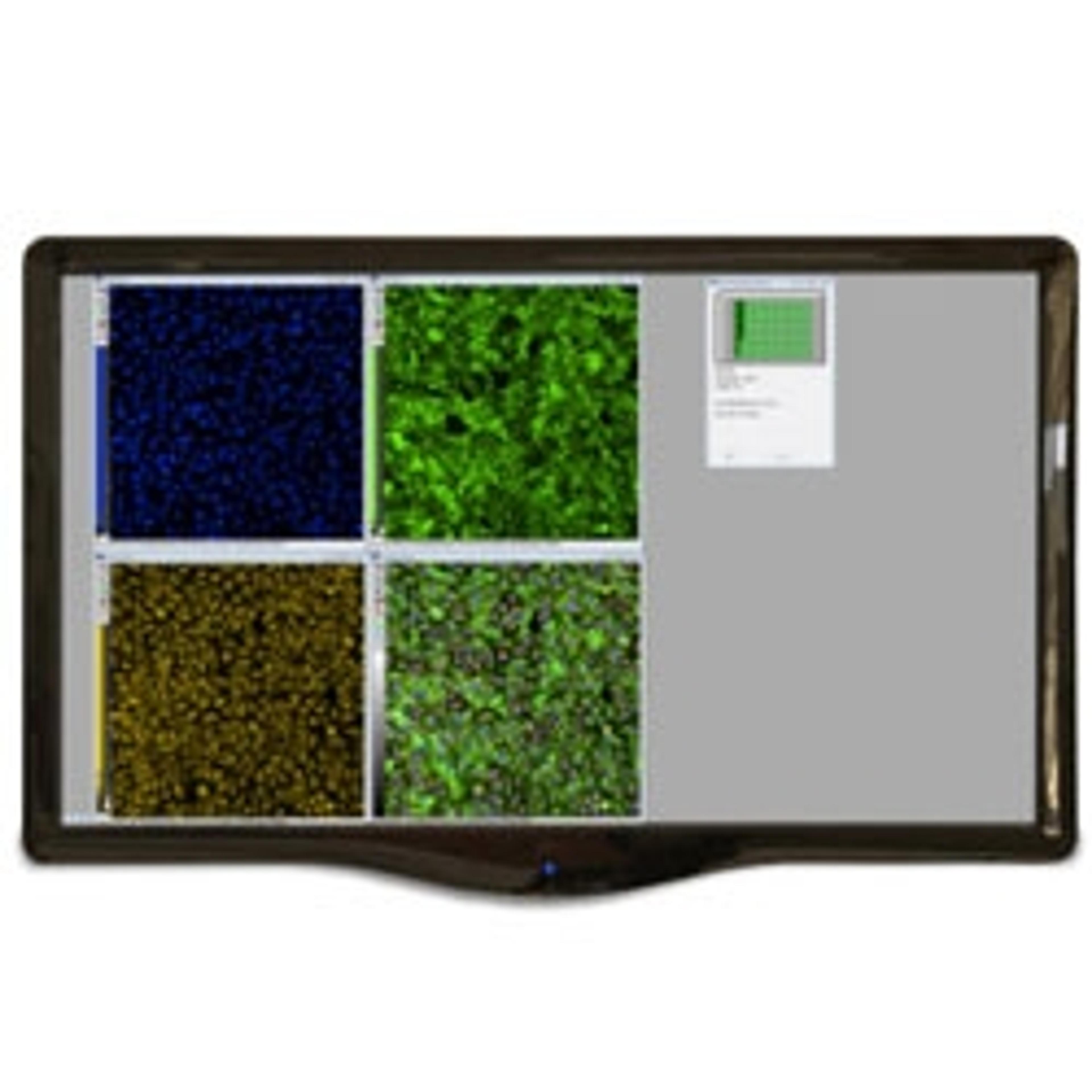
MetaXpress® High-Content Image Acquisition and Analysis Software is a comprehensive solution for high-content analysis featuring a tightly orchestrated and integrated workflow. The portfolio of application modules supports a range of needs from ease-of-use through to proprietary assay design. The software includes powerful and elegant tools for 2D and 3D imaging such as time lapse analysis. With the assistance of our acquisition setup wizard, you’ll be generating data in minutes.
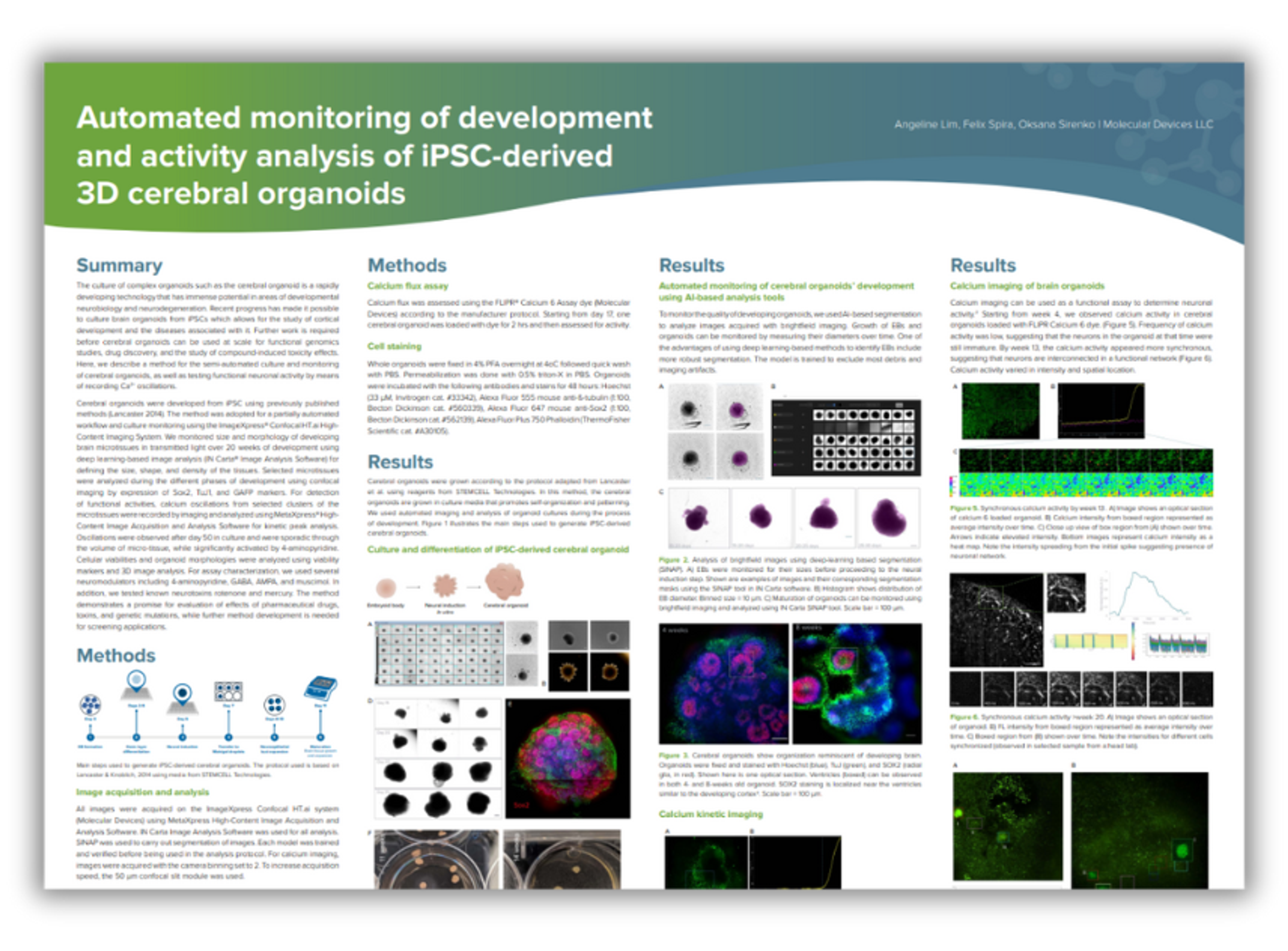
Automated monitoring of development and activity analysis of iPSC-derived 3D cerebral organoids
In this application note, Molecular Devices describes a method for semi-automated culturing and monitoring of cerebral organoids, as well as the testing of functional neuronal activity by means of recording Ca2+ oscillations.

High-throughput screening of 3D cell cultures with multiple high density scaffold-free spheroids for cancer toxicity studies
In this application note, Molecular Devices demonstrates the use of the Elplasia 96-well plates with a 3D culture workflow that includes spheroid generation, compound treatment, cytotoxicity assay, high-content imaging on the ImageXpress® Micro Confocal High-Content Imaging System, and 3D image analysis using MetaXpress® High-Content Image Acquisition and Analysis Software. The ability to easily generate multiple spheroids and to seamlessly integrate the workflow with high-content imaging promises to have significant applications in drug discovery and compound toxicology.
High-Content Assay for Morphological Characterization of 3D Neuronal Networks in A Microfluidic Platform
The focus of this application note is to develop a high-throughput 3D neurite outgrowth assay using iPSC-derived neurons developed in the microfluidic OrganoPlate® platform, with the goal of establishing 3D models for neurodegenerative diseases and neurotoxicology screens. The OrganoPlate® is a high-throughput platform that combines the most recent advances in 3D cell culture, Phaseguides™ and microfluidics. The OrganoPlate contains 96 tissue chips suitable for long-term culture of live cells, is amenable for screening purposes, and is compatible with standard laboratory equipment or automated systems, such as the ImageXpress® Micro Confocal High-Content Imaging System.
Analyze Experimental Results Using Curve Fitting with MetaXpress 6.5 Software
The new Curve Fitting Algorithms in the MetaXpress® 6.5 (MX 6.5) HighContent Image Acquisition and Analysis Software were designed to facilitate data visualization and the secondary analysis of data. The software allows you to generate a curve of best fit to a series of data points. With multiple functions, including 4-parameter logistic, and multiple curve outputs, such as EC50 and benchmark response, an array of graphs and curves can be generated to best represent data. Here, we show how to generate doseresponse relationship for defining the EC50 values of compounds using a toxicity assay as an example.
3D Analysis and Morphometric Characterization of Compound Effects on Cancer Spheroid Cultures
Cellular transformation/tumorigenicity assays using cultures of cells in semi-solid media (soft agar or Matrigel) has been well established for cancer research. The assay requires cells to grow in an anchorage independent manner, which is a hallmark of cancer cells. Compared with adherent cells grown in 2D monolayers, 3D growth conditions are believed to more accurately reflect the natural environment of cancer cells and span the gap between 2D cultures and animal studies. Importantly, 3D assays correlated better with tumorigenicity in animals, e.g. mouse xenografts. The growing interest in developing methods for personalized medicine, in which tumor cells from individual patients are tested for sensitivityto a panel of drugs , has led to a need for more relevant, time-sensitive studies.
High-Content 3D Toxicity Assay Using iPSC-Derived Hepatocyte Spheroids
There is increasing interest in exploring the use of three-dimensional (3D) spheroids for modeling developmental and tissue biology with the goal of accelerating translational research in these areas. As a result, the development of higher throughput quantitative assays using 3D cultures is an active area of investigation. In this study, we developed and optimized methods for the formation of 3D liver spheroids derived from human iPS cells as well as confocal imaging and analysis methods for toxicity assessment.
Increase Sensitivity in No-Wash Assays Using Confocal Imaging
Screening of hybridoma supernatants for antibodies directed against cell-surface antigens is an important step in the discovery and development of antibodies for vaccines or therapeutic use. The sensitivity of these assays is increased by taking advantage of the optical properties of confocal imaging such as utilizing the narrow depth of field to acquire high signal-to-background, crisp images with no out of focus light creating image blur.
Counting Cells with the Transmitted Light Analysis Module in MetaXpress
Label-free cellular assays are required for a multitude of biological applications that monitor the cell number, proliferation, health, confluency, and cytotoxicity. These applications necessitate efficient and robust transmitted light (TL) imaging and analysis capabilities providing precise segmentation for quantitation of cells and assessment in variety of cell responses and morphologies. Additionally, the coupling of high-contrast transmitted light imaging and fluorescent labeling and imaging is essential to numerous cellbased assays.
Apoptosis Detection Using EarlyTox Capase-3/7-D NucView 488 Assay Kit on ImageXpress Micro Systems
Apoptosis is an important mechanism signaling programmed death of cells in normal processes such as embryonic development, as well as in diseases including cancer and neurodegenerative conditions. This flexible assay can be performed using an ImageXpress® Micro High Content Imaging System and MetaXpress® Analysis Software for calculating the incidence of apoptotic cells in a well.