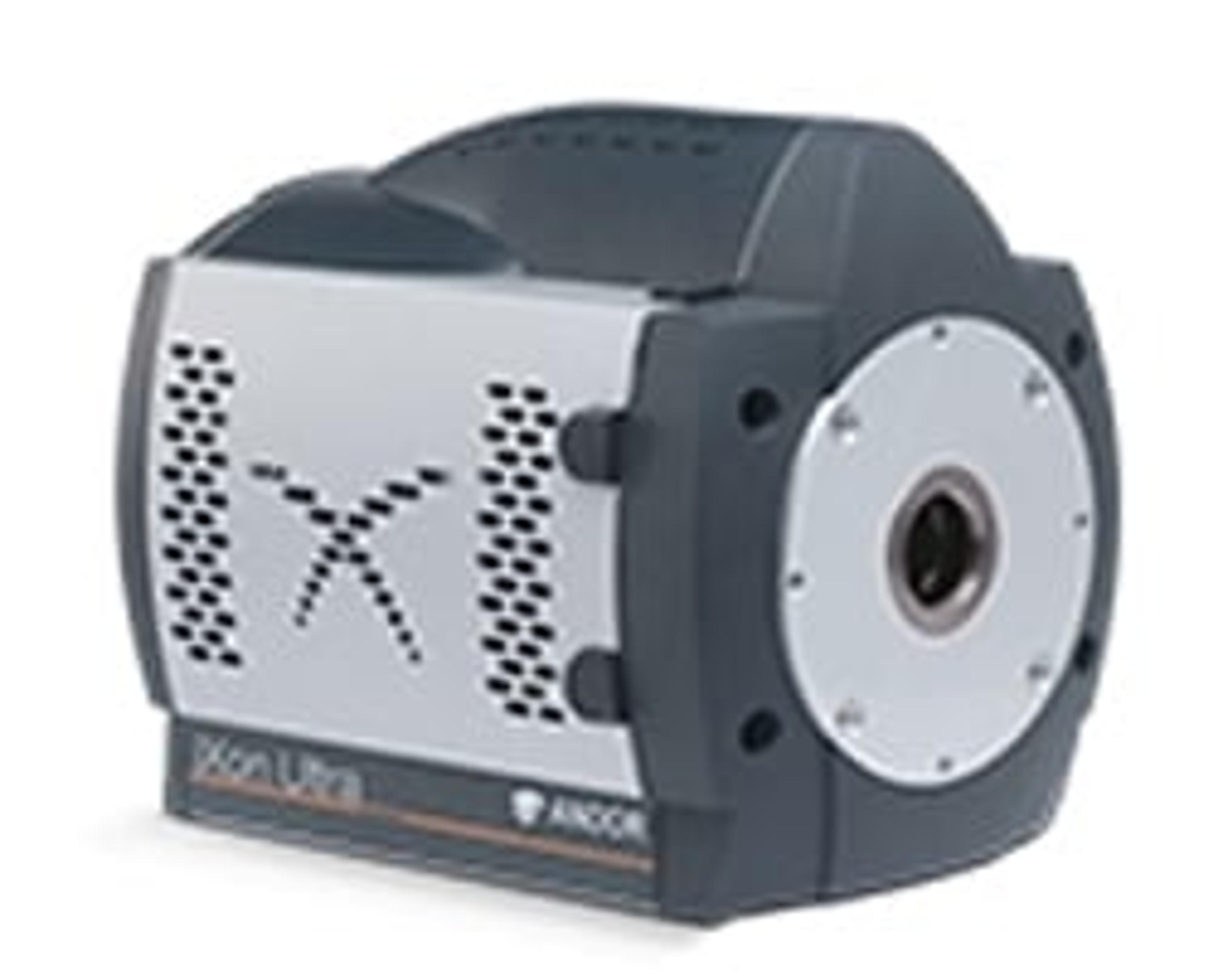
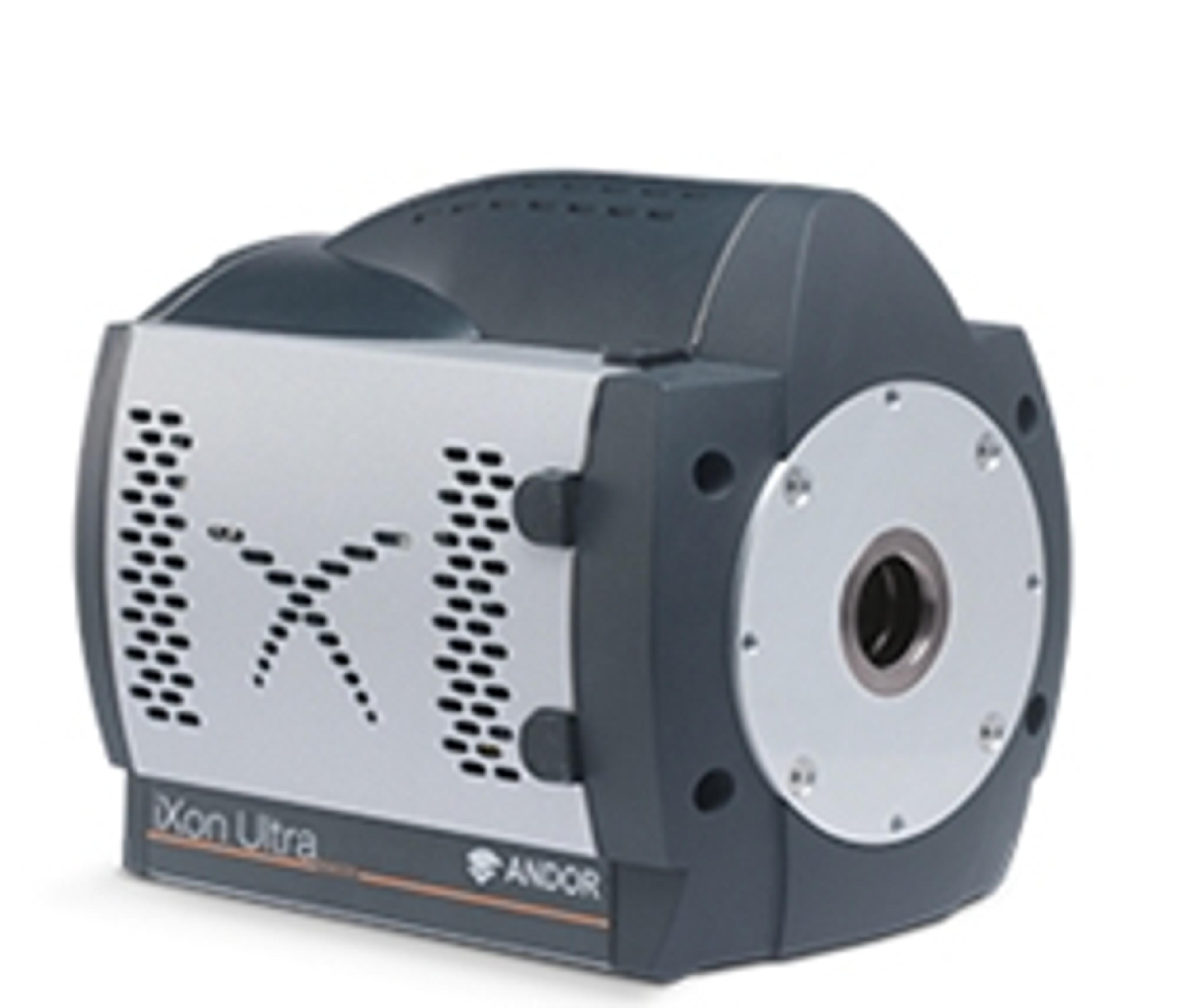
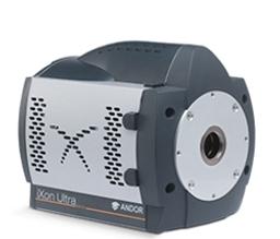
iXon Ultra 888 EMCCD Camera
The highly innovative iXon Ultra 888 is the world’s fastest Megapixel, Back-illuminated EMCCD camera, offering exceptional frame rates and single photon sensitivity across a large field of view. Building on a rich history of first to market innovation, the ‘supercharged’ iXon Ultra 888, represents a massive performance boost for the largest available EMCCD sensor, as well as the first USB3 enabled EMCCD camera. The iXon Ultr…


The supplier does not provide quotations for this product through SelectScience. You can search for similar products in our Product Directory.
Great image quality as always. USB 3 is super convenient. Pricey but worth it.
Fluorescence microscopy
USB 3 is very easy to use. SDK is also very well designed. My LabView code for the previous generation of iXon was easily converted to be used with the new camera. Image quality is great as before.
Review Date: 18 Aug 2017 | Oxford Instruments Andor
Overall the EMCCD camera is an excellent product.
Imaging Bragg scattered signal from periodically density modulated Bose-Einsten condensates
Service is excellent. So far the camera is working well as advertised with high reproducibility.
Review Date: 7 Nov 2016 | Oxford Instruments Andor
The highly innovative iXon Ultra 888 is the world’s fastest Megapixel, Back-illuminated EMCCD camera, offering exceptional frame rates and single photon sensitivity across a large field of view.
Building on a rich history of first to market innovation, the ‘supercharged’ iXon Ultra 888, represents a massive performance boost for the largest available EMCCD sensor, as well as the first USB3 enabled EMCCD camera.
The iXon Ultra 888 has been fundamentally re-engineered to facilitate 3x overclocking of the pixel readout speed to an unprecedented 30 MHz, whilst maintaining quantitative stability, accelerating the full frame rate performance to video rate. Furthermore, Andor’s unique ‘Crop Mode’ can be employed to further boost frame rates from a user defined sub-region, for example pushing a 512 x 512 sub-array to 93 fps and a 128 x 128 area to 697 fps.
With a 1024 x 1024 sensor format and 13 µm pixel size, the resolving power, field of view and unparalleled speed of the iXon Ultra 888 render it the most attractive and versatile EMCCD option for demanding applications such as single molecule detection, super-resolution microscopy, live cell imaging and high time resolution astronomy.
Features:
- 30 MHz readout delivering 26 fps at 1024 x 1024
- > 2.6x larger Field of View than ‘897’ model
- Optically Centered Crop Mode – Live Cell Super Resolution at 697fps
- Single Photon Sensitive
- EX2 Technology for wider QE response
- TE Cooling to -95°C

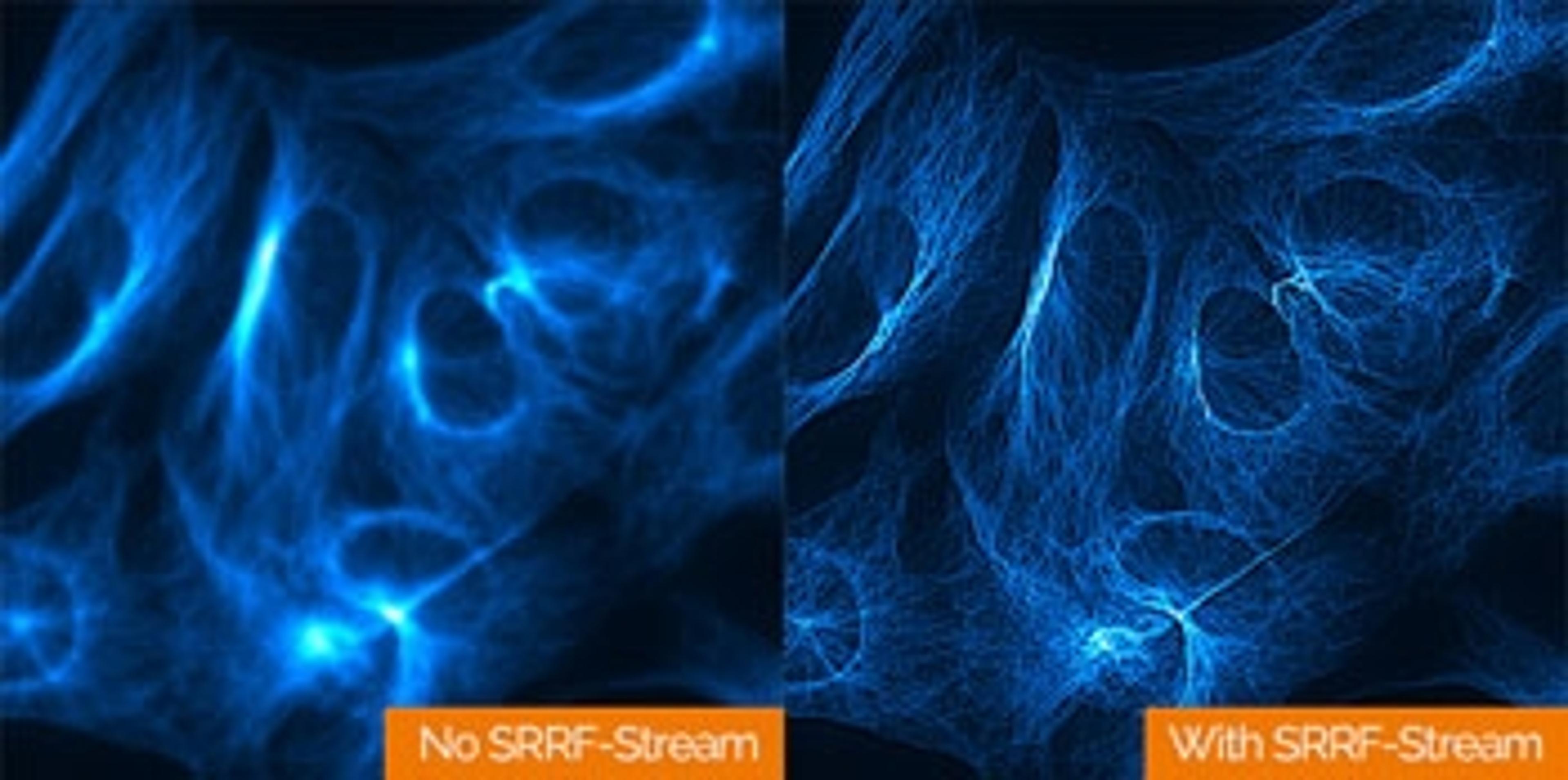
Discussing the Pros and Cons in Super-Resolution Imaging Techniques
Super-resolution microscopy™, also called nasoscopy, allows for observation of fluorescent samples at resolutions below the limit the diffraction of light imposes on any optical microscope. This limit, defined by Abbe, is at best ~200 nm laterally and ~500 nm axially, which is well above the resolution necessary to discriminate between different single molecules, or even an ensemble of molecules within a cellular compartment. Super-resolution approaches, by breaking this diffraction barrier, are therefore extremely helpful for the visualization of structural organization and the quantification of dynamic processes down to the molecular level. Applications include the study of membrane nanostructure, protein aggregation, nuclear machinery, molecular architecture of cell-cell interface, and synaptic transmission.
Discussing Pros and Cons in Super-Resolution Imaging Techniques
Super-resolution microscopy™, also called nasoscopy, allows for observation of fluorescent samples at resolutions below the limit the diffraction of light imposes on any optical microscope. This limit, defined by Abbe, is at best ~200 nm laterally and ~500 nm axially, which is well above the resolution necessary to discriminate between different single molecules, or even ensemble of molecules within a cellular compartment. Super-resolution approaches, by breaking this diffraction barrier, are therefore extremely helpful for the visualization of structural organization and the quantification of dynamic processes down to the molecular level. Applications include the study of membrane nanostructure, protein aggregation, nuclear machinery, molecular architecture of cell-cell interface, and synaptic transmission.
Observing Giant Atoms Towards Optically Imaging Rydberg Atoms with Single Particle Sensitivity with an EMCCD Camera
Considerable effort has been directed at observing the dynamics of a many-body quantum system with single particle resolution and sensitivity which would allow the realization of quantum simulators able to address fundamental questions about quantum and classical physics. Because of their exceptionally large dipole moments associated with their large size and long-range interactions, highly-excited (Rydberg) atoms in ultracold gases could be used to investigate quantum non-linear optical effects and dipole-mediated energy transport similar to the fundamental process in photosynthesis. This application note demonstrates a new approach to optically image Rydberg atoms based on a nondestructive and state-selective technique.
Andor launches Balor-X Camera for X-ray and Neutron imaging
Boasting a unique combination of 16.9 Megapixel sensor with rapid 54 fps full frame, ultra-low noise and high dynamic range, Balor is a versatile tool for high energy physics applications