HALO AI
Neural network driven tissue and cell classification for research & routine pathology

HALO AI classification of breast cancer H&E


Currently the best image viewer and analysis software on the market that I've used.
Image analysis
Currently the best image viewer and analysis software on the market that I've used.
Review Date: 8 Jun 2022
wonderful products, it is far beyond our expectations.
analyze tumor sections
Our lab both have the Halo Image Analysis System and the Halo AI, honestly speaking, the Halo system is beyond our expectations, especially the analysis process, annotation tools, the adjustable parameters, the texture and morphological recognition of nuclear and specific structure, and the after-sale care I strongly recommend you guys to choose it. it really worth the money.
Review Date: 7 Jun 2022
Great results, can't live without this instrument!
Analysis in tissue figure
Scientists can achieve lots of high-quality analysis data about figures by HALO software. Many functions of HALO are very helpful to study images, such as TMA, spatial analysis, high-Plex FL, and especially HALO AI. It is easy to operate. Its interface is simple, friendly, and convenient. Once we have problems, a Halo technician is able to help us to solve them in time.
Review Date: 1 Jun 2022
Great result. Nice tool for translational pathology research.
RNAscope and IHC analysis
We are interested in applying HALO AI in RNAscope and IHC analysis. Thanks to Yongtian ZHAO to give us wonderful training and support. We are improving and confident to apply it in the RNAscope assay development and scoring.
Review Date: 1 Jun 2022
Great
Halo for image research
Quick to use
Review Date: 1 Jun 2022
HALO AI is a collection of train-by-example classification and segmentation tools underpinned by advanced deep learning neural network algorithms. HALO AI classifiers can be trained to quantify tissue classes, to segment tissue classes for analysis with other HALO image analysis modules, to find rare events or cells in tissues, and to categorize cell populations into specific phenotypes.
SIMPLE & INTUITIVE WORKFLOW
HALO AI is fully integrated with the intuitive, easy-to-use HALO and HALO Link viewers and employs a simple three-step workflow. After defining what tissue classes or cell phenotypes you would like to segment, you train the neural network by drawing annotations – no computer programming or AI knowledge required. Trained classifiers can be applied to segment tissue and cells on any whole slide image or region of interest.
POWERFUL TISSUE SEGMENTATION
HALO AI now includes the option of three powerful neural networks – VGG, DenseNet and MiniNet. VGG, a well-known and more traditional network, was used to build the Indica Labs submission in the CAMELYON17 challenge and was the first neural network integrated with HALO AI. DenseNet is a more modern network capable of creating more robust classifiers at higher resolution compared to VGG. MiniNet, a custom network developed at Indica Labs, is more shallow than VGG or DenseNet, but can produce a solution quickly with limited training data and is therefore useful for testing new AI applications.
EXCEPTIONAL CELL CLASSIFICATION
Segment nuclei with the new Segmentation classifier. Utilize HALO AI’s pretrained networks for H&E, single IHC, or DAPI stained images for an out of the box solution. Or train your own nuclei segmentation network for a specific application (unique tissue or advanced staining protocols). Once nuclei are segmented, take it a step further using the Nuclei Phenotyper classifier to automatically assign cells into user defined phenotypes with a few quick training examples.

Introduction of a robust workflow for the whole-slide acquisition and co-registration of multiplex immunofluorescence tissue images
In this application note, explore how Indica Labs designed and tested an 8-plex immunophenotyping panel using the InSituPlex® approach to detect and classify T cells, macrophages, and tumor cell in non-small cell lung cancer and colorectal cancer FFPE tissue.
High impact publications with the HALO AI deep learning classifier add-on
HALO AI, the Deep Learning Classifier Add-on to the HALO® image analysis platform continues to advance research areas as diverse as infectious disease, metabolism, gastric pathology, neuroscience, myology, and oncology and immuno-oncology. In this application note, Indica Labs lists recent HALO AI publications according to research area and highlights which HALO AI network was used.
Depicting the cellular architecture of the tumor microenvironment by integrating hyperplex immunofluorescence and automated image analysis
The tumor microenvironment (TME) is emerging as an important factor that shapes the dynamic of tumor growth, heterogeneity, and response to therapies. In this application eBook, Indica Labs focuses on the phenotyping of cells across different tumor types on a tissue microarray (TMA) with an immuno-oncology panel encompassing 20 biomarkers. They interrogated their TME with the use of the COMET™ automated staining and imaging system, and HALO® and HALO AI image analysis platforms.
The power of artificial intelligence in the hands of the pathologists
In this white paper, Indica Labs demystifies AI with a bit of background, explains its applications in digital pathology and discusses how the HALO AI™ platform is making this powerful technology accessible to the pathology community.
RNAscope image analysis using HALO and HALO AI
In this application note, discover how the ISH module and FISH module available with the HALO® image analysis platform can be employed with HALO AI™ to quantitatively assess chromogenic and fluorescence RNAscope assays, respectively.
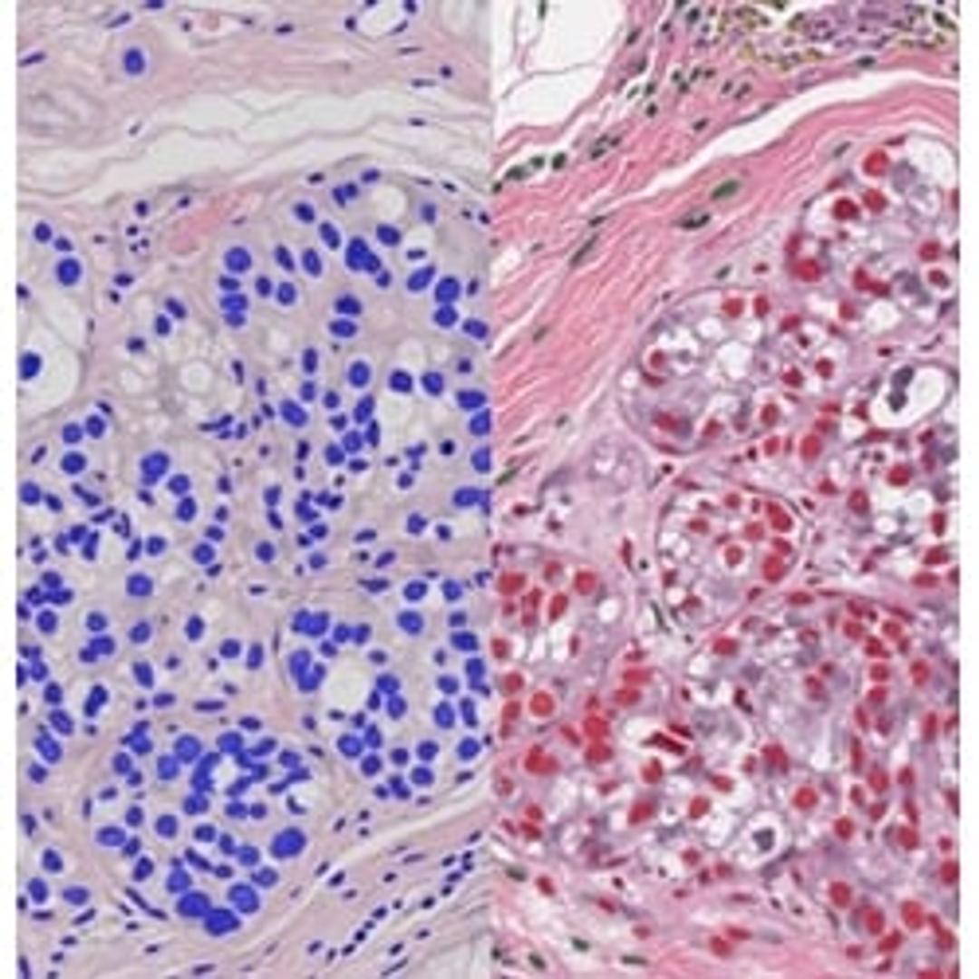
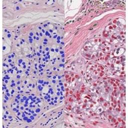
Quantification and characterization of glomeruli across diverse stains using HALO AI™
In this application note, Indica Labs highlights the potential for HALO® and HALO AI to quantify biomarkers, gene expression, and morphological changes in kidney disease, during development and in toxicological studies.
Qualitative and quantitative evaluation of the tissue microenvironment by high-resolution 17-plex immunofluorescence reveals distinct populations
In this application note, Indica Labs highlights how the use of Orion imaging combined with HALO image analysis provides a powerful and intuitive workflow for visualization and quantification of distinct microenvironment populations.
Sequential same slide multiplex immunofluorescence and H&E staining
In this application note, Indica Labs demonstrate a tissue-preserving workflow to generate H&E images from a slide that is previous stained and imaged in fluorescence on the CyteFinder® II HT Instrument.
High-impact publications with the HALO image analysis platform
Digital pathology with the HALO® image analysis platform continues to advance research areas as diverse as immunology, infectious disease, immuno-oncology, and neuroscience. Download the white paper below to see the caliber of publications that researchers are obtaining, using Indica's cloud-based software.
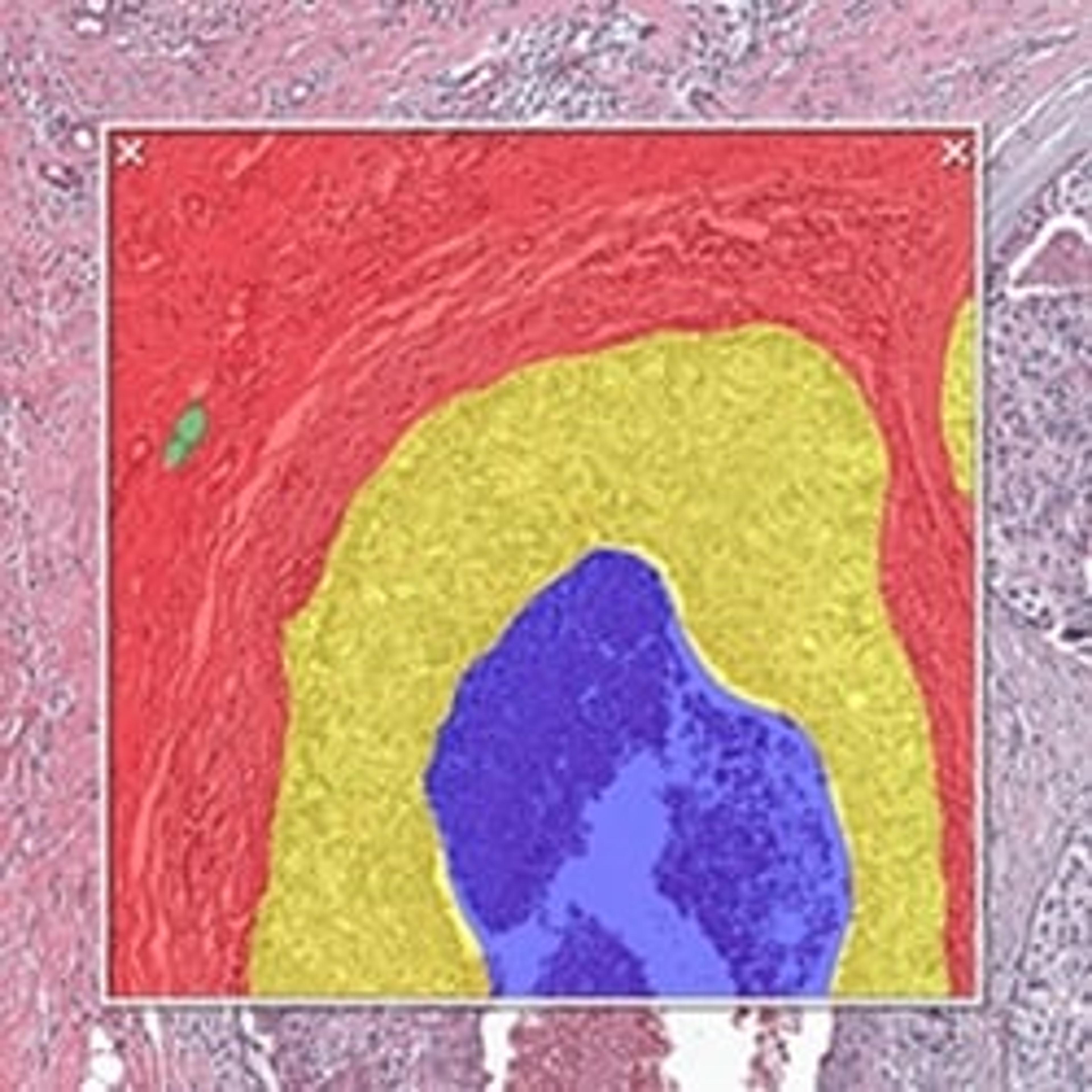
Robust identification of islets with variable morphology in H&E-stained pancreatic tissue using HALO AITM
In this application note, Indica Labs trains the HALO AITM VGG convolutional neural network (VGG-CNN) to identify islets within pancreatic tissue sections following H&E staining. It demonstrates how it is possible to build a robust classifier to accurately segment islets from surrounding exocrine tissue, irrespective of stain or morphological variability. This study highlights the potential for HALO AI to simplify the pathological evaluation of pancreatic tissue in metabolic research and toxicological pathology.
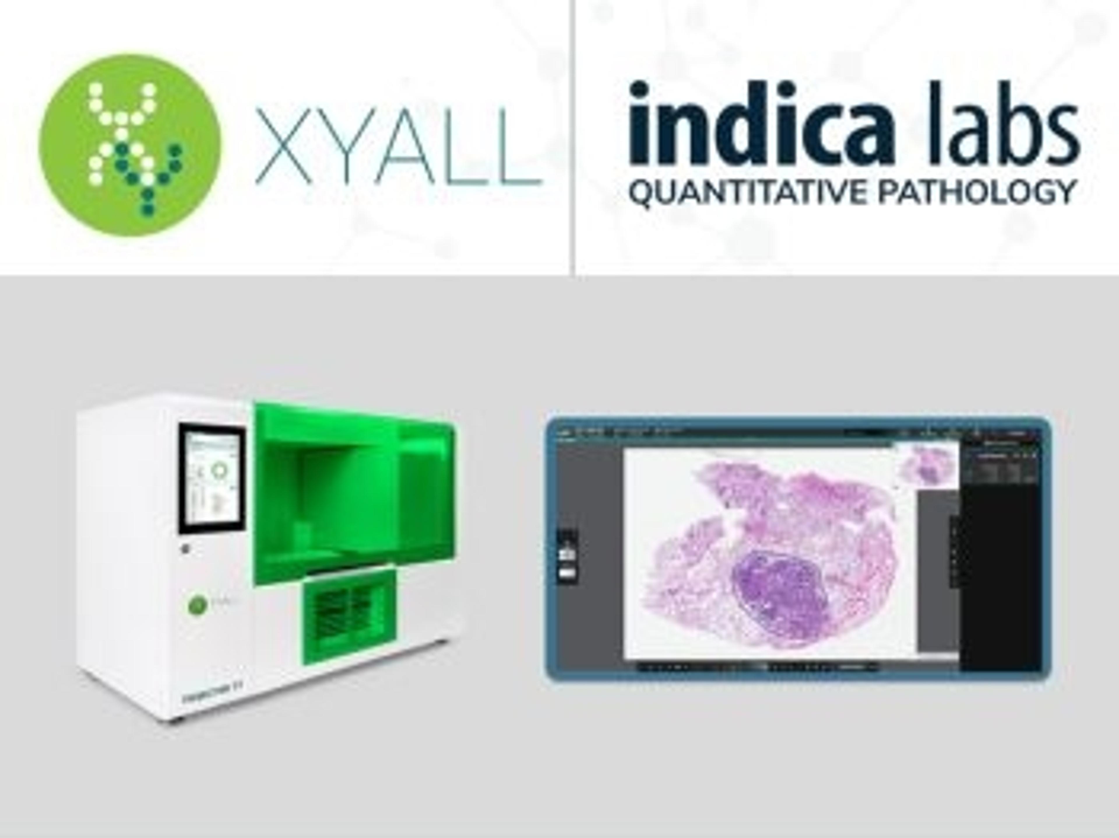
Xyall and Indica Labs forge global collaboration
The partnership is designed to bridge the gap between histopathology and molecular pathology
Indica Labs receives top honor in Albuquerque Journal ‘Flying 40’ list of top tech firms in New Mexico
The company received the award due to its staggering growth rate over the past few years
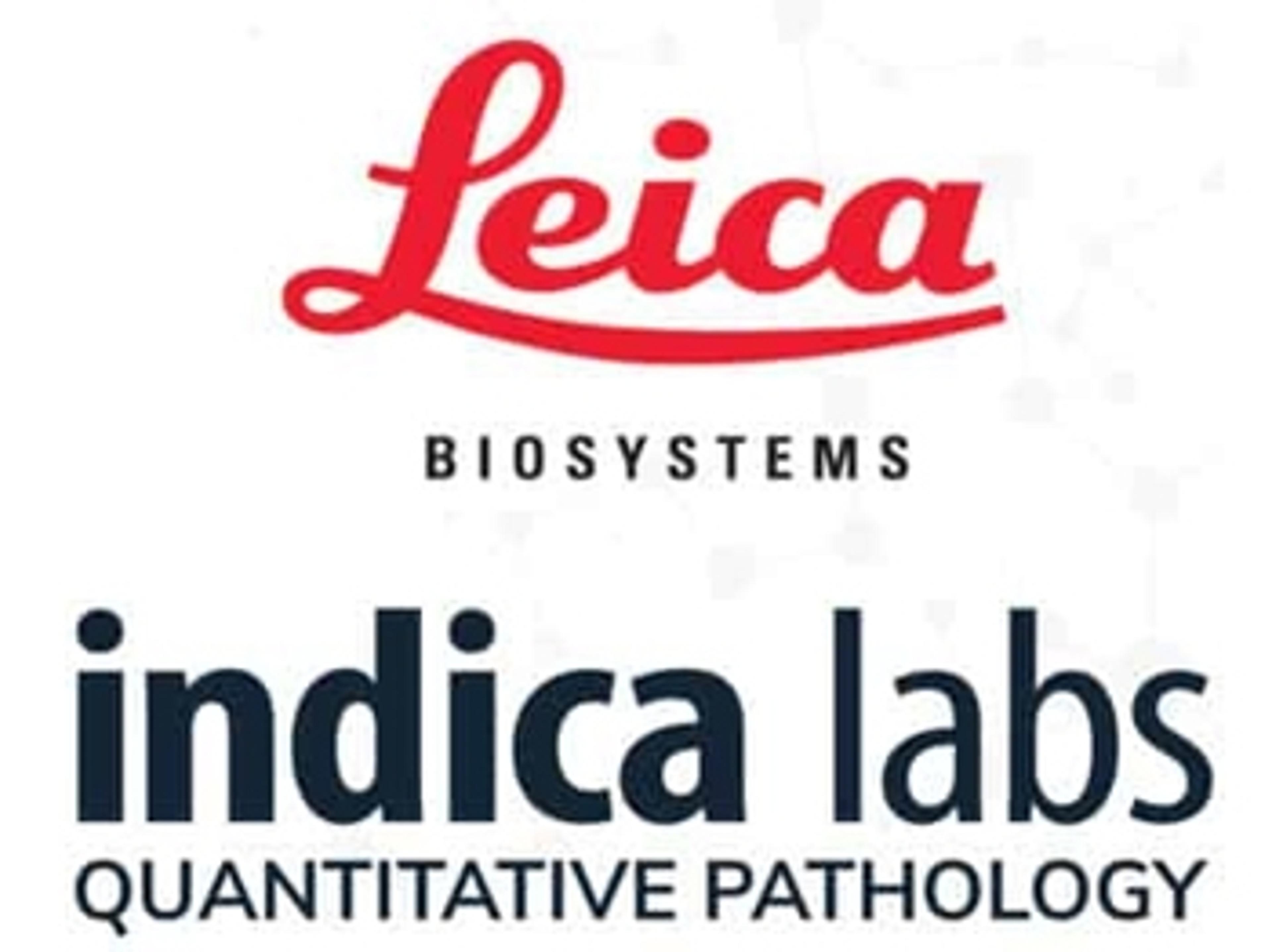
Leica Biosystems announces partnership with Indica Labs to deliver integrated digital pathology workflow solutions for mutual customers
The collaboration is focused on delivering compatible digital pathology workflow solutions

Indica Labs announces collaboration with The Industrial Centre for Artificial Intelligence Research in Digital Diagnostics (iCAIRD)
The collaboration aims to develop an AI-based algorithm for the automated reporting of lymph node status in colon cancer

Indica Labs announces launch of cloud-based digital pathology deployment at NCI
Indica Labs is pleased to announce the formal launch of its software within the National Cancer Institute (NCI)
Indica Labs and Ibex partner to deliver AI-powered clinical workflows for digital pathology
The seamless integration of AI into digital pathology workflows aims to improve quality and efficiency of cancer diagnosis