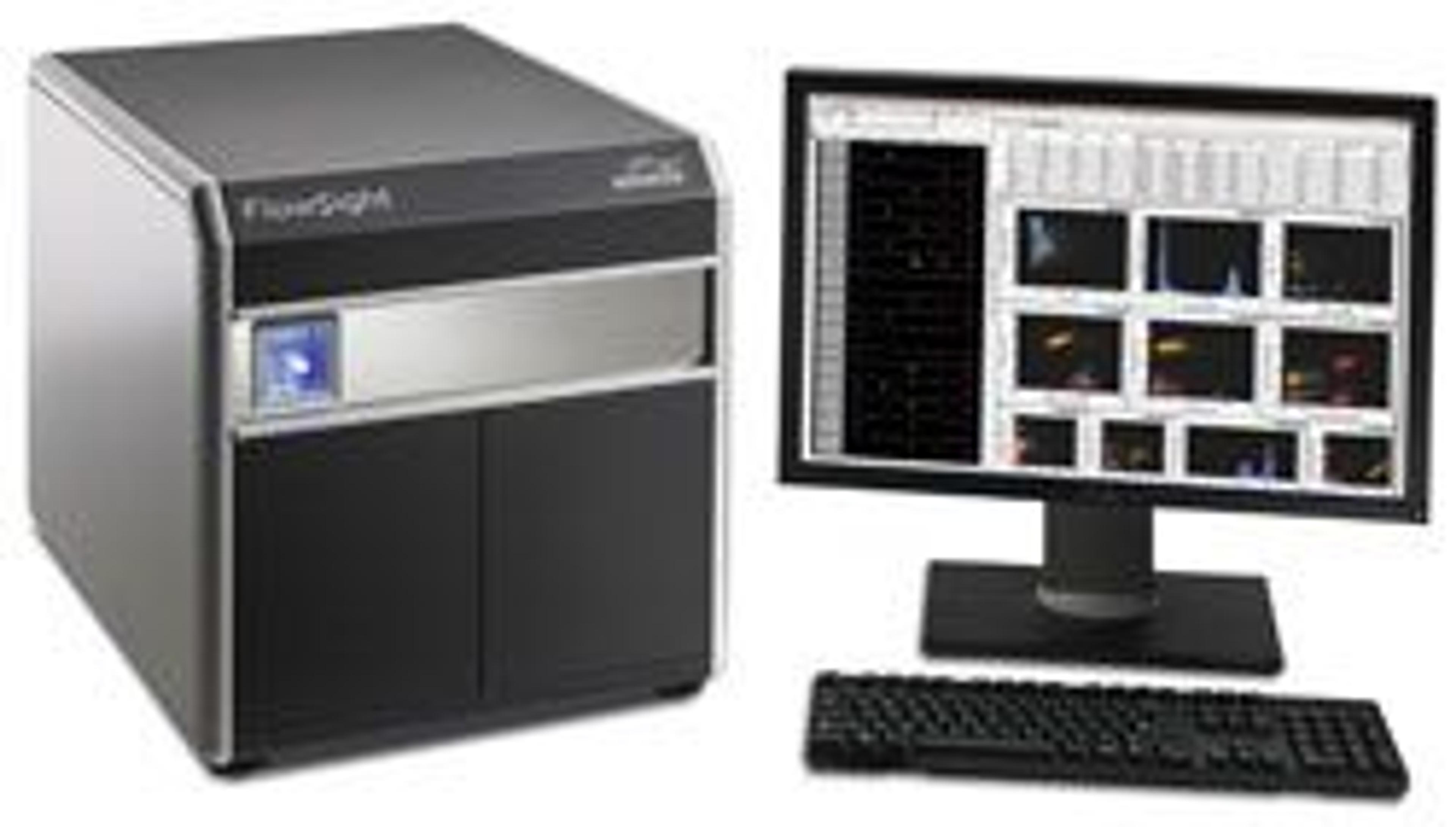
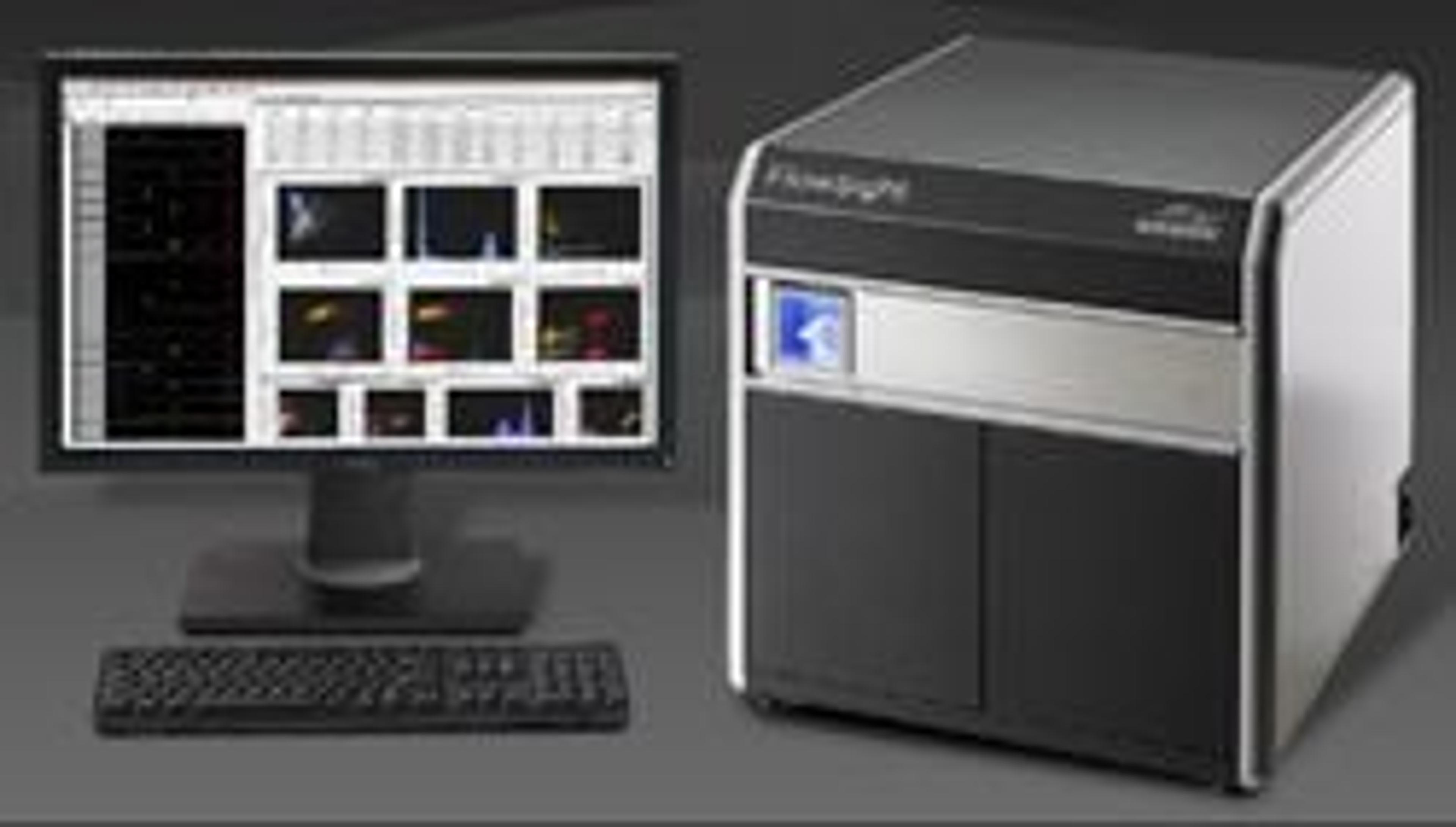
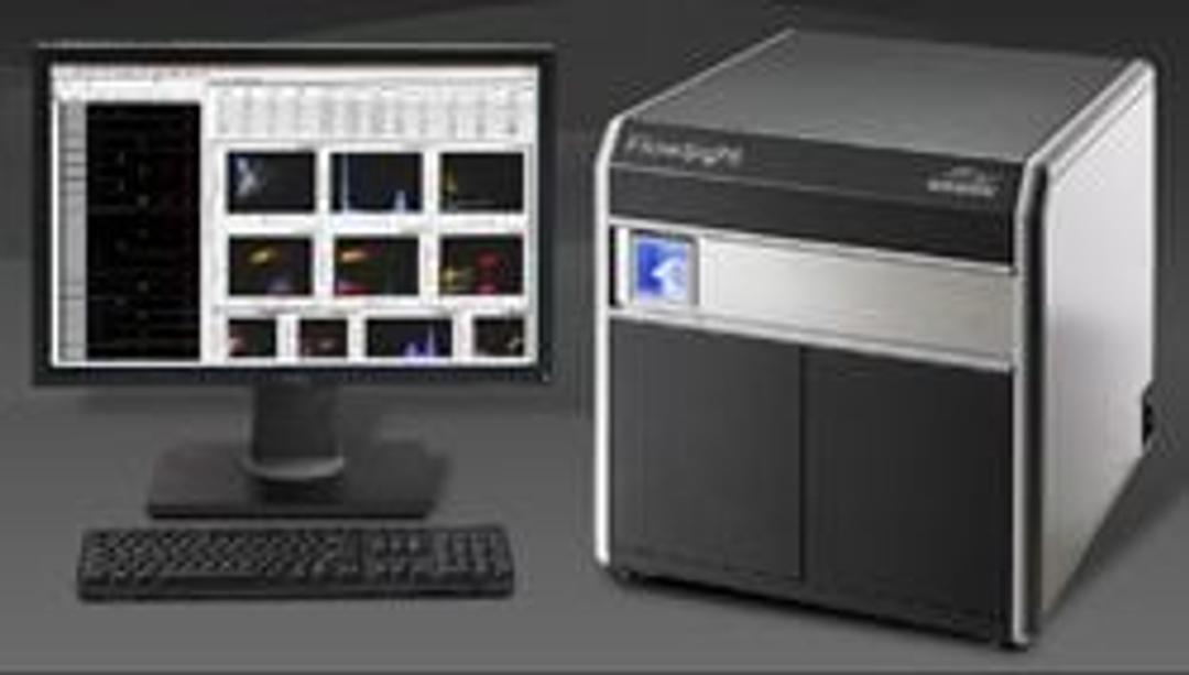
Flowsight® Imaging Flow Cytometer
The FlowSight flow cytometer offers high performance in a small package. Its innovative design increases signal and minimizes noise to provide unmatched fluorescence sensitivity. Twelve standard detection channels simultaneously produce brightfield, darkfield and up to ten channels of fluorescence imagery of every cell. With these unique capabilities, the FlowSight enables a broad range of applications. Intuitive: Easy-to-use,…


The supplier does not provide quotations for this product through SelectScience. You can search for similar products in our Product Directory.
Great for cellular immunology and signaling network
Translational Research, cellular immunology
It gives very high quality results with less amount of samples that is not possible with any other instrument.
Review Date: 3 Aug 2017 | Merck
The FlowSight flow cytometer offers high performance in a small package. Its innovative design increases signal and minimizes noise to provide unmatched fluorescence sensitivity. Twelve standard detection channels simultaneously produce brightfield, darkfield and up to ten channels of fluorescence imagery of every cell. With these unique capabilities, the FlowSight enables a broad range of applications.
Intuitive: Easy-to-use, with imagery for every cell:
The FlowSight operates like a conventional flow cytometer but also provides imagery of every cell. Powerful and intuitive analysis software seamlessly links quantitative data to imagery:
- Click on a dot in any plot to see the corresponding cell imagery.
- Click on a bin in any histogram to view all the cells in that bin.
- Draw gates on dot plots and view the resulting populations to validate results.
Affordable: Designed and priced for every lab:
The FlowSight is powerful enough for the core lab but sized and priced for any lab. The system can be factory configured or field upgraded with up to four excitation lasers (405, 488, 561, 642 nm), a 96-well plate AutoSampler, and a powerful quantitative image processing option. Whether in a base configuration or fully-optioned, the FlowSight sets a new standard of value.

The Immunology of Cancer
In this infographic, learn about the interactions between the immune system and cancer cells and how this understanding has led to breakthroughs in cancer vaccines and other novel immunotherapies. Detection reagents and instrument platforms have become increasingly sophisticated and sensitive for detection of key tumor antigens and associated immunology targets.
The Art and Science of Experimental Design with Fluorophores
Imaging cytometers are at the confluence of advances in flow cytometry and light microscopy. The ability to image tens of thousands of fluorescently labeled cells results in data that are both visually striking and highly quantitative. In this infographic, learn how Amnis® technology combines lasers and optics to accommodate hundreds of fluorophores and fluorescent reagents. Build an expert palette for your own memorable discoveries with imaging flow cytometry.
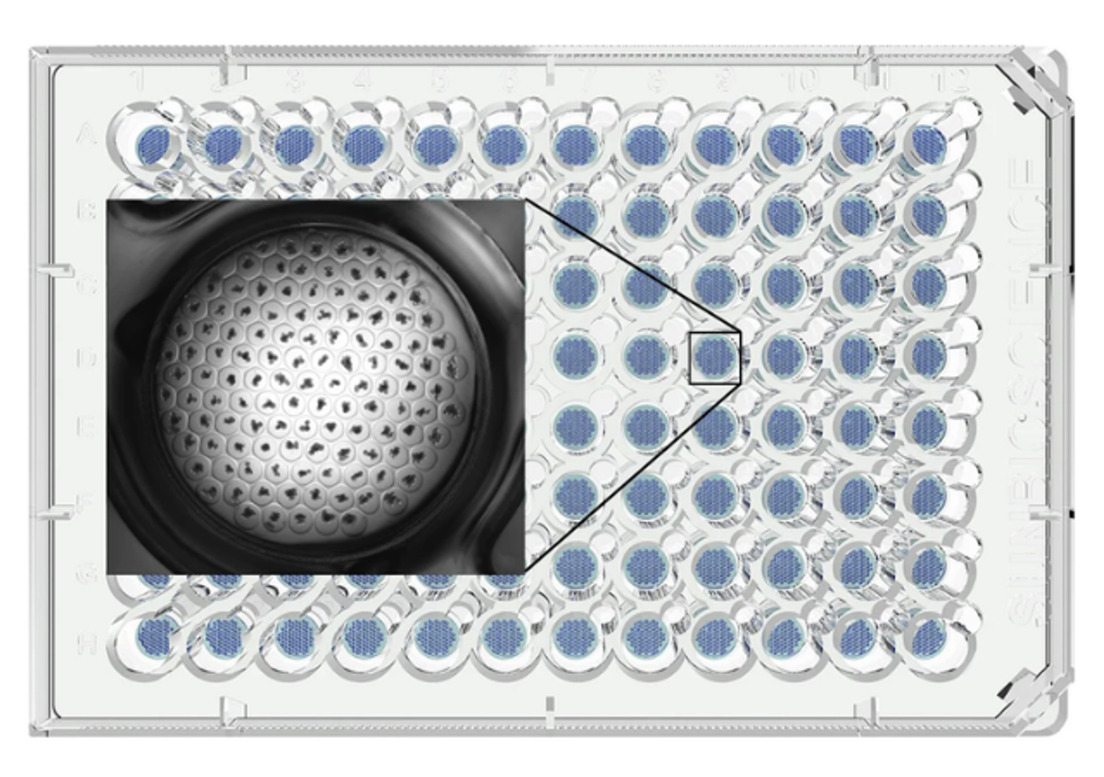
Measuring Cell Cycle and Mitotic Index with the FlowSight
The cell cycle is a series of orchestrated events that leads to cell replication. It is characterized by three interphase stages (Gap 1, S Phase, and Gap 2) and a dividing stage called mitosis. Mitosis is further divided into four primary phases: prophase, metaphase, anaphase and telophase. Progression of cells through the cell cycle is highly regulated through feedback loops that control cell growth via cyclin-dependent kinases (CDKs). Activation of CDKs mark the progression from one stage of the cell cycle to the next and therefore offer opportunities to intervene in cancer and other cell proliferative disorders. A detailed assessment of the cell cycle and mitosis are essential to the development of CDK-targeted therapies. This application note describes the use of the FlowSight for cell cycle analysis via the classical assessment of DNA content as well the calculation of the mitotic index via the assessment of DNA condensation in nuclear images.
EMD Millipore Products Enable Customer Success
Many EMD Millipore products are used by Brian McFarlin at the University of North Texas. Dr. McFarlin has maintained his relationship with EMD Millipore because of our innovative products, outstanding customer service, and because the technologies developed by EMD Millipore in just the last 5 years have accelerated his research as shown in this video.

MilliporeSigma Can Advance Neuroscience Research with Biomolecules and Supporting Technologies
Visit MilliporeSigma at Neuroscience 2016, San Diego, USA, November 12-16, booth 2813