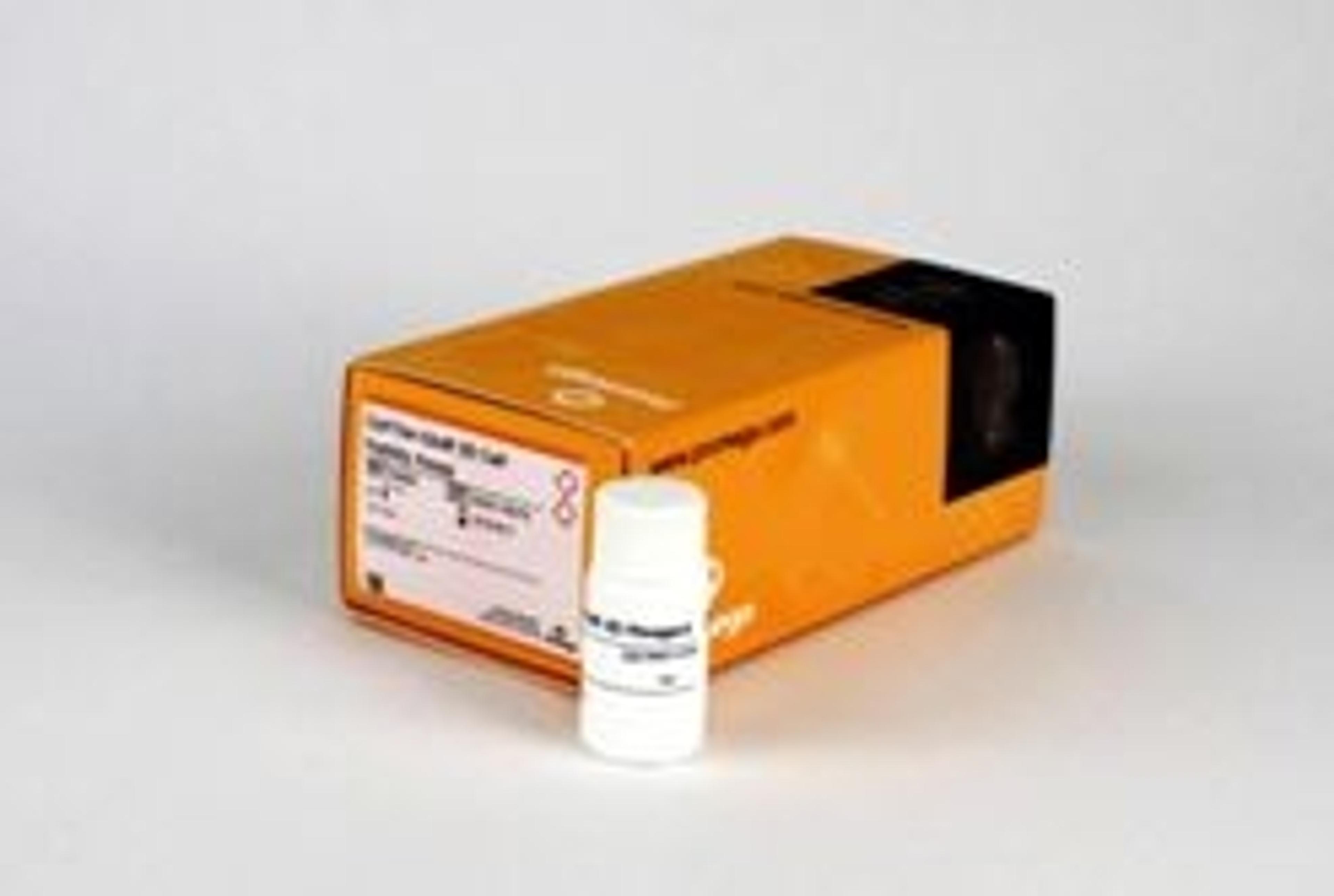
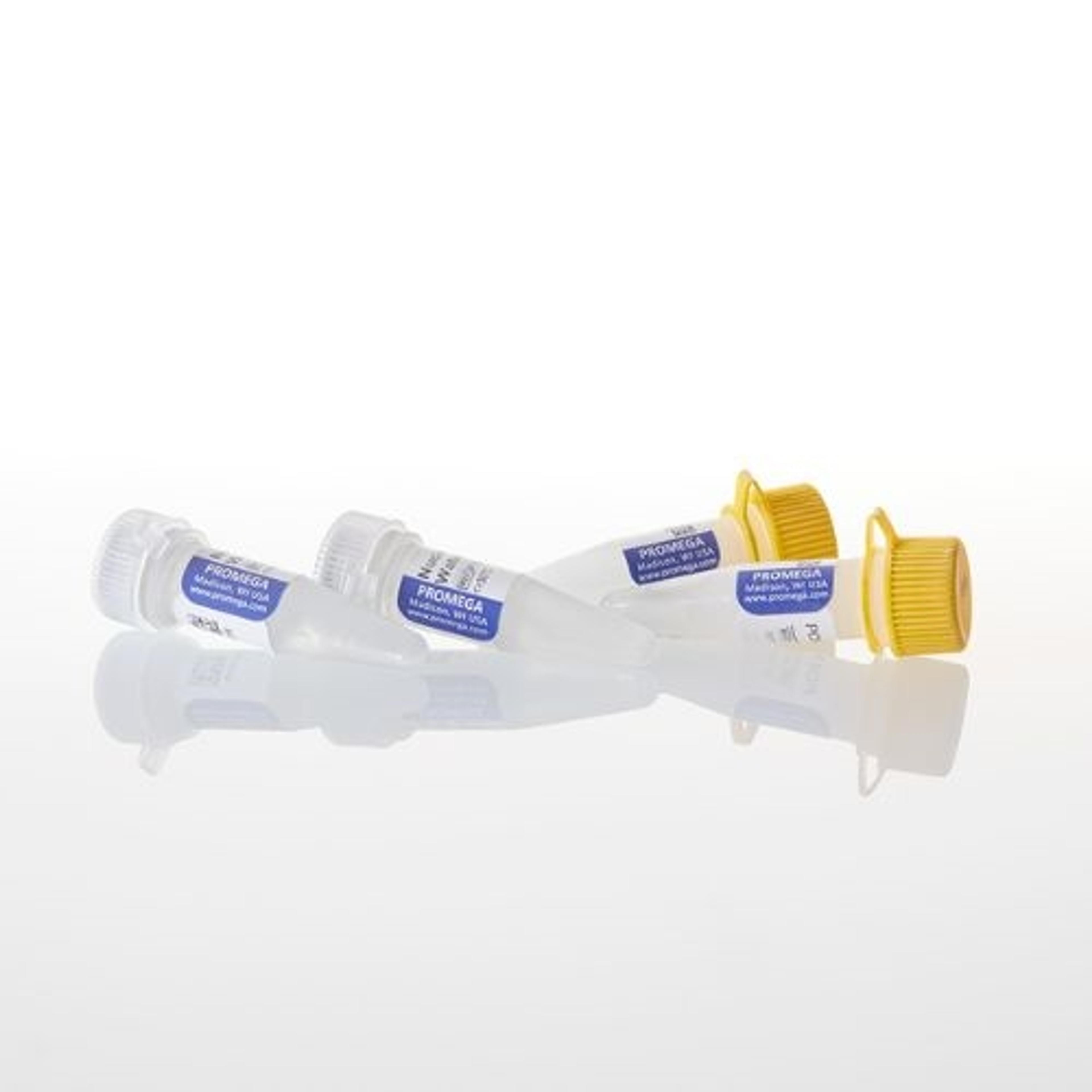
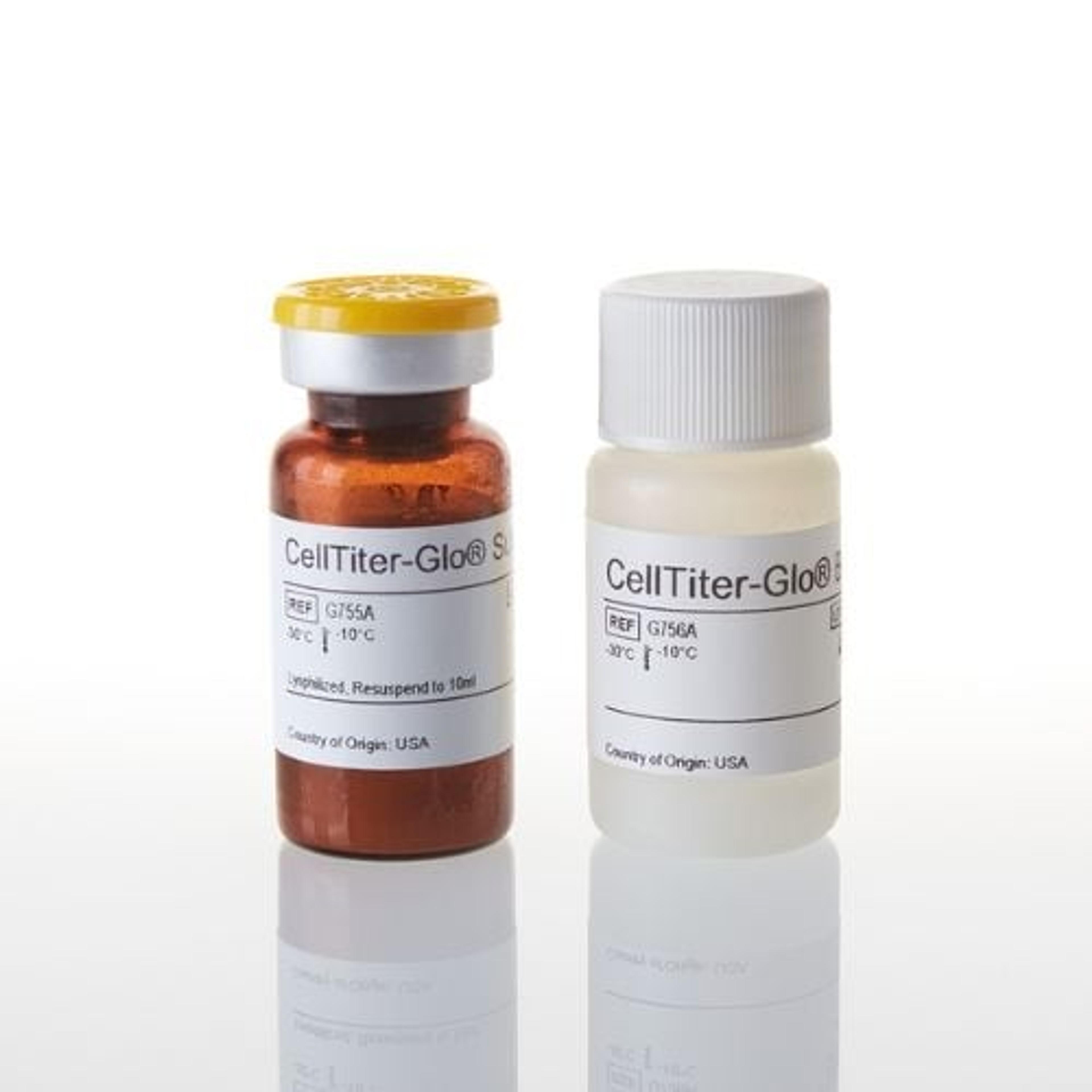
CellTiter-Glo® 3D Cell Viability Assay
The CellTiter-Glo® 3D Cell Viability Assay is a homogeneous method to determine the number of viable cells in 3D cell culture based on quantitation of the ATP present, which is a marker for the presence of metabolically active cells. This ready-to-use reagent is based on the original CellTiter-Glo® Luminescent Cell Viability Assay chemistry and eliminates the need to combine buffer with lyophilized substrate when prepa…


Receive your quote directly from the manufacturer.
Easy to use, consistent results.
Intestinal organoids
Extremely easy to handle, consistent, and reproducible results. Can prepare standard control when purchasing standard ATP.
Review Date: 25 Oct 2019 | Promega Corp.
Most used reagent in our lab currently.
Measure viability in 3D tumour assay
Easesy to use - defrost and add directly to the cells, leave for only 15mins and then read. Simple and reliable.
Review Date: 27 Jun 2019 | Promega Corp.
Highly recommended.
Cell viability
Easy to use, very robust, but expensive. Will recommend to others.
Review Date: 17 Apr 2018 | Promega Corp.
Excellent product, use it almost on a daily basis.
Spheroid and tumoroid toxicity.
CellTiter Glo 3D is very easy to use and works very efficiently, yielding high quality and reproducible results. When we moved to 3D cell culture technologies, there weren't many products available for efficient use in these systems, so we were very happy to be able to use the product early on.
Review Date: 22 Mar 2018 | Promega Corp.
Best cell assay for 3D cell culture!
Cell viability assessment of spheroids
CellTiterGlo3D is the only assay that gives reproducible results on spheroid cultures. The results are always very precise, so that one can detect even minor changes in cell viability, which is not detectable with other products.
Review Date: 22 Apr 2016 | Promega Corp.
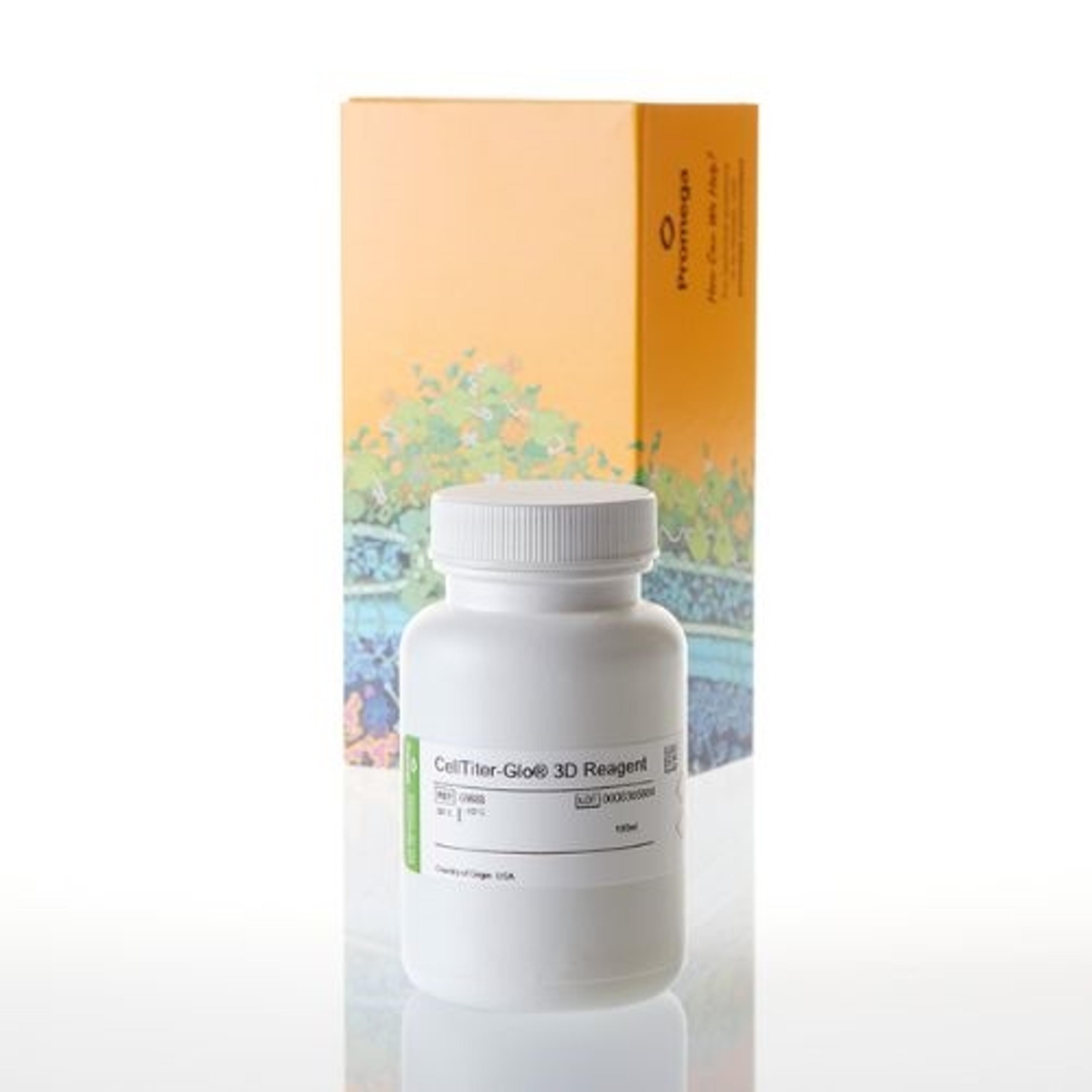
The CellTiter-Glo® 3D Cell Viability Assay is a homogeneous method to determine the number of viable cells in 3D cell culture based on quantitation of the ATP present, which is a marker for the presence of metabolically active cells.
This ready-to-use reagent is based on the original CellTiter-Glo® Luminescent Cell Viability Assay chemistry and eliminates the need to combine buffer with lyophilized substrate when preparing reagent. The CellTiter-Glo® 3D Cell Viability Assay is formulated with more robust lytic capacity and is designed for use with microtissues produced in 3D cell culture.
The homogeneous assay procedure involves addition of a single reagent (CellTiter-Glo® 3D Reagent) directly to cells cultured in serum-supplemented medium. Cell washing, removal of medium and multiple pipetting steps are not required. This assay is compatible with multiwell-plate formats, making it ideal for automated high-throughput screening (HTS). The CellTiter-Glo® 3D Assay has been used successfully with 3D microtissue cell culture produced via hanging-drop plates, ultra-low attachment plates, Matrigel®-coated plates, agarose-coated plates, cultures suspended in methylcellulose and Alvetex® plates.
Features & Benefits:
- Robust Penetration into Microtissues - Improved lytic capacity allows use over a broad range of microtissue sizes compared to other viability assay methods.
- Ready-to-Use Reagent - No mixing of components required; simply thaw, equilibrate to room temperature and "add-mix-incubate-measure". Convenient for HTS applications.
- Fast Results - Data can be recorded in 30 minutes or less after adding reagent, quicker than when using colorimetric or fluorometric viability assays.
- Superior Sensitivity - The signal-to-background ratio of this assay applied to microtissues is much greater than that of standard colorimetric and fluorometric assays.
- Flexible Format - The assay can be used with various multiwell formats (96-well and regular or low-volume 384-well). Data can be recorded by luminometer, CCD camera or other imaging devices capable of reading luminescence in multiwell plates.
- Glow-Type Signal - Stable luminescent signal half-life >3 hours, depending on cell type and culture medium used, allows batch mode or consecutive processing of multiple plates.
Applications:
- Cell viability assays with 3D microtissue cell culture or standard cell culture samples.
- Cytotoxicity assays with 3D microtissue cell culture or standard cell culture samples.

Modifications Required for ATP and Caspase Detection Assays Applied to 3D Cell Spheroids
This scientific poster investigates whether cell viability and apoptosis assays designed for 2D monolayers of cells would work effectively with 3D spheroid models. Cell viability assay reagents that measure ATP contain detergent to lyse cells and release ATP. Some commercial reagents have been found to have a low recovery of ATP from spheroids compared to acid extraction accepted as the gold standard. Reformulation of ATP assay reagent to contain higher amounts of detergent, increasing mixing time using a plate shaker, and using a longer incubation time in the presence of lytic reagents resulted in improvements in extraction of ATP from large 3D cell spheroids.
Compound Profiling for Anti-Cancer Activity using NCI-60 Cell Lines and Tecan’s Fluent Laboratory Automation Solution
Tecan has re-invented automation with Fluent, a unique instrumentation concept built around the application-specific needs of cell biology and drug discovery laboratories. Fluent breaks new ground, delivering greater capacity and increased speed; the platform provides superior throughput and walk-away time, making it easier to get more done, more effectively. This application notes describes the successful automation of a cell viability assay using a selection of NCI cell lines which are commonly employed in cancer drug discovery.
Maximize Informational Content from Cell-Based Assays Using Multi-Parameter Mechanistic Toxicity Profiling
Testing compounds of interest in the biological context of the cell is a necessary step to identify toxic liability or confirm mechanism of action. This poster shows a mechanistic toxicity profiling panel that has been created in 384-well format consisting of assays used together to evaluate cell viability, cytotoxicity, and apoptosis. The assays consist of detection chemistries for live and dead cell protease activity, ATP content, membrane integrity, and caspase-3/7 activity. Select assays can be multiplexed together to enable more compounds to be analyzed per plate.
Validation of In Vitro Assays to Measure Cytotoxicity in 3D Cell Cultures
This poster presents the results of experiments designed to improve performance of assays for 3D spheroids. The lytic capacity of assay reagents was enhanced by optimizing the detergent concentration and the parameters for physical disruption necessary to extract the desired markers. Improved protocols and reagents are described to measure ATP as a cell viability marker, caspase-3/7 activity as an apoptosis marker, glutathione as a marker of oxidative stress and HIF-1 promoter driven expression of luciferase as a hypoxia marker.
Design and Validation of Bioluminescent Assays for 3D Cell Culture Models
This poster demonstrates the development of an improved reagent formulation for bioluminescent detection of ATP for measuring cell viability.
A Bioluminescent Cell Viability Assay Optimized for 3D Microtissues
This poster reports on the CellTiter-GloTM 3D, a bioluminescent ATP detection assay for measuring cell viability with an optimized protocol and an improved formulation that has been expressly designed to measure the viability of 3D microtissues.
Verifying Performance of Cell Viability and Apoptosis Assays Applied to 3D Microtissues
The approach of this poster is to investigate whether increasing detergent concentrations to increase lytic capacity or providing physical disruption of samples, would improve the results of cell viability and apoptosis assays applied to microtissues grown using 3D culture models.