Azure Sapphire FL Biomolecular Imager
Designed for flexible choice in detection chemistry and samples, the Sapphire FL brings precise quantitation of nucleic acids and proteins.


Flexibility with uncompromising performance – From in vitro
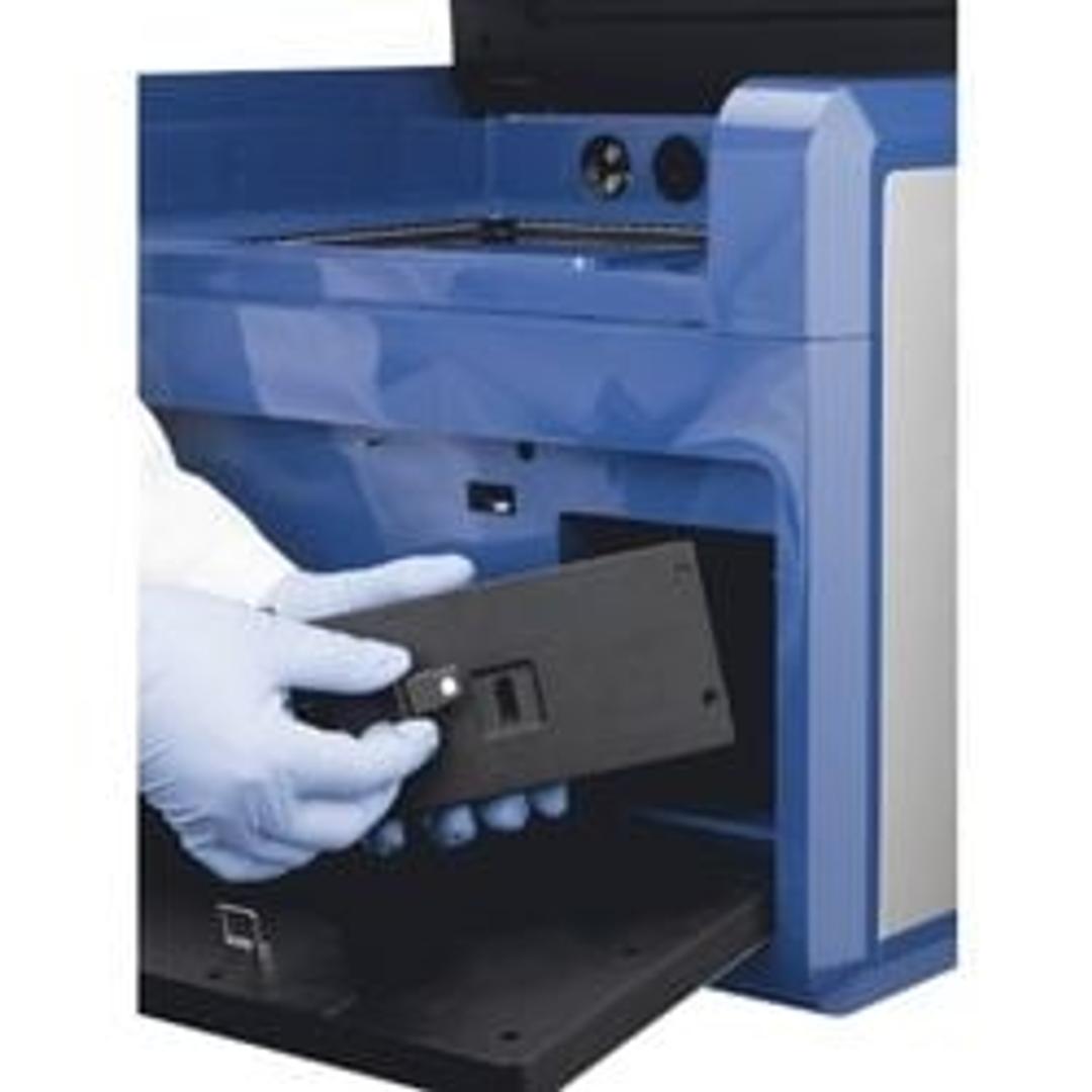
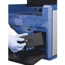
Laser module filter ready to insert CLIENT

Receive your quote directly from the manufacturer.
Great imager
Biomedical sciences
Our university did not have the ability to image florescent proteins. We now have the Azure Sapphire and have had great success detecting low signal florescent molecules. The software is very easy to use and we get great images.
Review Date: 27 May 2026 | Azure Biosystems
The Sapphire FL is the ultimate biomolecular imager for FLEXIBILITY. With customizable and user-changeable laser and filter modules, the Sapphire FL easily adapts to a lab’s changing needs and advancing research. The Sapphire FL offers customizable and user-changeable optical modules, 5–1000 μm resolution scans, a Z-plane range from -1.0 to +6 mm, 5 anesthesia ports for imaging living animals, chemiluminescence detection through the Chemiluminescence Module, and much more.
- Innovation-driving performance: high resolution imaging and wide depth of field enable imaging of many sample types.
- Imaging without compromise: one true workhorse system images fluorescence, NIR, phosphor, and chemiluminescent* samples down to 5 microns
- 4 cm clearance: allows for imaging of samples with depth, like tissues and small animals.
- 5 anesthesia ports: enables in vivo imaging of small animals. Compatible with standard anesthesia kits.
- Large 25x25cm field of view: Scan as many as six membranes or six 96-well plates at a time to save time.
- Customizable and easily upgradeable: pick the modules that support your research. Easily swap lasers and filters for expanded dye flexibility.
- Broad dynamic range: distinguish subtle differences in expression to image both strong and weak banks without experiencing saturation. EDR extends dynamic range to 24 bits of data.
- Whole slide imaging: Screen slides before microscopic analysis by imaging multiple slides at a resolution of 5 microns. The adjustable focal plane enables scanning of thick samples.
Brochures

Explore the Sapphire FL Biomolecular Imager
In this product brochure, learn more about the Azure Biosystems Sapphire™ FL biomolecular imager. Discover customizable and user-changeable laser and filter modules, making the Sapphire FL easily adaptable to a lab’s changing needs and advancing research. The Sapphire FL offers 5–1000 µm resolution scans, a Z-plane range from -1.0 to +6 mm, five anesthesia ports for imaging living animals, chemiluminescence detection through the Chemiluminescence Module, and much more.
In vivo imaging and monitoring of RFP+ tumors using the Sapphire FL
In vivo fluorescent imaging of model organisms such as mice is particularly relevant to cancer research. It can be applied in the study of tumor progression, and in assessing the effectiveness of anti-tumor treatments over time. Azure Biosystems describes the in vivo imaging of tumors in living mice using the Sapphire™ FL Biomolecular Imager, including tumor measurement and tracking tumor growth over time. Discover how attributes of the Sapphire FL make this type of imaging possible, such as integrated anesthesia ports, the ability to accommodate specimens with depth up to 4 cm, and the ability to image near-infrared fluorescence and chemiluminescence in addition to fluorescent imaging.
Sapphire FL imaging of multiplex fluorescent immunohistochemistry slides
Immunohistochemistry (IHC) is a widely used technique for identifying antigens in tissue specimens on slides. While IHC slides are typically imaged using a microscope, fluorescent scanners like the Sapphire™ FL Biomolecular Imager are useful for rapid triage and whole-slide documentation. Azure Biosystems demonstrates the use of the Sapphire FL Biomolecular Imager to image a multiplex fluorescent IHC experiment. Using standard optical modules, the Sapphire FL is used to detect two antigens in a tissue section of mouse lung. Discover how using the Sapphire FL for rapid triage allows researchers to quickly select the best slides for microscopic analysis. In addition, images of whole slides (compared to small sections of slides captured by microscopes) can supplement microscope images and assist in documentation.
Imaging dyes with large Stokes shifts on the Sapphire FL
Significant work has gone into developing fluorescent dyes with larger Stokes shifts for biological applications. Stokes shifts greater than 80 nm can minimize crosstalk between the excitation light and detection of emitted fluorescence during imaging, leading to better signal-to-noise ratios and better imaging. Explore how the Sapphire FL Biomolecular Imager provides the flexibility needed to image dyes with large Stokes shifts. Its modular excitation lasers and emission filters can be easily mixed and matched to achieve non-standard pairings compatible with specialty dyes. Azure Biosystems demonstrates the use of this mechanism on the Sapphire FL to image Western blots detected with antibodies labeled with MegaStokes dyes.
Unlock the power of In-Cell Westerns with total cell staining and advanced imaging
In-Cell Westerns (ICW) have emerged as a powerful and versatile technique, facilitating the study of cellular behaviors at a molecular level. The ICW assay is a fluorescence-based immunodetection technique that combines the principles of immunofluorescence and Western blotting, allowing researchers to probe specific target proteins within whole cells. Azure Biosystems conducted an experiment looking into the potential of this technique in the study of intricate cellular responses. Discover how combined with the Sapphire™ FL Biomolecular imaging system with high sensitivity and minimal cross-talk, ICW becomes a dynamic and versatile tool, capturing subtle variations in protein expression and treatment response.
Multicolor fluorescent imaging of cultured cells on the Sapphire FL Biomolecular Imager
Most cell culture experiments require that the viability of cells under specific experimental conditions be determined. It is important to confirm that culture conditions are optimized and not unnecessarily stressful, potentially leading to cell death. A common assay to measure cell viability is a dual fluorescence assay in which cells are stained with two nucleic acid–binding dyes: acridine orange (AO) and propidium iodide (PI). Azure Biosystems demonstrates using the Sapphire™ FL Biomolecular Imager to image cells stained with AO/PI and to visualize individual live and dead cells. The Sapphire FL has the capacity to image one sample under up to three different fluorescence channels simultaneously, allowing differential staining assays like AO/PI to be carried out quickly and simply.
BiFC assay for determination of in-vitro protein-ligand interaction
Bimolecular fluorescence complementation (BiFC) assay is a method used to directly visualize protein-protein interaction in cells. Azure Biosystems and the Malchiodi Lab, University of Buenos Aires, Argentina, explore the application of the BiFC assay to the study of in vitro interaction between the Zika virus NS4B protein and its ligand, TBK1. Discover how the Sapphire Biomolecular Imager makes it possible to detect bimolecular fluorescence complementation spots from transfected adherent cells grown in clear plastic well plates.
Imaging CFP-expressing cells with the Sapphire FL
There is some evidence that green fluorescent protein (GFP) can be more toxic to cells than cyan fluorescent protein (CFP) or yellow fluorescent protein (YFP). This suggests that CFP or YFP might be better options when making stable clones for tracking cells in long-term in vivo studies. Azure Biosystems demonstrates imaging of CFP-expressing cultured cells with the Sapphire™ FL Biomolecular Imager using the Sapphire’s 450 Standard Optical Module. Discover how features of the Sapphire FL make it an ideal choice for imaging cells and model organisms expressing CFP or other fluorescent reporter proteins.
Fluorescent IHC imaging of tumor tissue arrays on the Sapphire FL
Tissue microarrays offer a high-throughput approach to histology, immunohistochemistry (IHC), fluorescence in situ hybridization (FISH), and other experiments carried out on tissue sections. They are particularly valuable for studying tumors and cancer biology, as they facilitate comparisons among different sample types. Tissue microarrays are typically imaged under a microscope, one sample at a time. Azure Biosystems describes how tissue microarrays were stained with fluorescent stains or probed with antibodies in a two-color IHC experiment, and the Sapphire™ FL Biomolecular Imager was used to capture images of the entire array. These whole-slide images can be used to catalog slides and provide an initial survey of staining success before proceeding with higher-resolution imaging.
Easy screening of autophagy-associated acidic compartments
There are several techniques and strategies for estimating autophagy, though most of them are cumbersome and not compatible with in vivo experimental designs. Acridine orange (AO) is a cell-permeable fluorescent dye that accumulates in acidic compartments such as lysosomes and autophagic and endosomal vesicles, allowing their detection in live cells. Azure Biosystems demonstrates screening for autophagy activity in cells stained with AO acidotropic fluorescent dye and detected with the Azure Biosystems Sapphire™ FL Biomolecular Imager. With its ability to carry out multiplex fluorescence imaging, and to image multi-well plates including 24-well plates, the Sapphire FL can be part of a quick and easy in vivo evaluation of autophagy induction in cell culture using an AO staining approach.
Scanning microarrays with the Sapphire FL Biomolecular Imager
Microarrays have a broad range of research and clinical applications, and have been used extensively to study gene expression and to carry out genotyping. Imaging microarrays requires a scanner with sufficient resolution, appropriate light sources, a wide dynamic range, and the ability to reproducibly position and focus on a microarray slide. A multifaceted imaging system such as the Sapphire™ FL Biomolecular Imager provides the ability to scan microarrays among its large catalog of compatible applications. Discover how the Sapphire FL scans microarrays and generates high-resolution images ready for downstream analysis.