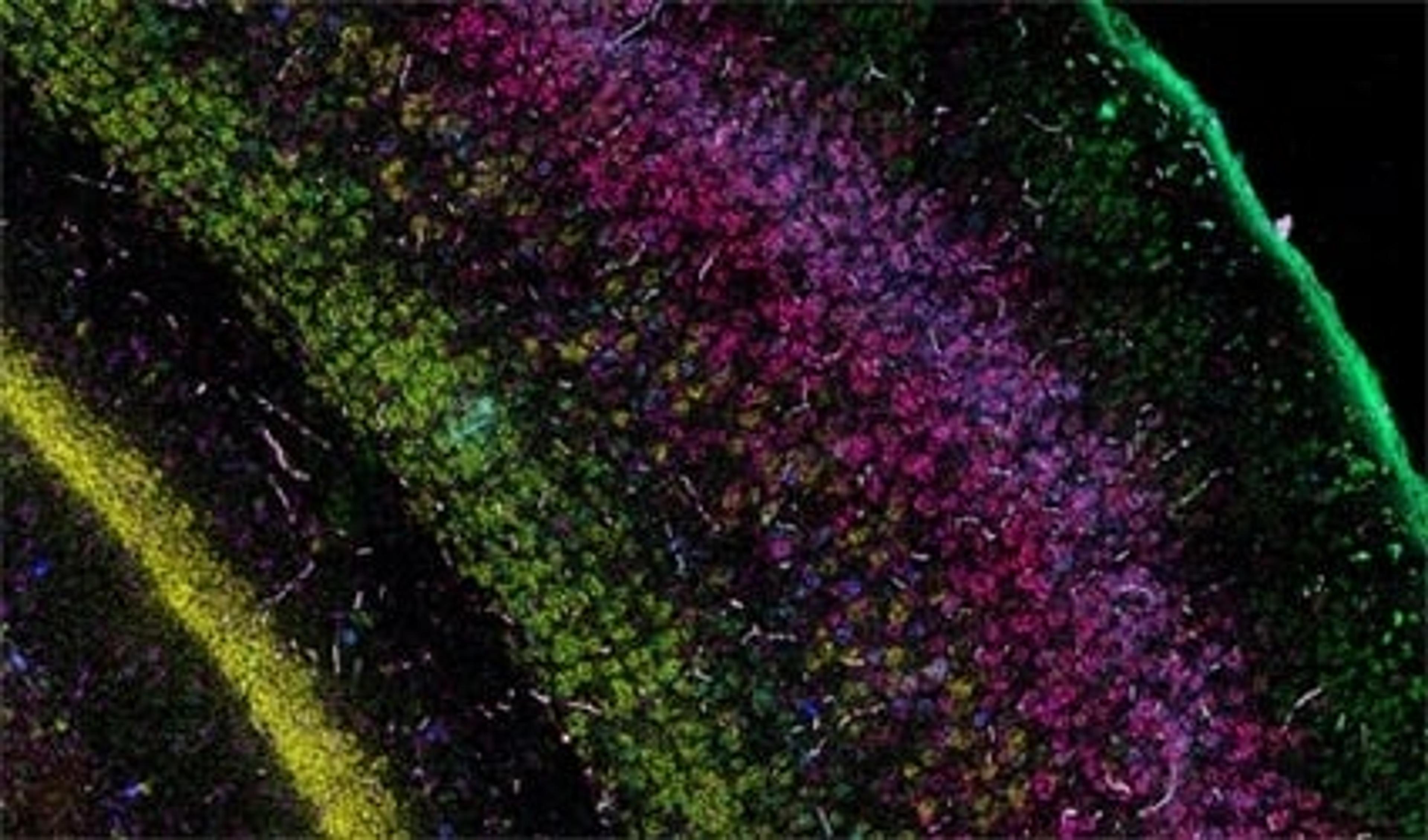
‘Google Maps’ for the human body – How spatial genomics is revolutionizing our understanding of disease

Discover how the Wellcome Sanger Institute is helping to develop a physical atlas of all human organs

18 Feb 2024

Charlie Carter
Life Sciences Editor

Editorial article

The Human Cell Atlas is an international consortium with the shared ambition of discovering and documenting the rich diversity of cell types across different human tissues. Understanding cell-type diversity throughout the human body allows researchers to explore how these cell types function and dysfunction in human health and disease states.

Human Cell Atlas
The Human Cell Atlas is a global initiative of the Wellcome Sanger Institute, that aims to create comprehensive reference maps of all human cells.

“A lot of the disease symptoms that span many different organs arise from the same specific cell types in the human body,” explains Dr. Omer Bayraktar, junior group leader at the Wellcome Sanger Institute, a leading player in the consortium. Bayraktar highlights that neurological disorders illustrate this phenomenon incredibly well, due to the rich diversity of both neuronal and non-neuronal cell types in the brain. Examples of this include the dopaminergic neurons that are affected in Parkinson's disease and the interneurons in schizophrenia.

In this exclusive SelectScience® interview, we find out more about the Wellcome Sanger Institute’s contribution to the Human Cell Atlas, including the development of cutting-edge technologies to further understanding of the human brain and the role of cell diversity in neurological disease.

From genes to organs: Creating a 3D map of the entire human body

The first wave of the project makes extensive use of single-cell RNA sequencing (scRNA-seq) technologies, enabling researchers to see the molecular diversity and gene expression patterns within previously unstudied human tissues in a high-throughput fashion. However, through use of this dissociative transcriptomic method, the spatial context of cells within the tissue of interest is lost. Of the method, Bayraktar states: “This is a good tool for achieving an unbiased first look at the different cell types across different organs, but you have no idea where the different cell types are coming from in your tissues of interest.”

The current focus now takes this first-wave information and locates the identified cell types and expressed genes within the intact architecture of a specific organ. Bayraktar shares that the aim is to create, as many Human Cell Atlas labs like to say, “the Google Maps of the human body”. The project combines the strengths of anatomy and cell morphology with molecular profiling, to create a three-dimensional atlas to help identify where different cell types are located in the tissue, as well as what genes and proteins are being expressed.

This spatial interrogation of gene expression is known as spatial genomics, and despite the promise of this emerging field, the current methods available are still in their infancy.
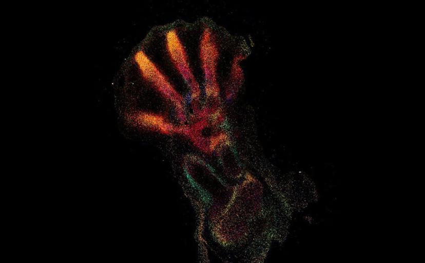
High-multiplexed smFISH reveals the spatial distribution of distinct cell types across the developing human hindlimbs

Sensitive, robust, and versatile spatial genomic methods

When looking for imaging solutions for spatial genomics, Bayraktar’s lab favors commercially accessible technology that offers robust levels of detection and readily works on very many different tissue types: “We want more convenient ways to study our different tissues, instead of having to invest heavily on developing new technologies or troubleshooting these on each new tissue of interest.”

RNA in situ hybridization (RNA ISH) is one such imaging method that allows for the detection of the expression of chosen target genes of interest. Bayraktar and his lab use single-molecule fluorescence ISH (smFISH) and other RNA ISH techniques to perform highly sensitive, spatially resolved mapping of gene expression with single-cell resolution to validate the observations that come from their scRNA-seq studies.

“In our first study, we used low multiplex RNA ISH to map the previously unknown diversity of astroglial cell types across layers of the mouth and the human cerebral cortex,” says Bayraktar. “What RNA ISH allows us to do, at single-cell resolution, with very high transcriptomic sensitivity, is quantify the expression of our target genes of interest with spatial resolution.”

Scalability for the future study of human disease

Bayraktar highlights that the first wave of the Human Cell Atlas was very successful due to the highly scalable nature of scRNA-seq and transcriptomic methods. Due to the degree of cell-type diversity in human organs, there is still the need for this type of scalable platform for spatial genomic applications. “To chart the Human Cell Atlas of the entire body, we need access to large-scale platforms that can apply spatial genomic methods to human organs, of a large number of individuals, and ultimately the entire human body,” explains Bayraktar.

To be able to tell apart the different cell types across different human tissues, you need to be able to assay the expression of hundreds of genes simultaneously. The most robust RNA ISH platforms that are currently available address much lower levels of multiplex RNA detection. Bayraktar highlights the Wellcome Sanger Institute’s high-throughput spatial genomic initiative, aiming to “push the current level of multiplexing, taking it from imaging four genes to imaging several hundred genes simultaneously, and create scalable platforms that can be applied to many different tissue sections, organs, and ultimately the entire human body.”

Once these scalable spatial genomic platforms have been developed, they can be harnessed to identify the diversity and location of cell types throughout the body and relate this to gene expression patterns in disease states. The study of selective long-term cell durability, in disorders such as Down’s Syndrome or amyotrophic lateral sclerosis (ALS), is just one of the applications that Bayraktar and his lab hope to address. However, the ultimate aim is to apply this kind of technology to inform our understanding of a wide range of neurological, neurodevelopmental, and neurodegenerative disorders.
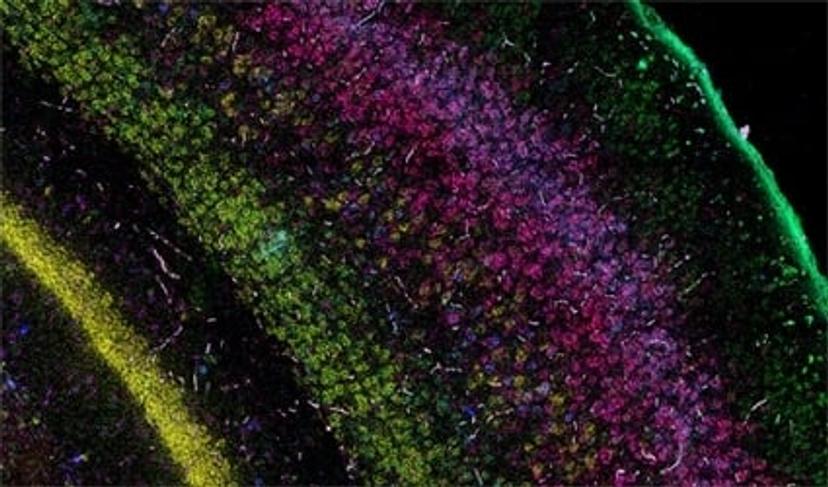
RNAscope® Multiplex Fluorescent assays are ideal for simultaneous detection studies of any genes in nearly any tissue type.

Looking for an RNA ISH solution for your gene expression studies?

Advanced Cell Diagnostics’s Pharma Assay Services provides RNAscope and BaseScope in situ hybridization (ISH) assay services to support pre-clinical and clinical studies for pharma and biotech partners globally. Tissue sectioning, ISH staining, high-resolution full slide scanning, scoring, and image analysis are performed by a dedicated team of highly trained specialists, scientists, and board-certified pathologists. With direct access to the developers of the technology, the Pharma Assay Services team provides unparalleled expertise in ACD’s ISH platform and delivers fast, high-quality data designed to meet your study objectives and timelines.

Links

Tags

Cell / Tissue CultureCell culture or tissue culture is used to study the biology of cells or tissues and to isolate cellular products in an environment which can be manipulated and well defined. Accurately control your culture environment with bioreactors or culture incubators, bind your cells to a surface or together with an extracellular matrix. Distinguish cell types with differential media or proliferate cells with certain characteristics using selective media. Enrich your media with supplements such as growth factors, sera and vitamins. Find the best cell and tissue culture products, kits and equipment in our peer-reviewed product directory: compare products, check customer reviews and receive pricing direct from manufacturers.Cell Lines Stem Cells and Primary CellsPrimary cell cultures, established cell lines and stem cells are vital for <i>in vitro</i> and <i>ex vivo</i> experimentation. High-quality cells, optimized for your applications, alongside optimized cell substrates, growth medium and supplements, are critical for experimental success. Explore a range of cells suitable for your applications, including isogenic cell lines, competent cells, induced pluripotent stem cell (iPSC)-derived cell lines, fungal/bacterial/mammalian cell lines, stem cells and cancer cell lines. Find the best cells for your research in our peer-reviewed product directory: compare products, check customer reviews and receive pricing direct from manufacturers.Data AnalysisData analysis hardware and software is available to make data processing straight-forward yet powerful. Data software can be used for math and stats, technical graphing and image analysis. In addition, software is available for specific data analysis of electrophoresis, densitometry, ELISA and DNA sequencing.Cellular PathologyCellular Pathology deals with the microscopic analysis of tissue samples and cells. Sample preparation and processing includes fixation, staining, sectioning and slide mounting, using equipment such microtomes and cryostats. In choosing immunohistochemistry and immunocytochemistry kits, consider chromogens, staining method, antibodies, microscopes and imaging.Cell-Based AssaysCell-based assays are used to monitor the presence, quantity and activities of a desired cellular analyte including drug molecules or biomarkers. This can reveal information on cell health (apoptosis, cytotoxicity, viability and proliferation assays), cell metabolism, cell migration and cell signaling mechanisms. Find the best cell-based assay products, kits and equipment with our peer reviewed product directory: compare products, check customer reviews and receiving pricing direct from manufacturers.DNA / RNA Extraction and PurificationPurified DNA and RNA are required for numerous downstream molecular biology applications. Consequently, the importance of high-quality DNA/RNA extraction and purification equipment cannot be underestimated. Many purification kits are available and are typically optimized for nucleic acid type and source, including plasmid DNA, genomic DNA, mRNA, RNA and viral nucleic acid purification kits. Automated extraction and purification of nucleic acids can be implemented with magnetic bead separator instruments or high-throughput purification workstations. Find the best DNA/RNA extraction and purification equipment in our peer-reviewed product directory: compare products, check customer reviews and receive pricing direct from manufacturers.DNA SequencingDNA sequencing, such as sanger sequencing, is a biological technique that determines the precise order of nucleotide bases in a fragment or template of DNA. DNA sequencers and genetic analyzers are based on capillary electrophoresis, where labeled DNA fragments are electrophoretically separated by size as they migrate through a polymer. Find the best DNA sequencing products, including DNA sequencing kits, genomic libraries and genetic identity kits in our peer-reviewed product directory: compare products, check customer reviews and receive pricing direct from manufacturers.Biopharmaceutical AdvancesBiopharmaceutical advances follow the development of pharmaceuticals derived from biotechnology, also known as biotechnology medicines. Biopharmaceuticals may be produced from cell lines, plants, or microbial cells. Important considerations of biopharmaceutical use include application, cost, production process and purification.In Vivo Imaging Systems<i>In vivo</i> imaging systems, including pre-clinical imaging systems and medical imaging systems are used to non-invasively visualize and capture images of live animals and plants. Monitor the natural processes or diseases of your subjects using small-animal pre-clinical imaging systems, including single photon positron emission tomography (SPECT), positron emission tomography (PET), computed tomography (micro-CT), magnetic resonance imaging (MRI), X-ray radiography, ultrasound, fluorescence and bioluminescence imagers. Multimodal systems and software solutions are also available for correlative analysis of organ, tissue, cell, or molecular-level processes. Find the best in vivo imaging products in our peer-reviewed product directory: compare products, check customer reviews and receive pricing direct from manufacturers.Drug DeliveryDrug Delivery refers to dosage form, route of administration, formulations, technologies, and systems for transporting a pharmaceutical compound in the body as needed to safely achieve its desired therapeutic effect. Drug delivery is often approached via the biopharmaceutical or small molecule drug's formulation, but it may also involve medical devices or drug-device combination products. Considerations include instrumentation, software and services. Scale-upScale-Up is generally defined as the process of transferring the results of the discovery phases of the drug discovery process, obtained on a laboratory scale, to the pilot plant and finally to production scale. Scale-Up uses reactors and pump systems as well as reagents, standards and buffers. Often Scale-Up is provided as a service by Contract Manufacturing Organizations (CMOs). DNA / RNA QuantificationDetection and quantification of nucleic acids is important in molecular biology, cloning, expression, forensics and clinical diagnostics. Nucleic acids can be detected by labeling with colorimetric, fluorescent or radio labels and using in situ hybridization kits to identify specific sequences. Multiple nucleic acids can be detected and quantified at once using RNA / DNA detection beads or RNA / DNA microarrays. Find the best DNA / RNA Quantification products in our peer-reviewed product directory: compare products, check customer reviews and receive pricing direct from manufacturers.Gene Expression and Molecular CloningMolecular cloning is a set of techniques that utilizes vectors to transfer recombinant DNA into host cells and is an essential tool for investigating the expression of genes and proteins in bacterial or mammalian cells. A variety of vectors optimized for gene cloning and expression in a range of host organisms are available, alongside competent cells for genetic replication. Here, you can explore a range of molecular tools, high-quality genomic and cDNA libraries, premade clones, transformation and transfection reagents and mutagenesis or gene expression detection assays and expression arrays. Find the best gene expression and molecular cloning products in our peer-reviewed product directory: compare products, check customer reviews and receive pricing direct from manufacturers.NeuroscienceNeuroscience research investigates the neurological mechanisms underlying behavior, neurodegenerative diseases, and other brain conditions. Learn about the innovative technologies for bioimaging, electrophysiology, cell culture, chromatography and other techniques used in this field.NeurobiologyNeurobiology studies the biology of the nervous system, including the structure and function of neurons, glial cells, and their interactions. It is a key field in understanding neurological disorders such as Alzheimer�s, Parkinson�s, and schizophrenia. Research in neurobiology is essential for developing therapies for these conditions. Browse our peer-reviewed product directory to find the best neurobiology research tools, compare products, check reviews, and get pricing directly from manufacturers.
‘Google Maps’ for the human body – How spatial genomics is revolutionizing our understanding of disease