
How digital microscopy is extending access to hematology expertise
Learn how the NED® Digital Microscope can support real-time remote hematology review and more connected peripheral blood smear workflows
30 Sept 2026
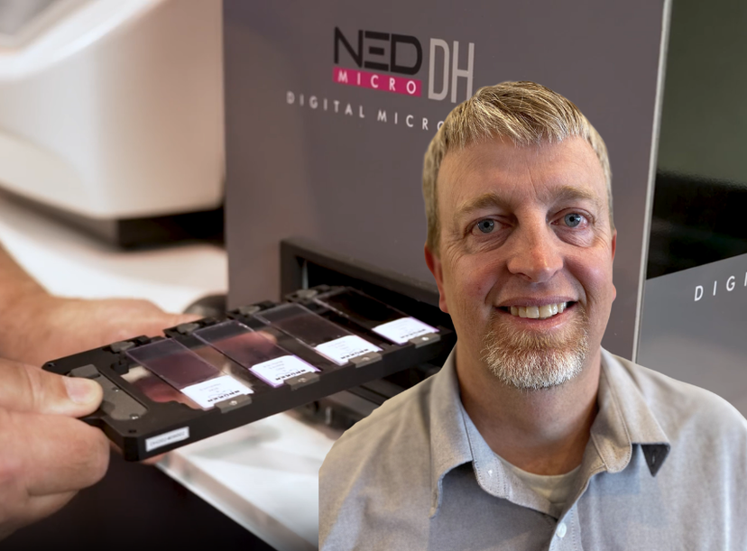
Bryan Keisel, Service and Applications Support Manager at Bruker Microbiology & Infectious Diagnostics/ ELITechGroup Inc.
A peripheral blood smear cannot travel instantly between laboratory sites, but the expertise needed to review it can. For laboratories working across remote locations, sharing specialist knowledge has traditionally meant transporting slides, sending selected images or waiting for an experienced colleague to reach the microscope. Digital hematology offers another option: giving specialists remote access to the original slide in real time.
Bryan Keisel, Service and Applications Support Manager at Bruker Microbiology & Infectious Diagnostics/ ELITechGroup Inc. in Logan, Utah, has experienced laboratory automation from both sides of the instrument.
After approximately 12 years in hospital laboratories, primarily in molecular diagnostics, he spent another decade supporting clinical laboratories as a field service engineer. His work has taken him from small physician-office laboratories to major reference centers and introduced him to everything from manual bench testing to large automated track systems.
“In every laboratory, regardless of size, technologists are being asked to do more with less,” Keisel says.
That pressure is particularly apparent when an automated hematology analyzer flags a sample for manual review. At that point, the workflow becomes dependent not only on the availability of a microscope, but also on access to someone with the experience to interpret what it reveals.
Why peripheral blood smear review still depends on specialist expertise
Automated analyzers handle much of the routine workload in modern hematology laboratories, but some specimens still require closer investigation. Unusual cell populations, flagged results, body fluids, and bone marrow samples may need a technologist or specialist to examine cellular morphology directly.
The challenge is that experienced staff are not distributed evenly across every laboratory site. Keisel has seen some laboratories struggle to transfer morphology knowledge and expertise to newer technologists, while smaller hospitals may also lack immediate access to a pathologist or hematopathologist when an unusual finding appears.
“If you had that expertise on site, it would resolve a lot of questions,” he says. “But having a hematopathologist on call can be very expensive for a small hospital or hospital system.”
Physical location therefore becomes part of the workflow. A slide may need to be transported to a central laboratory, or a static image sent to a specialist who cannot examine the surrounding fields.
Remote digital microscopy gives specialists control of the slide
Traditional second opinions often require two people to share the same microscope. One person moves away from the eyepiece, another takes their place, and both hope the relevant cell remains in view.
“You ask somebody to come and take a look at it, and hopefully the microscope hasn’t been bumped, and the focus is still where it was,” Keisel explains.
NED® Digital Microscope changes that interaction by displaying the smear digitally and allowing an authorized remote user to navigate the specimen. Instead of reviewing only a preselected image, the specialist can move between fields and examine the wider cellular context.
“The colleague on the other side of the world can take control and drive the instrument,” Keisel says. “They can move around and look at the whole slide. They get a better picture than they would from one static image.”
Keisel likens the navigation to moving around a digital map. This could enable a central hematology team to support peripheral laboratories, allow colleagues to examine an unusual case together or help an experienced professional guide someone still developing morphology skills.
Remote digital microscopy gives specialists the whole-slide context, rather than limiting them to a single image of the cell in question.
Bryan Keisel Bruker Microbiology & Infectious Diagnostics/ELITechGroup Inc.
Consistent blood smear staining supports digital hematology
Remote access does not compensate for inconsistent specimen preparation. The cellular features presented on screen still depend on the quality of the original smear and stain.
Keisel recalls how differently slides could appear when staining was performed manually. “Everyone does it a little differently,” he says. “When you go to read the slide, that difference can be reflected in what you see.”
Bruker’s Aerospray® slide stainers are designed to standardize this upstream stage. Laboratories can configure the staining appearance to suit local preferences, including the balance between red and blue tones, then apply those settings consistently.
This connection between preparation and review is central to the workflow. Aerospray supports the production of a familiar stained slide, while NED® Digital Microscope provides the digital environment in which it can be examined and shared.
How the NED Digital Microscope complements automated hematology analyzers
The NED® Digital Microscope and Aerospray® stainers are not intended to take over the high-volume work performed by automated hematology analyzers. Keisel instead sees the digital microscope as a flexible addition for specimens that leave the routine pathway.
The system can support screen-based white blood cell differential review, allowing users to navigate the slide and assign cells to relevant categories. Images can be retained for documentation, teaching or further review, including image stacks that present a cell at different focal planes.
“It does not just save one image,” Keisel explains. “It saves a stack, so you can move through different focal planes and see the different components of the cell.”
Viewing smears on screen may also provide a more comfortable alternative for professionals who spend long periods at a conventional microscope.
Digital hematology implementation requires laboratory IT support
Remote control introduces practical questions around network access, cybersecurity, and data storage and governance. Keisel says pathologists often respond positively to the possibilities for consultation and education, but hospital IT teams may understandably require reassurance about connectivity.
“There is a balance between allowing users to collaborate and the hospital wanting everything secure,” he says.
Early involvement from IT could therefore be as important as instrument selection when introducing remote microscopy across a laboratory network.
Making hematology expertise easier to reach
The defining value of NED® Digital Microscope is not complete diagnostic automation. Automated analyzers remain central to high-throughput testing, while qualified professionals remain responsible for morphology review and interpretation.
Instead, digital microscopy changes how those professionals can reach the slide. It can allow a peripheral laboratory to consult a central specialist, help colleagues examine an uncommon finding together, and turn live specimens into shared teaching material.
The slide may remain in one laboratory, but the expertise surrounding it can extend much further.
Learn more about the NED® Digital Microscope and how it can support remote hematology review.
Related products
Request Quote for All Products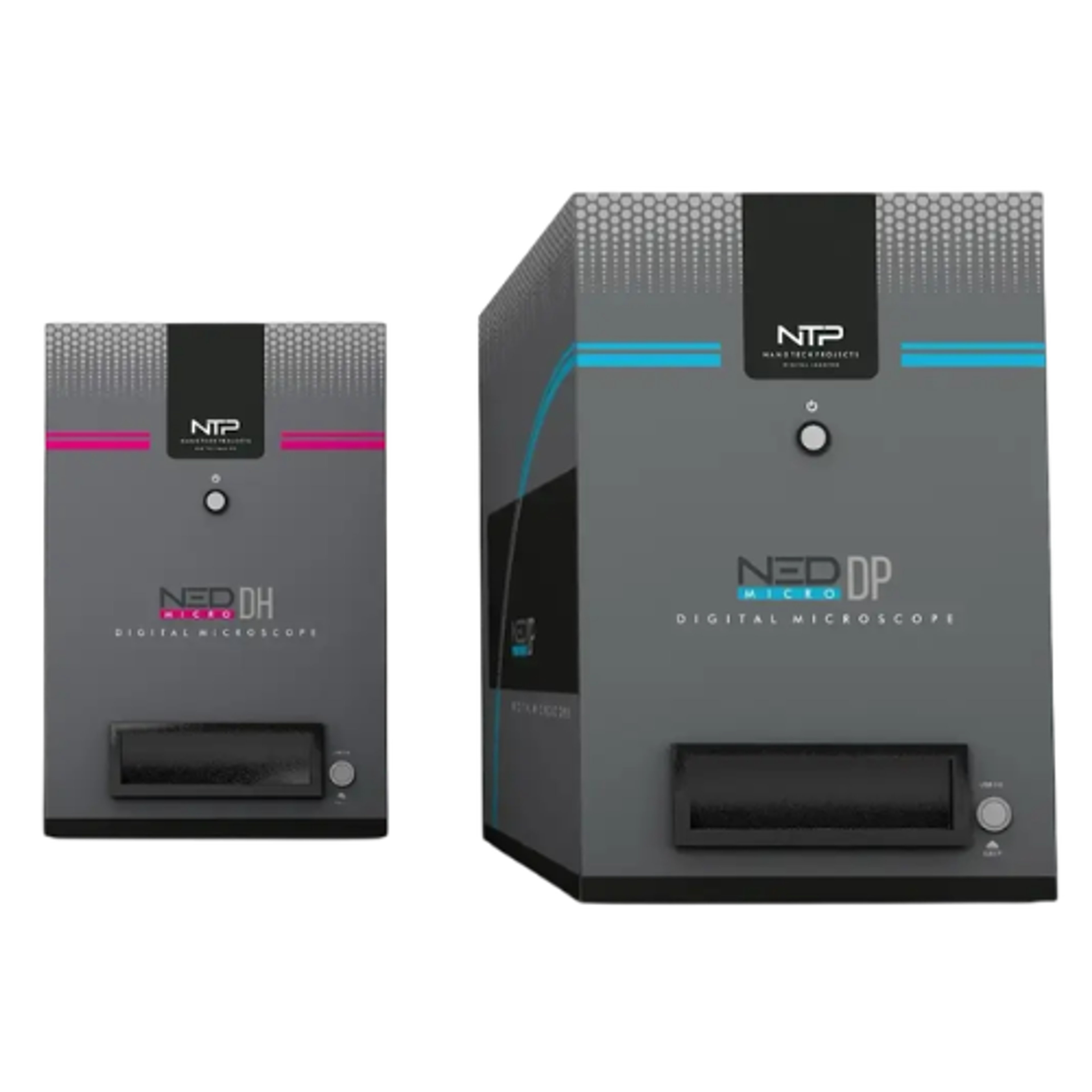
NED® Digital Microscope
Bruker Biomedical SystemsExperience NED Digital Microscope – where speed, accuracy, and collaboration meet.
Links
Tags
Frequently asked questions
Show frequently asked questions
How does the NED Digital Microscope support remote peripheral blood smear review?
The NED® Digital Microscope lets authorized specialists remotely control the instrument, navigate the whole slide, examine multiple fields, and assess cellular context beyond a single static image. This helps peripheral laboratories access hematology expertise in real time.
Why is consistent blood smear staining important in digital hematology?
Digital review depends on the quality of the original smear and stain. Bruker’s Aerospray® slide stainers standardize staining while allowing laboratories to configure preferred red and blue tones, helping produce familiar, consistent slides for review with the NED® Digital Microscope.
How does the NED Digital Microscope complement automated hematology analyzers?
Automated hematology analyzers handle high-volume routine testing, while the NED® Digital Microscope supports specimens requiring morphology review. It enables screen-based white blood cell differentials, cell categorization, remote consultation, and image-stack storage across different focal planes for documentation, teaching, or further review.