How AI-assisted digital workflows are transforming morphology review in the clinical laboratory
Guest editorial by Eric Pabon, MBA, Senior Manager, Global Product Marketing, Hematology, Beckman Coulter
6 Oct 2026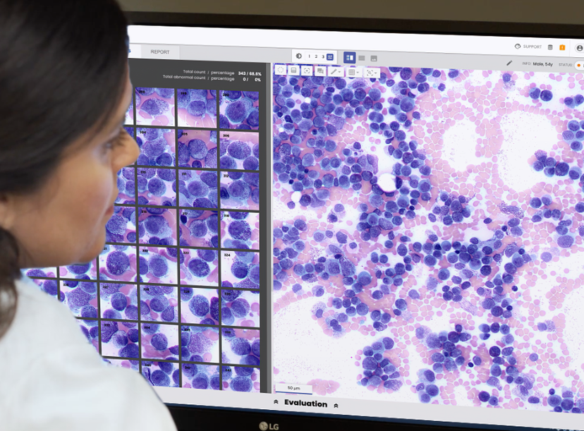
The Scopio Labs X100 & X100HT solution combines AI-assisted cell classification with full-field bone marrow aspirate review.
Key takeaways
- AI-assisted digital microscopy can support bone marrow aspirate (BMA) review while keeping final interpretation in the hands of morphology experts.
- In a real-world time-and-motion study, a digital BMA workflow reduced time spent examining cells by approximately 46%. Overall analysis time remained comparable to manual microscopy.
- Algorithm performance is only one part of AI adoption. Technologies must also fit into existing laboratory practices and systems.
Where AI fits in pathology
Pathology laboratories are managing larger, more complex caseloads with limited workforce capacity¹. AI is being explored to assist with defined, repetitive, and time-intensive tasks, including cell and tissue image analysis, quantification, and pattern recognition.
Bone marrow aspirate (BMA) review is one area where these tools may be useful. The work requires specialized morphological expertise, and examining the required number of cells can be time-consuming.
Any new technology also has to work within existing laboratory practices and systems. Training, validation, interoperability, data privacy, and regulatory oversight all factor into implementation².
Why BMA review is a candidate for digitalization
Bone marrow evaluation plays an important part in diagnosing and monitoring leukemias, lymphomas, and other hematologic disorders. Bone marrow biopsies provide information about marrow architecture, while aspirate review enables detailed assessment of individual cell morphology and relative cell populations.
BMA review is also one of the more demanding morphology tasks performed in the laboratory, in part because of the number of cells that must be examined.
International Council for Standardization in Hematology (ICSH) guidelines recommend counting at least 500 cells across at least two smears³. This supports standardized reporting but requires considerable time and concentration from the morphology expert. Traditional microscopy involves repeatedly moving between the microscope and a manual or electronic counting device as hundreds of cells are reviewed.
Digital microscopy can reduce some manual steps in BMA review. The Scopio Labs X100 Full-Field Bone Marrow Aspirate (FF-BMA) Application* uses computational photography to reconstruct high-resolution full-field images equivalent to 100× oil immersion. Within this digital workflow, AI-assisted analysis supports tasks such as region-of-interest identification and cell pre-classification. The morphology expert then reviews the digital images and AI-assisted classifications and remains responsible for final interpretation⁴.
What the real-world workflow study found
A prospective time-and-motion study conducted in a hospital laboratory in Northern Italy compared traditional manual microscopy with the digital BMA workflow5. Data collection began approximately two months after implementation, allowing time for training and integration into routine laboratory operations. The laboratory typically processed four to six BMA tests per day. The study included 11 observed analyses: two performed with manual microscopy and nine with the digital application.
Despite the limited number of observations at a single site, the study offers a real-world look at how digital review may affect laboratory workflow.
Overall analysis time was similar between the two approaches: 29 minutes, 21 seconds with manual microscopy compared with 28 minutes, 14 seconds with the digital approach⁵. The larger difference was seen in the time spent examining cells.
The 500-cell count was the most time-intensive component of manual analysis, accounting for 68.5% of total review time. With the digital workflow, it accounted for 38.5%⁵. Time spent reviewing the 500 cells, abnormal cells, and megakaryocytes fell from 20 minutes, 7 seconds with manual microscopy to 10 minutes, 51 seconds with digital review, a reduction of approximately 46%⁵.
That time savings did not fully carry through to the overall analysis. At the study site, the digital workstation was in a separate area and not connected to the hospital network, adding transfer and data-entry steps. The authors estimated that integration with the laboratory information system (LIS) could reduce reporting time by a further 39%⁵.
The physical demands of manual microscopy
Researchers also tracked the physical movements involved in the BMA review.
During one 12-minute observation period, researchers recorded 139 head and neck movements and more than 600 finger key presses. The morphology expert also reported finger stiffness, headaches, and neck and back strain during manual microscopy. Digital review consolidated much of the analysis into a single interface, reducing these repetitive actions⁵.
The study did not formally measure ergonomic outcomes, so further research would be needed to determine the broader impact of digital review on repetitive movements and physical strain. More research would be needed to determine whether the same findings are seen across other laboratories⁵.
Evaluating the full workflow
At this site, the digital approach reduced cell-examination time by about 46%. Overall analysis time remained similar because transfer and data-entry steps added time elsewhere. The digital approach also reduced repetitive movements associated with manual microscopy, and the morphology expert remained responsible for final review and interpretation.
The findings suggest that the value of digital microscopy depends on more than what happens during cell analysis. Integration with the LIS, reporting processes, and routine laboratory practice can determine how much of that time savings is ultimately realized.
Beyond time savings, digital review may also support greater workflow standardization, facilitate remote consultation, and help laboratories manage increasing workloads amid ongoing workforce pressures. These broader considerations matter when evaluating where AI-assisted digital microscopy may add value in the laboratory.
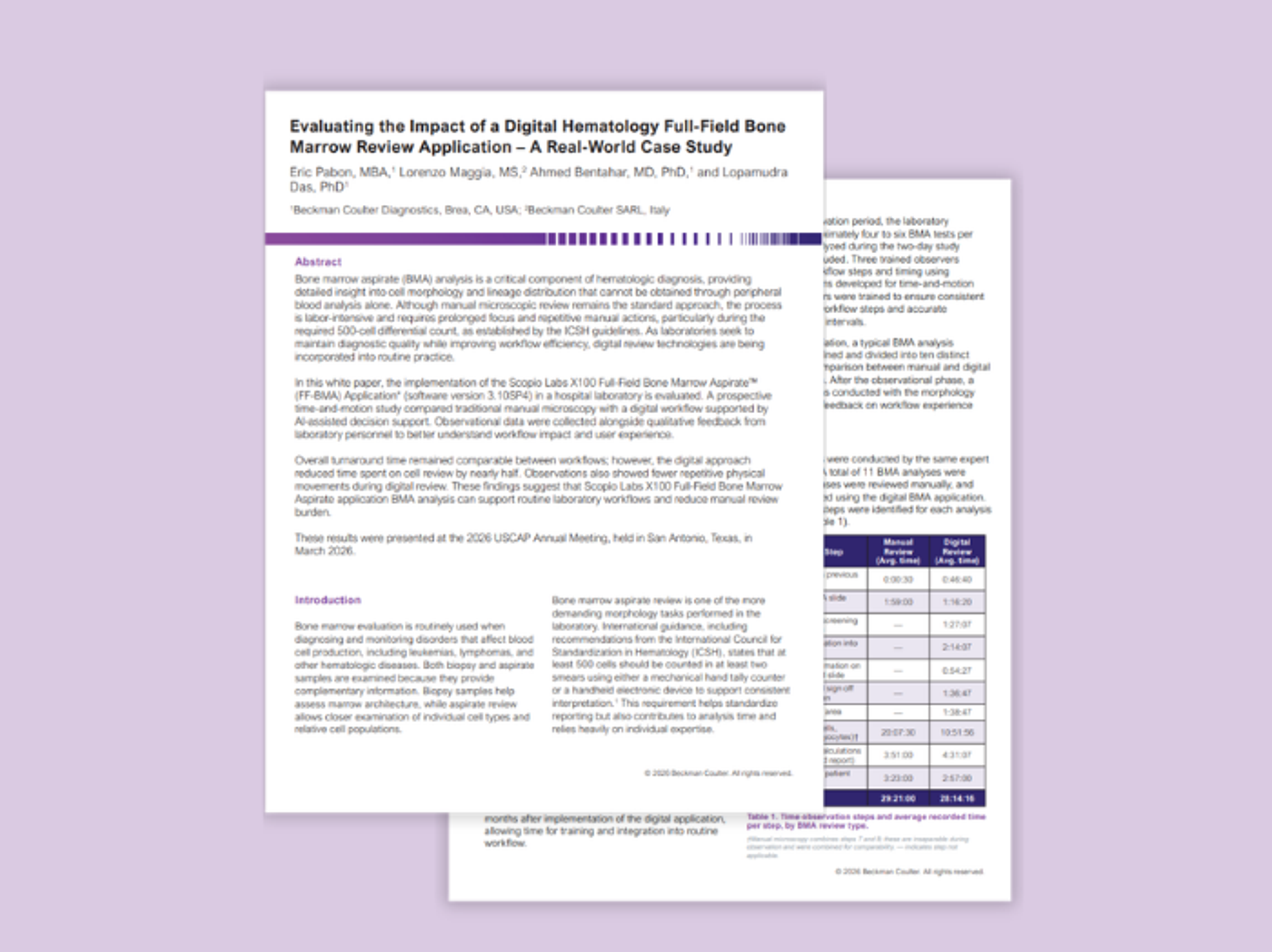
To explore the complete study methodology, workflow comparison, and findings, download the full whitepaper, Evaluating the Impact of a Digital Hematology Full-Field Bone Marrow Review Application - A Real-World Case Study.
* Not available in all countries. BMA is pending submission and clearance by the United States Food and Drug Administration; not yet available for in vitro diagnostic use in the United States.
References
1. Arora K, Moore L. The pathologist shortage crisis has an AI solution. Medical Laboratory Observer. August 13, 2026.
2. Fahim YA, Hasani IW, Kabba S, Ragab WM. Artificial intelligence in healthcare and medicine: clinical applications, therapeutic advances, and future perspectives. Eur J Med Res. 2025;30:848. doi:10.1186/s40001-025-03196-w.
3. Lee SH, Erber WN, Porwit A, Tomonaga M, Peterson LC; International Council for Standardization in Hematology. ICSH guidelines for the standardization of bone marrow specimens and reports. Int J Lab Hematol. 2008;30(5):349-364. doi:10.1111/j.1751-553X.2008.01100.x.
4. Bagg A, Raess PW, Rund D, et al. Performance evaluation of a novel artificial intelligence-assisted digital microscopy system for the routine analysis of bone marrow aspirates. Mod Pathol. 2024;37(9):100542. doi:10.1016/j.modpat.2024.100542.
5. Pabon E, Maggia L, Bentahar A, Das L. Evaluating the Impact of a Digital Hematology Full-Field Bone Marrow Review Application – A Real-World Case Study. Beckman Coulter Diagnostics; 2026. 2026-15389.