University of California Irvine redefines mass spectrometry imaging with the Shimadzu iMScope QT
19 Jan 2026

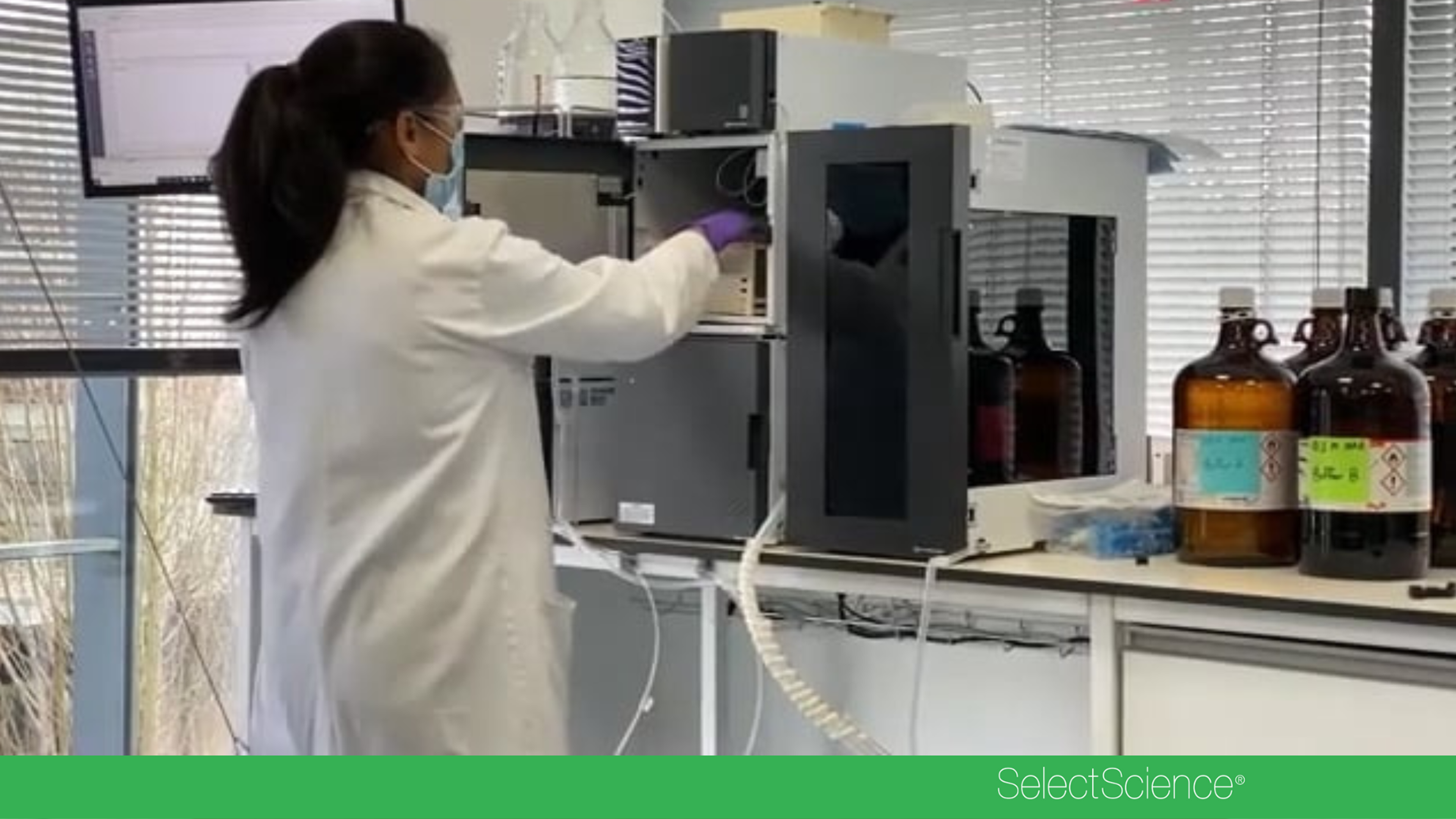
Mass spectrometry imaging (MSI) is rapidly reshaping spatial biology by allowing scientists to map metabolites, lipids, and proteins directly in tissues with unprecedented clarity. In this SelectScience interview, Dr. Felix Grun, Director of the Mass Spectrometry Facility at University of California Irvine, shares how the iMScope QT from Shimadzu is transforming MSI by enabling high-resolution, high-speed spatial metabolomics and lipidomics across diverse research areas. With its atmospheric pressure MALDI design, built-in optical microscope, intuitive workflow, and powerful ImageReveal software, the Shimadzu iMScope QT is accelerating scientific discovery and opening new possibilities in spatial biology and multiomics research.
About the company
Shimadzu Corporation
Shimadzu Corporation was founded in Kyoto, Japan in 1875 and has been supplying cutting-edge analytical and measuring instruments for a broad range of applications for over 150 years. Key markets include pharmaceuticals, life sciences, food & beverages, chemicals, petrochemicals, environmental, material science, and forensics/toxicology. Products include chromatographs (HPLC/UHPLC/SFC, GC), mass spectrometers (GC-MS/MS, LC-MS/MS, MALDI, ICP-MS), spectrophotometers (FT-IR, Fluorescence, UV-VIS-NIR), atomic spectrometers (AA, ICP), X-ray spectrometers (ED-XRF, XRD, XRF), thermal analyzers, Total Organic Carbon (TOC) analyzers, particle size analyzers, material testing machines, and balances, etc.
FAQs
How is the Shimadzu iMScope QT advancing mass spectrometry imaging (MSI) for spatial metabolomics and lipidomics research?
The Shimadzu iMScope QT advances mass spectrometry imaging by delivering high-resolution, high-speed spatial metabolomics and lipidomics directly in tissue samples. Its atmospheric pressure MALDI design, integrated optical microscope, intuitive workflow, and ImageReveal software enable scientists to map metabolites, lipids, and proteins in tissues with unprecedented clarity, accelerating discovery in spatial biology and multiomics research.
What role does the Shimadzu iMScope QT play in spatial biology and multiomics, according to Dr. Felix Grun at the University of California Irvine?
According to Dr. Felix Grun, Director of the Mass Spectrometry Facility at the University of California Irvine, the Shimadzu iMScope QT is transforming spatial biology and multiomics by enabling detailed MSI-based mapping of metabolites, lipids, and proteins in tissues. Its combination of high-resolution imaging, high-speed data acquisition, and powerful ImageReveal software opens new possibilities for spatial biology studies and multiomics workflows across diverse research areas.
Why is atmospheric pressure MALDI with a built-in optical microscope important in the Shimadzu iMScope QT for MSI applications?
Atmospheric pressure MALDI combined with a built-in optical microscope in the Shimadzu iMScope QT is important because it supports high-quality, high-resolution MSI while simplifying the experimental workflow. This design allows researchers to visually inspect tissue morphology and precisely correlate optical images with mass spectrometry data, improving the clarity and accuracy of spatial maps of metabolites, lipids, and proteins in tissue-based spatial biology and multiomics research.
Related products
Request Quote for All Products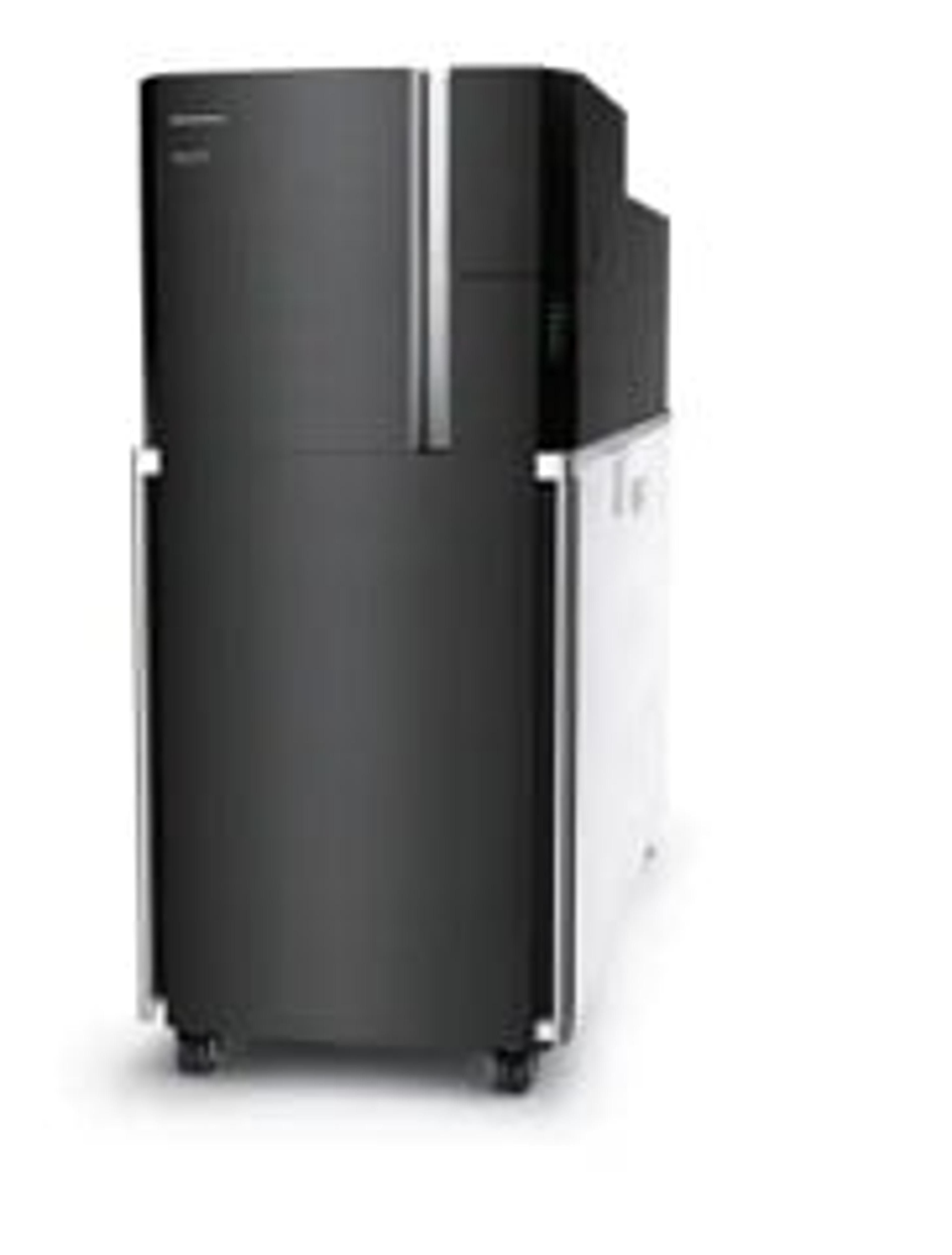
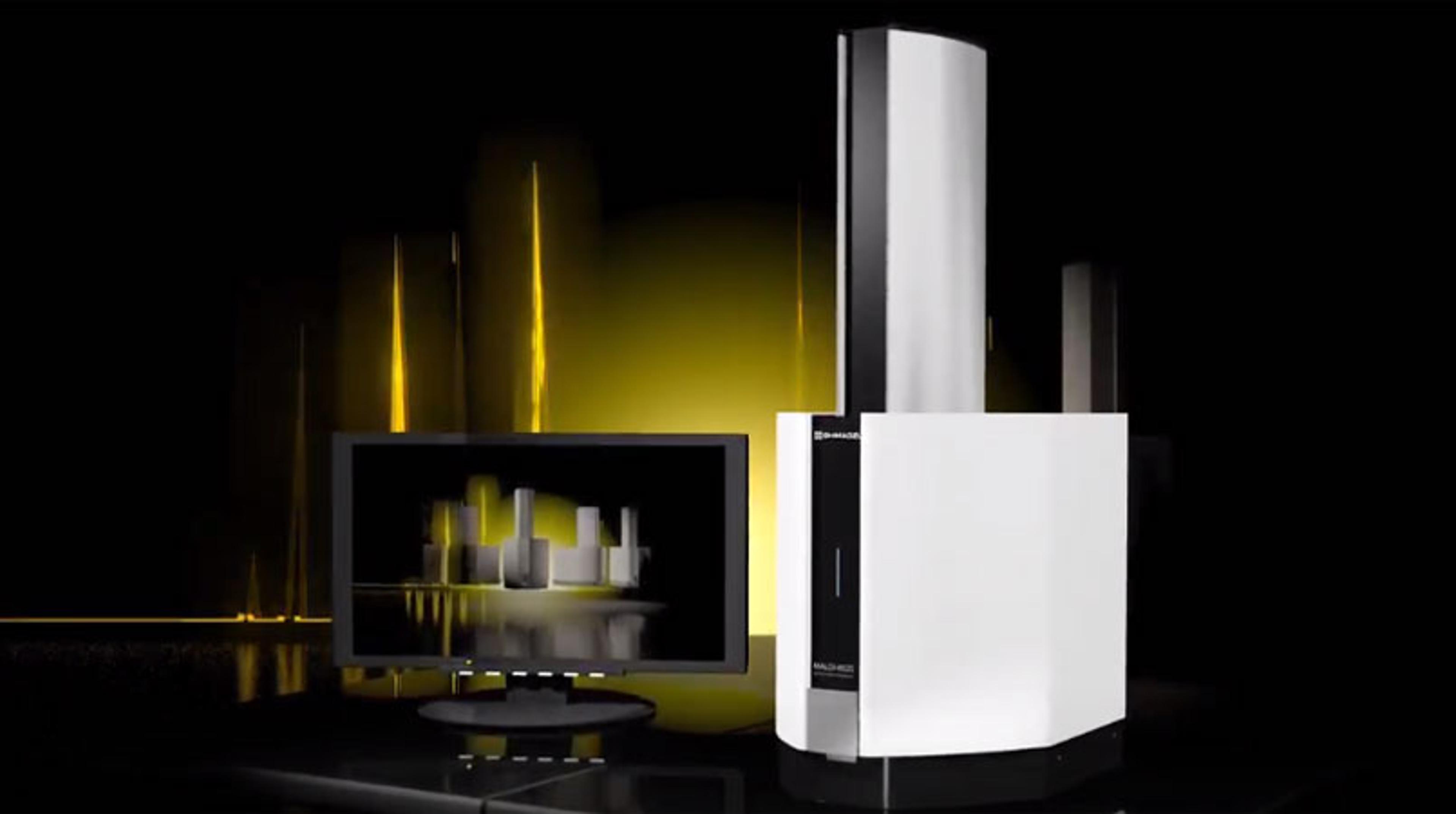
iMScope QT
Shimadzu CorporationThe iMScope QT inherits the concept of a mass spectrometer equipped with an optical microscope from the iMScope series, the iMScope QT is also Shimadzu's fllagship model for MS imaging with a Q-TOF MS. The iMScope QT boasts not only fusion with morphology studies but also excellent speed, sensitivity, and spatial resolution, clearing the way to next-generation mass spectrometry imaging.