Enumeration of Live, Circulating Tumor Cells Using SmartFlare™ Probes and an Amnis® Imaging Flow Cytometer
17 Feb 2015Circulating tumor cells (CTCs) are released into the bloodstream from primary and metastatic cancers and are potentially useful for early cancer detection, diagnosis, and treatment. In this study, an Amnis ImageStream®X Mark II imaging flow cytometer was employed along with SmartFlare™ fluorescent RNA detection probes to collect imagery from large numbers of WBCs that were spiked with live SKBR-3 human breast cancer cells.
Related products
Request Quote for All Products
Image Stream®X Mark II Imaging Flow Cytometer
MerckThe revolutionary ImageStream®X Mark II Imaging Flow Cytometer combines the speed, sensitivity, and phenotyping abilities of flow cytometry with the detailed imagery and functional insights of microscopy. This unique combination enables a broad range of applications that would be impossible using either technique alone. This instrument produces multiple high-resolution images of every cell directly in flow, including brightfield and darkfield (SSC), and up to 10 fluorescent markers with sensitivity exceeding conventional flow cytometers. Compared to its predecessor, the new ImageStream®X Mark II Imaging Flow Cytometer offers a streamlined workflow, greater flexibility, and optimizations for rare cell applications. Taken together, the capabilities of the ImageStream®X Mark II make it superior for traditional flow applications while greatly expanding the scope of flow cytometry. Applications include the study of cell-cell interactions, phagocytosis, apoptosis and autophagy, the characterization of circulating tumor cells, and many others.
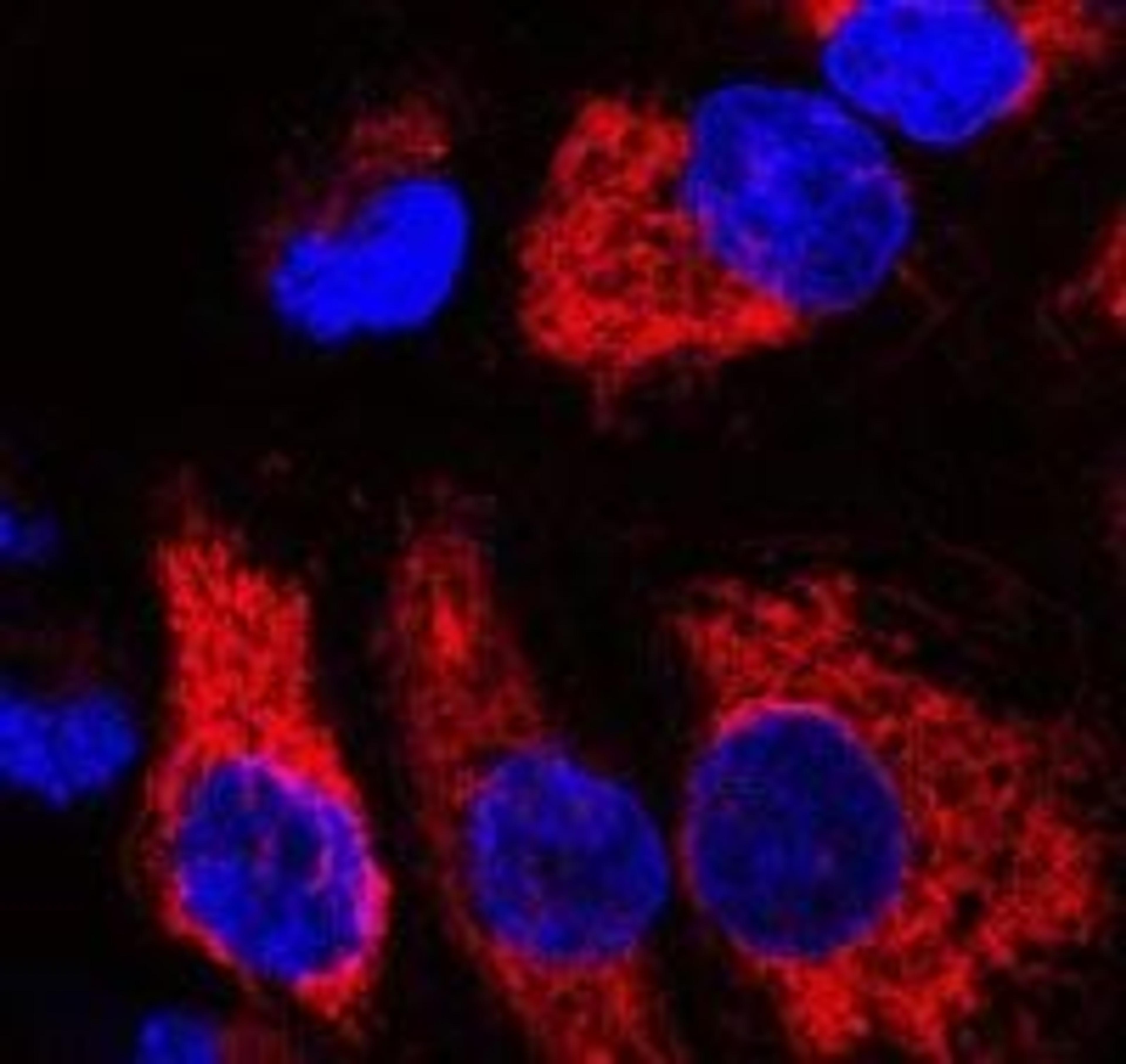
SmartFlare RNA Detection Probes
MerckSmartFlare Probes work by recognizing specific native RNA sequences within living cells, therefore there are many possible sequences that you could design to recognize your target of interest. Instead of lysed cells, switch to live cells. And while you’re at it, eliminate sample preparation and transfection steps all together. Live cell RNA detection is now possible, in a single incubation step using inert nanoparticle technology to specifically detect native RNA. And when you’re done, the probes exit the cells allowing you to perform downstream analyses with the same, unperturbed cells. Make the smart change!SmartFlare RNA Detection Probes are fueled by the NanoFlare technology developed by renowned chemist Dr. Chad Mirkin.What's Smart? Detect RNA expression in live cells for real time, physiologically relevant data Eliminate laborious, costly sample preparation Easy analysis on the fluorescent detection platform of your choice Nanoparticle based technology that allows cells to be used for downstream assays