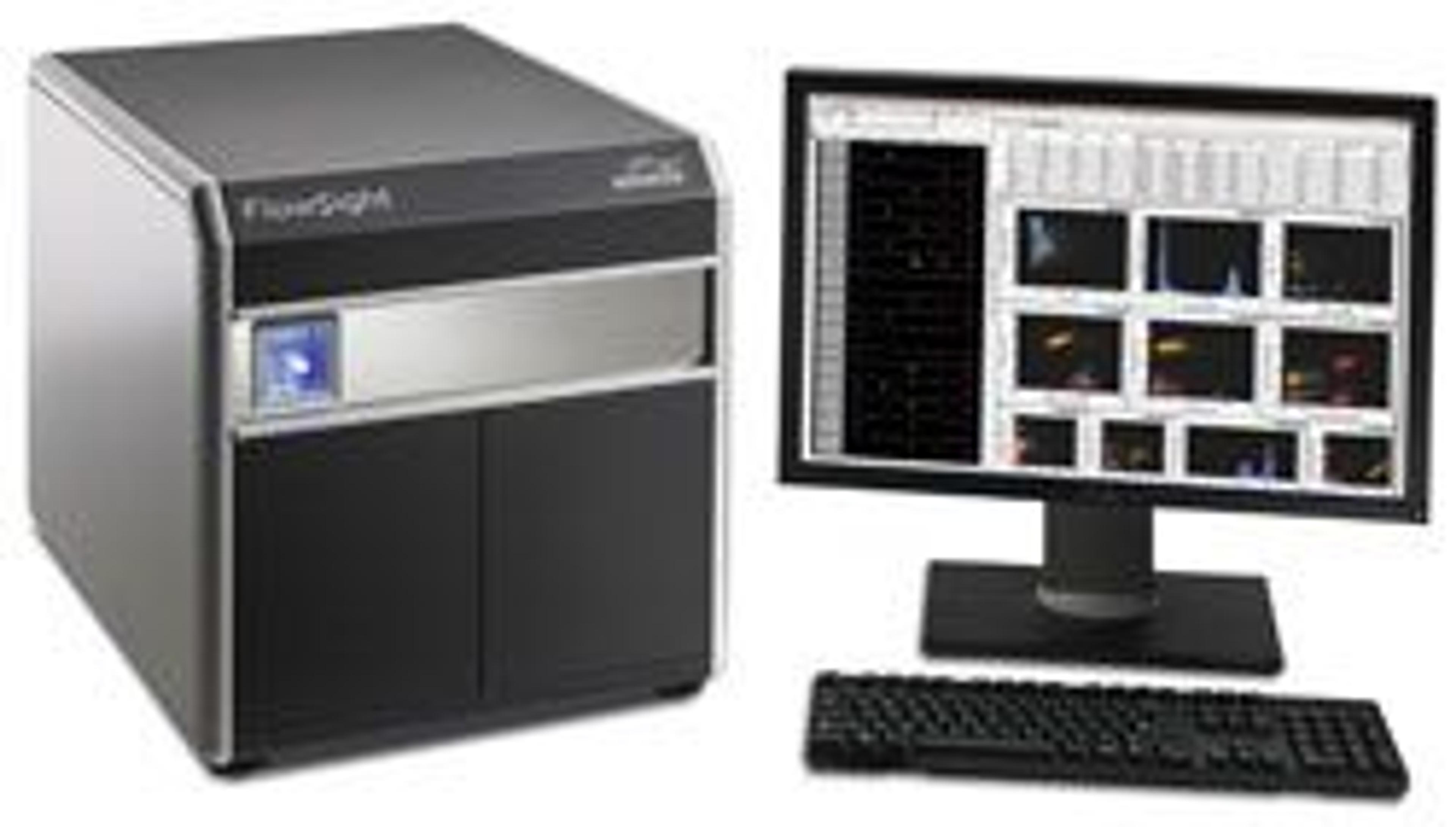
Image Stream®X Mark II Imaging Flow Cytometer
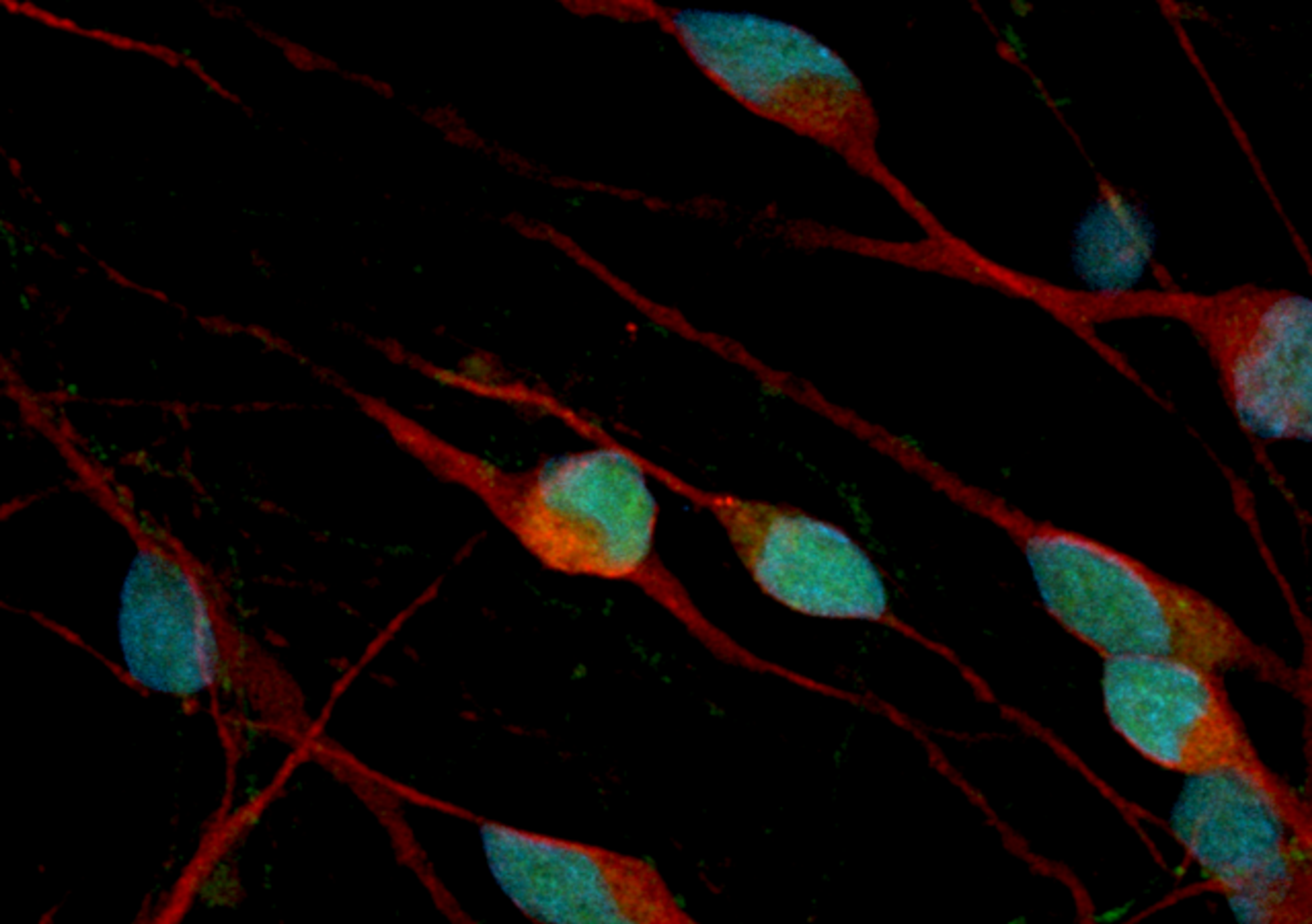
The revolutionary ImageStream®X Mark II Imaging Flow Cytometer combines the speed, sensitivity, and phenotyping abilities of flow cytometry with the detailed imagery and functional insights of microscopy. This unique combination enables a broad range of applications that would be impossible using either technique alone. This instrument produces multiple high-resolution images of every cell directly in flow, including brightfie…


The supplier does not provide quotations for this product through SelectScience. You can search for similar products in our Product Directory.
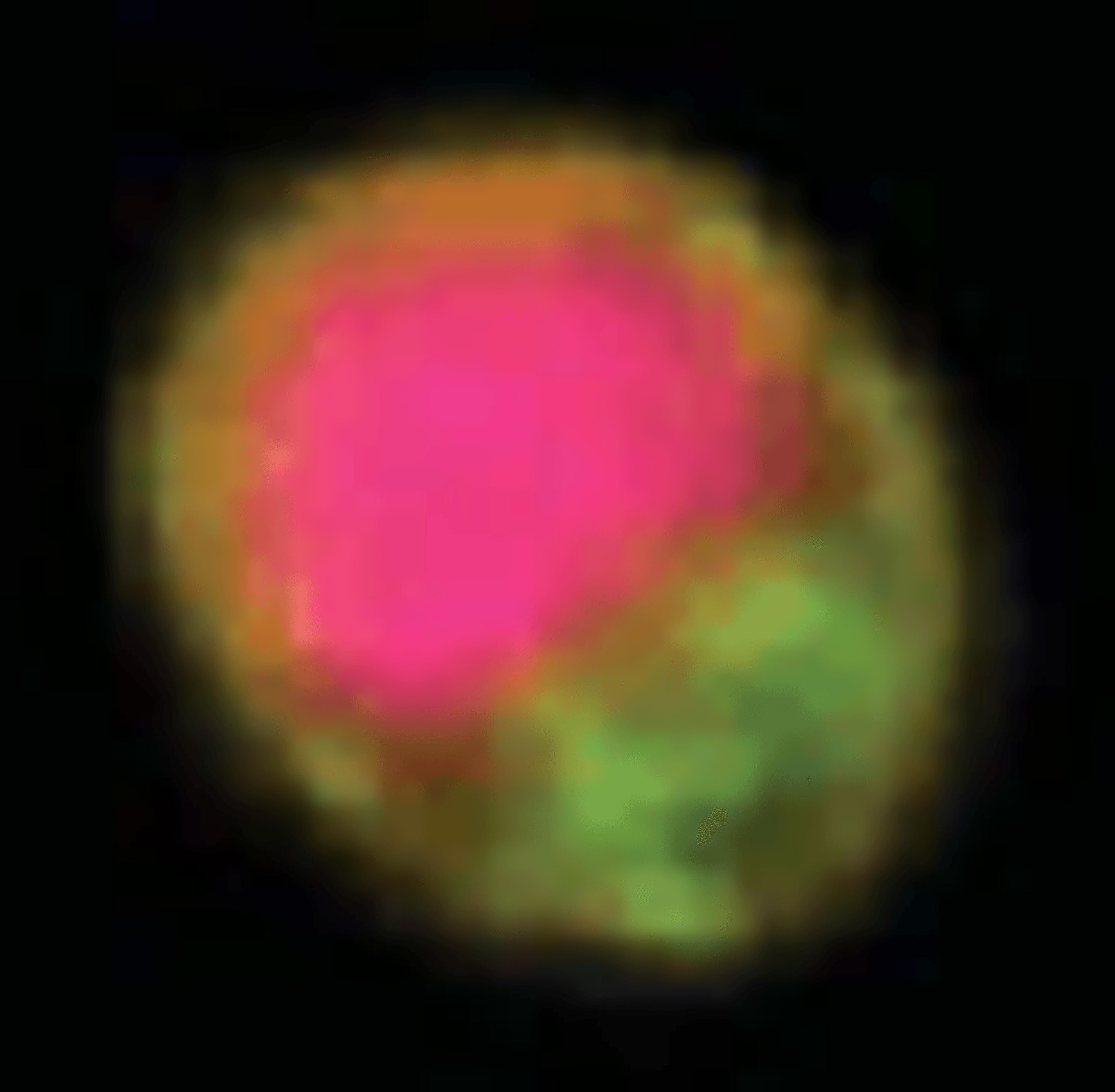
In flow cell imaging has great resolution.
Review Date: 28 Jun 2011 | Merck
The revolutionary ImageStream®X Mark II Imaging Flow Cytometer combines the speed, sensitivity, and phenotyping abilities of flow cytometry with the detailed imagery and functional insights of microscopy. This unique combination enables a broad range of applications that would be impossible using either technique alone.
This instrument produces multiple high-resolution images of every cell directly in flow, including brightfield and darkfield (SSC), and up to 10 fluorescent markers with sensitivity exceeding conventional flow cytometers. Compared to its predecessor, the new ImageStream®X Mark II Imaging Flow Cytometer offers a streamlined workflow, greater flexibility, and optimizations for rare cell applications.
Taken together, the capabilities of the ImageStream®X Mark II make it superior for traditional flow applications while greatly expanding the scope of flow cytometry. Applications include the study of cell-cell interactions, phagocytosis, apoptosis and autophagy, the characterization of circulating tumor cells, and many others.

The Immunology of Cancer
In this infographic, learn about the interactions between the immune system and cancer cells and how this understanding has led to breakthroughs in cancer vaccines and other novel immunotherapies. Detection reagents and instrument platforms have become increasingly sophisticated and sensitive for detection of key tumor antigens and associated immunology targets.
The Art and Science of Experimental Design with Fluorophores
Imaging cytometers are at the confluence of advances in flow cytometry and light microscopy. The ability to image tens of thousands of fluorescently labeled cells results in data that are both visually striking and highly quantitative. In this infographic, learn how Amnis® technology combines lasers and optics to accommodate hundreds of fluorophores and fluorescent reagents. Build an expert palette for your own memorable discoveries with imaging flow cytometry.
Enumeration of Live, Circulating Tumor Cells Using SmartFlare™ Probes and an Amnis® Imaging Flow Cytometer
Circulating tumor cells (CTCs) are released into the bloodstream from primary and metastatic cancers and are potentially useful for early cancer detection, diagnosis, and treatment. In this study, an Amnis ImageStream®X Mark II imaging flow cytometer was employed along with SmartFlare™ fluorescent RNA detection probes to collect imagery from large numbers of WBCs that were spiked with live SKBR-3 human breast cancer cells.
Quantitation of γ-H2AX Spots on the ImageStream
This application note shows ImageStream methods for quantifying DSB foci using automated γ-H2AX and 53BP1 spot counting, both in tumor cell lines and in primary murine CD8+ T cells. We also show quantification of MDC and γ-H2AX colocalization at DSB foci following irradiation.
Phagocytosis and Quantitation of Internal Zymosan, L. monocytogenes, M. tuberculosis, and Anthrax Spores
This application note shows that ImageStream-based analysis provides an objective measurement of pathogen internalization as well as quantitation of internal pathogens or pathogen material (zymosan particles, anthrax spores, M. tuberculosis, L. monocytogenes) within cells of the innate immune system.
Oceanography on the ImageStream: Diatoms and Other Phytoplankton
This application note shows an ImageStream-based analysis that combines morphology and fl uorescence-based classification of various phytoplankton species (including diatoms, dinofl agellates and cyanobacteria) grown in culture or collected from seawater.
Multiplexing Signal Transduction Measurement on the ImageStream for High Content Analysis and High Throughput
In this application note nuclear translocation of NF-κB is measured in THP1 cells on a per cell basis in an automated and quantitative manner in up to 64 samples simultaneously.
Identification and Measurement of Bacterial Size Using the ImageStream
This application note describes two bacterial studies performed using the ImageStream high speed imaging cytometer. By combining high speed image collection with quantitative image based measurements, the ImageStream enables accurate identification and classification of bacteria within heterogeneous samples.
FRET Analysis of Protein-Protein Interaction and Redox Sensor Folding Using the ImageStream
This application note shows FRET measurement with different fluorphore combinations and experimental applications using the ImageStream imaging cytometer, which enables quantitative FRET measurement directly from images of a large numbers of cells per sample.
Morphologic and Location-Based Classification of Differentiating Erythroid Lineage Cells using the ImageStream
In this application note human CD34-positive early hematopoietic cells were cultured to promote differentiation into erythroid lineage cells. During the differentiation program cells were sampled at 10, 13, or 16 days followed by staining with a nuclear dye and for expression of CD71 and glycophorin A, then analyzed immunophenotypically and morphologically on the ImageStream.