Quantitation of γ-H2AX Spots on the ImageStream
6 Feb 2011This application note shows ImageStream methods for quantifying DSB foci using automated γ-H2AX and 53BP1 spot counting, both in tumor cell lines and in primary murine CD8+ T cells. We also show quantification of MDC and γ-H2AX colocalization at DSB foci following irradiation.
Related products
Request Quote for All Products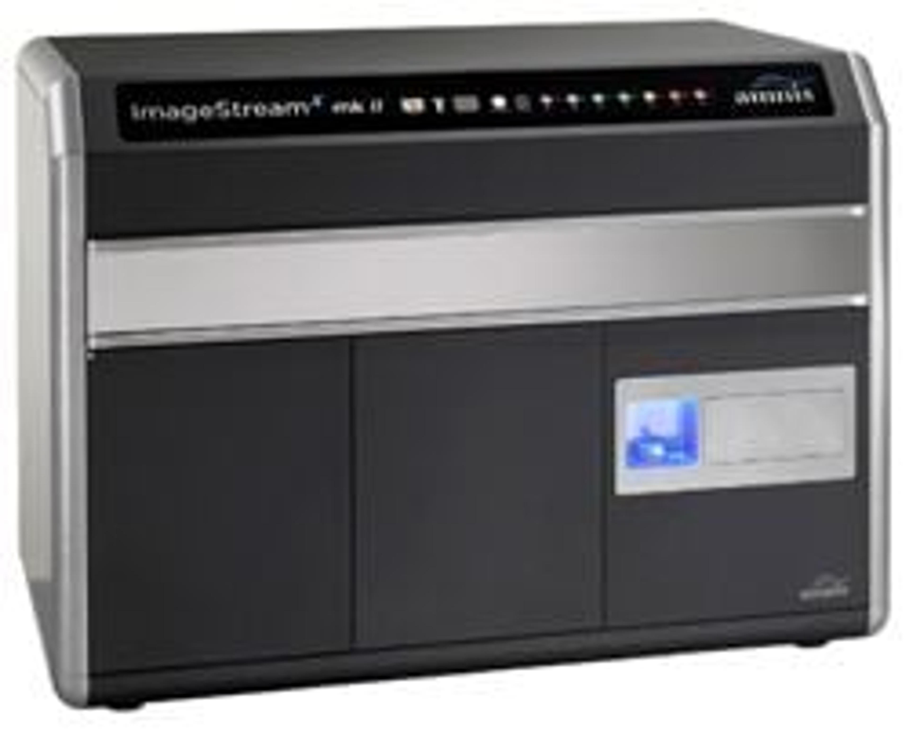
Image Stream®X Mark II Imaging Flow Cytometer
MerckThe revolutionary ImageStream®X Mark II Imaging Flow Cytometer combines the speed, sensitivity, and phenotyping abilities of flow cytometry with the detailed imagery and functional insights of microscopy. This unique combination enables a broad range of applications that would be impossible using either technique alone. This instrument produces multiple high-resolution images of every cell directly in flow, including brightfield and darkfield (SSC), and up to 10 fluorescent markers with sensitivity exceeding conventional flow cytometers. Compared to its predecessor, the new ImageStream®X Mark II Imaging Flow Cytometer offers a streamlined workflow, greater flexibility, and optimizations for rare cell applications. Taken together, the capabilities of the ImageStream®X Mark II make it superior for traditional flow applications while greatly expanding the scope of flow cytometry. Applications include the study of cell-cell interactions, phagocytosis, apoptosis and autophagy, the characterization of circulating tumor cells, and many others.