ResourceLife Sciences
Automated Imaging and Analysis of 3D Tumor Spheroids, Cancer Stem Cell Colony Formations
8 Mar 2019This application notes details information regarding the Celigo S which is a fully automated imaging system that has been developed for the analysis of tumorspheres, embryoid bodies and cancer stem cell colonies. It has been used in the creation of 3D tumor spheroid-based functional assays for target validation and drug evaluation.
Related products
Request Quote for All Products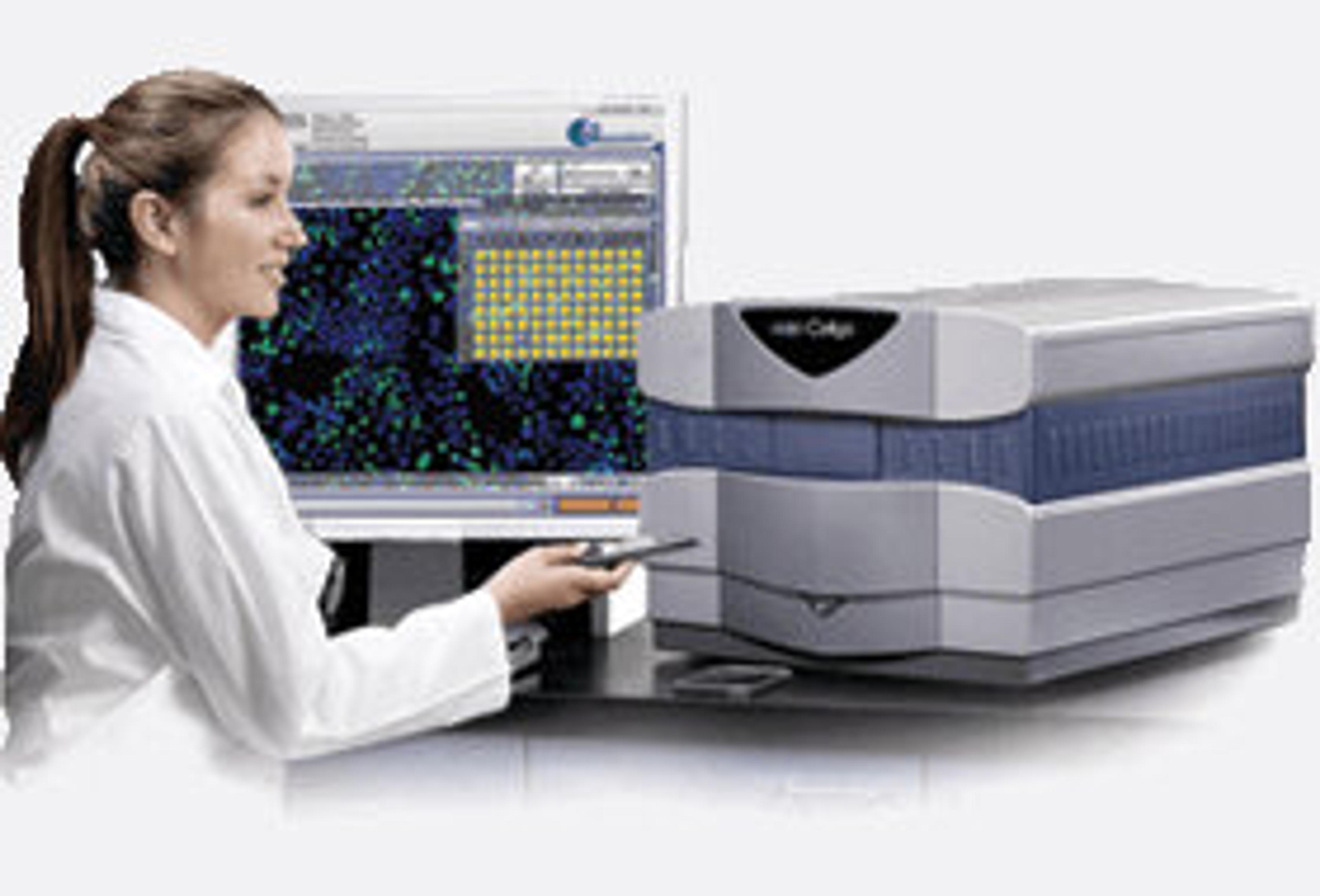
Celigo S Image Cytometer
A micro-well, plate-based multi-channel bright field and 3 fluorescent channels imaging cytometer for 2D and 3D culture using both adherent and suspension cells.
Tags
Flow Cytometry / Cell CountingFlow cytometers are used to count, sort and examine multiple characteristics of cells. Other cell analysis equipment includes image cytometers, cell counters, fluorescence-activated cell sorters (FACS), magnetic-activated cell sorters (MACS), and a range of flow cytometry assay kits. Flow cytometers can reveal information on cell viability, cell proliferation, apoptosis and cell cycle progression, as well as identify cell populations and intracellular or cell-surface molecules. Additionally, some flow cytometers, known as FACS, have an additional sorting function after analysis. Cell counters and image cytometers count live and dead cell populations and can also conduct cell proliferation assays. Find the best flow cytometers, cell counters and cell sorters in our peer-reviewed product directory: compare products, check customer reviews and receive pricing direct from manufacturers.Cell-Based AssaysCell-based assays are used to monitor the presence, quantity and activities of a desired cellular analyte including drug molecules or biomarkers. This can reveal information on cell health (apoptosis, cytotoxicity, viability and proliferation assays), cell metabolism, cell migration and cell signaling mechanisms. Find the best cell-based assay products, kits and equipment with our peer reviewed product directory: compare products, check customer reviews and receiving pricing direct from manufacturers.Cancer CellsCancer cells are abnormal cells that divide uncontrollably, leading to the formation of tumors and the spread of cancer. Studying cancer cells is crucial for developing new treatments and understanding tumor biology. Explore cancer cell research products in our peer-reviewed product directory; compare products, check reviews, and get pricing directly from manufacturers.Cell ImagingCell imaging can be achieved using a number of techniques including confocal microscopy, transmission electron microscopy, atomic force microscopy, and light sheet microscopy.TumorsTumor research focuses on understanding abnormal cell growth that leads to cancer. Identifying biomarkers, studying tumor microenvironments, and developing targeted therapies are critical for advancing cancer treatment. Early detection and personalized treatment options are key to improving outcomes for patients. Browse our peer-reviewed product directory to explore tools for tumor research, diagnostics, and cancer therapies; compare products, read customer reviews, and get pricing directly from manufacturers.Cell Based Automation