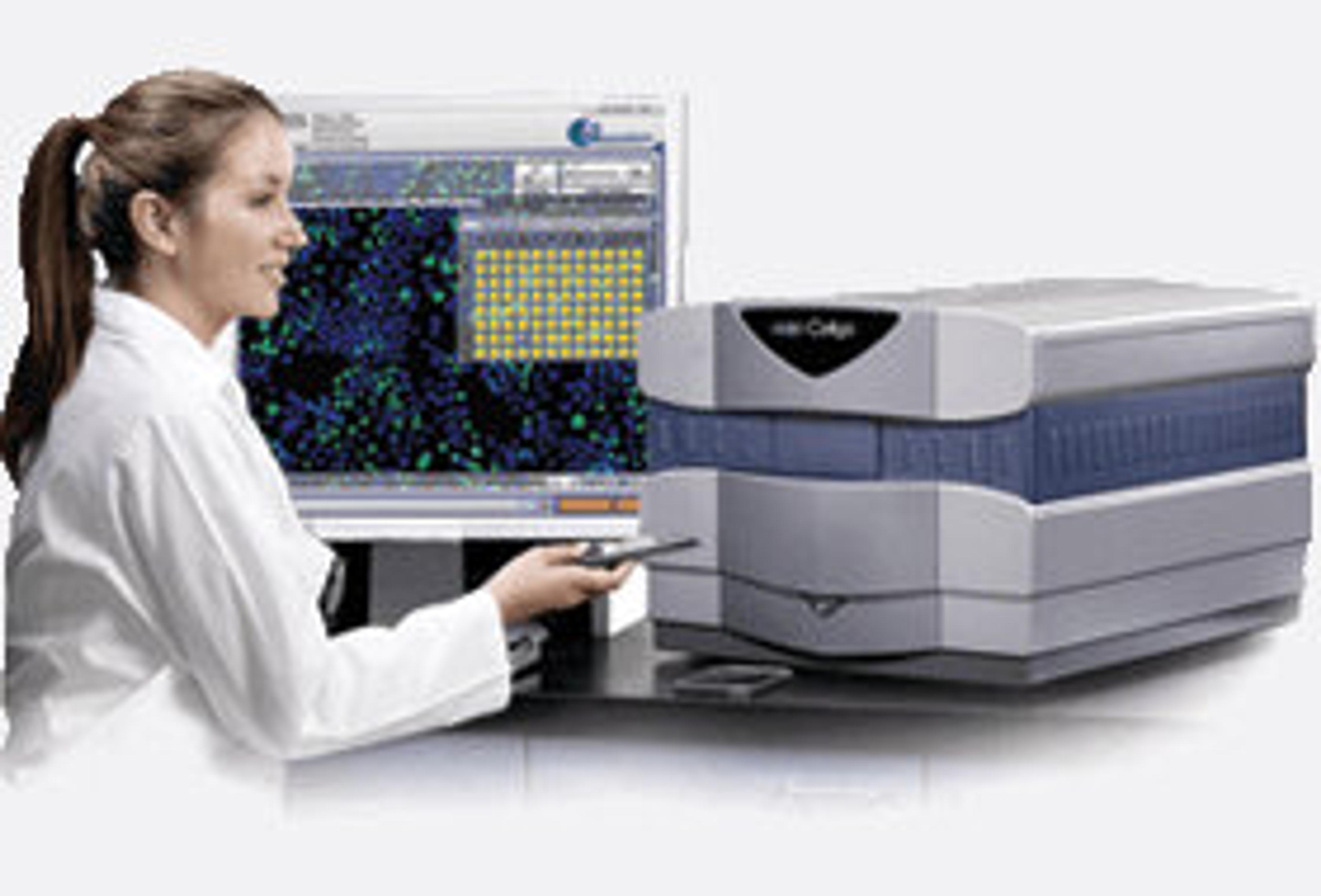
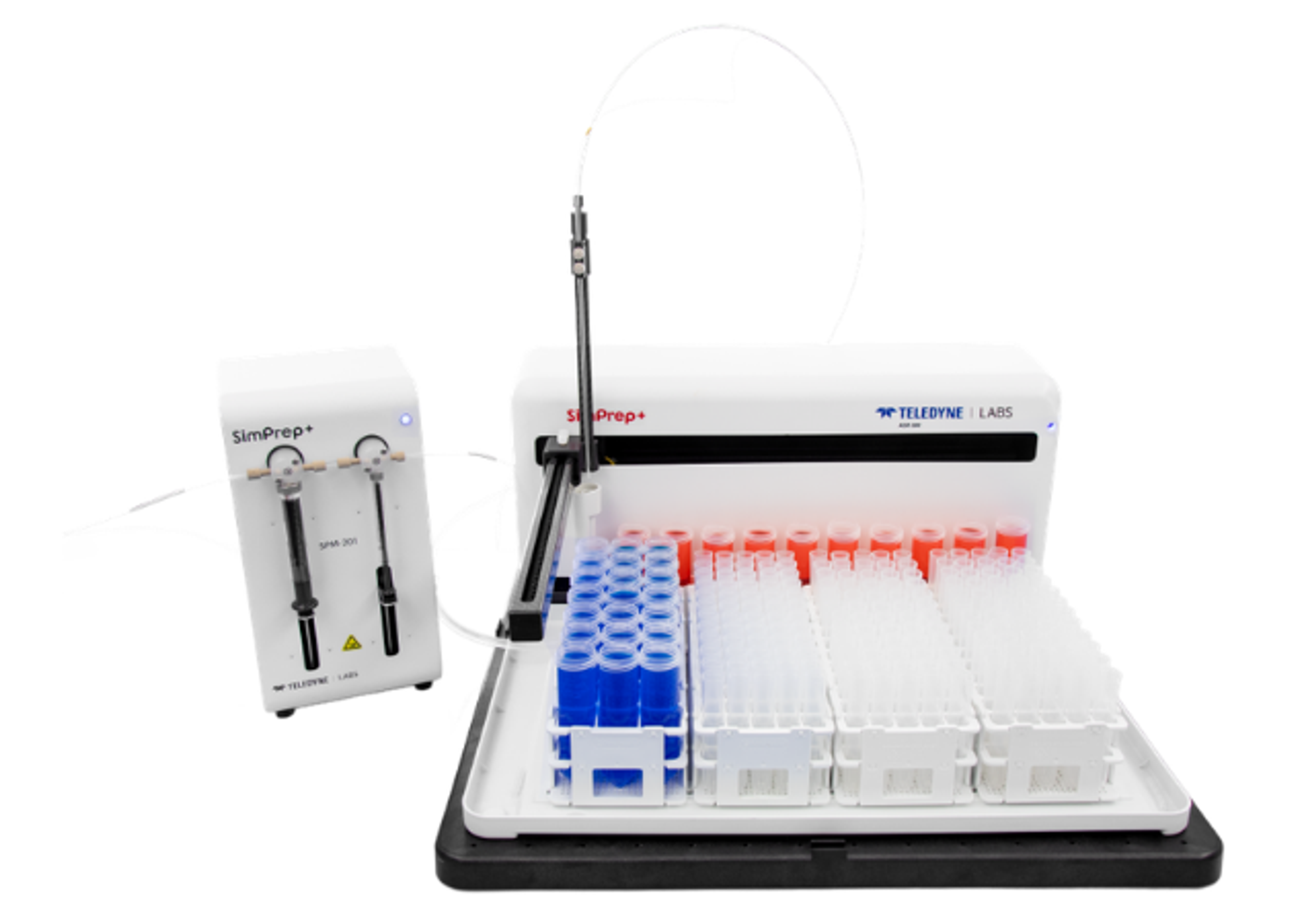
Celigo S Image Cytometer
A micro-well, plate-based multi-channel bright field and 3 fluorescent channels imaging cytometer for 2D and 3D culture using both adherent and suspension cells.

Plate-based imaging cytometer

The Celigo S Image Cytometer offers best-in-class bright field imaging capabilities with 3 channels of fluorescent imaging: Blue (Ex 377nm / Em 470nm), Green (Ex 483nm / Em 536nm) and Red (Ex 531nm / Em 629nm). It is ideal for multiplexed assays and provides high-speed, fully automated imaging and quantification of suspension, adherent, tumor spheroids, iPSC and cancer stem cell colonies. The system images every cell in every well, without any well edge effect. A 96-well plate will take less than 5 minutes to image. System is compatible with automation platforms for full integration.

Rapid, Label-Free Counting and Characterization of Live Embryoid Bodies
This application note explores the benefits of a fast, automated method for embryoid body (EB) analysis. The Celigo™ Colony Counting: Embryoid Body Application by Nexcelom Bioscience quickly and accurately analyzes EB populations to assess the number, size, and shape of live EBs within multi-well plates.
Automated Imaging and Analysis of 3D Tumor Spheroids, Cancer Stem Cell Colony Formations
This application notes details information regarding the Celigo S which is a fully automated imaging system that has been developed for the analysis of tumorspheres, embryoid bodies and cancer stem cell colonies. It has been used in the creation of 3D tumor spheroid-based functional assays for target validation and drug evaluation.
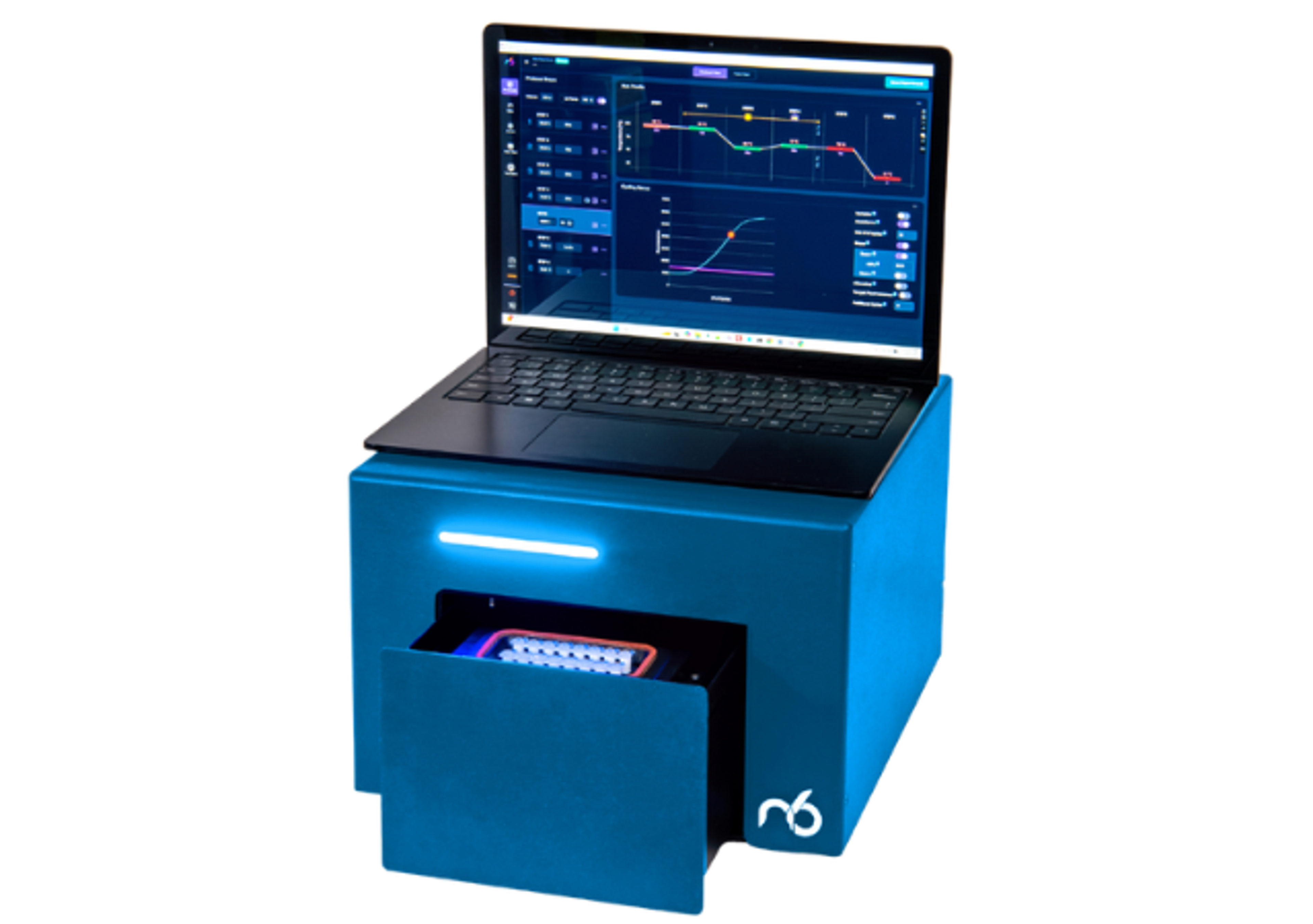
Automated Cell Growth Tracking for Cytotoxicity and Proliferation
The Growth Tracking application automatically integrates label-free cell counts of the same well/flask from different time points to provide direct measurement of growth rates and doubling times, which is also a good overall assessment of cell health.
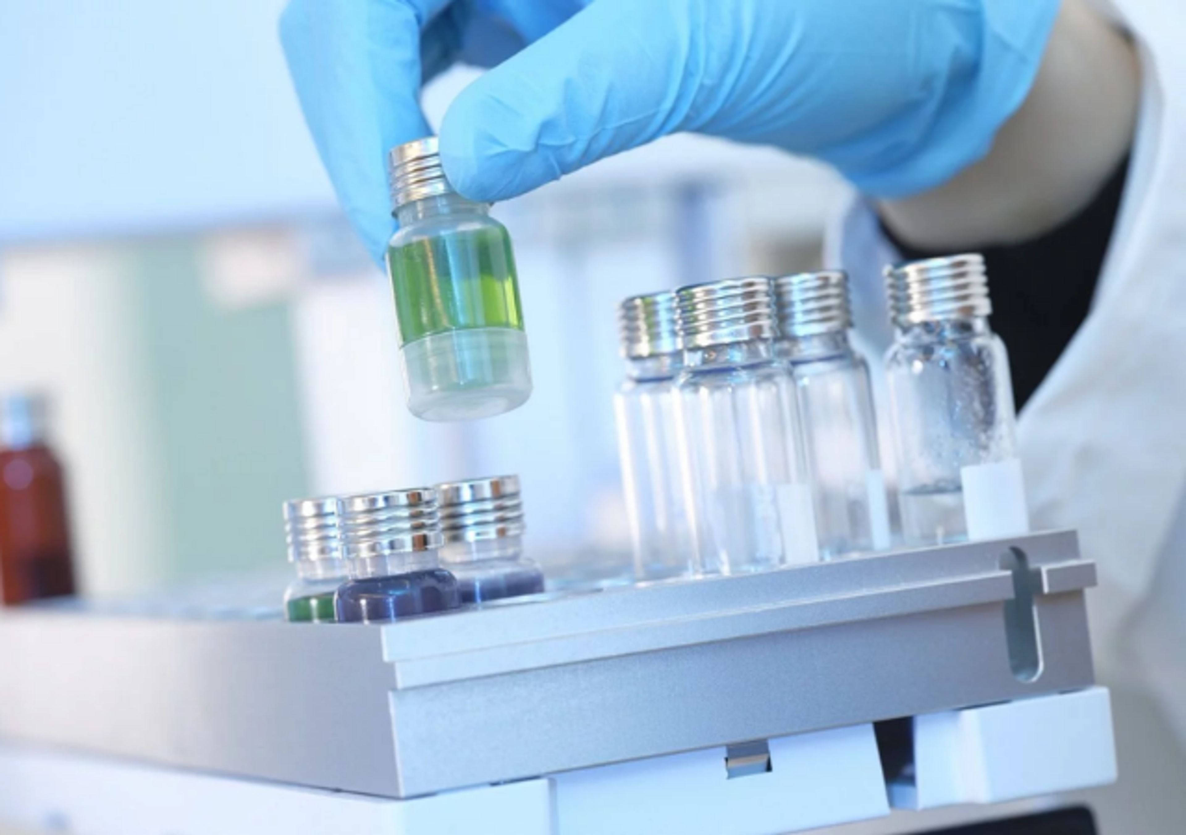
Cell Line Development - Single Cell Detection, Clonal Validation, Transfection
The process of developing a cell line to produce a specific protein or antibody involves multiple stages, all of which can be greatly aided by Celigo imaging cytometer.