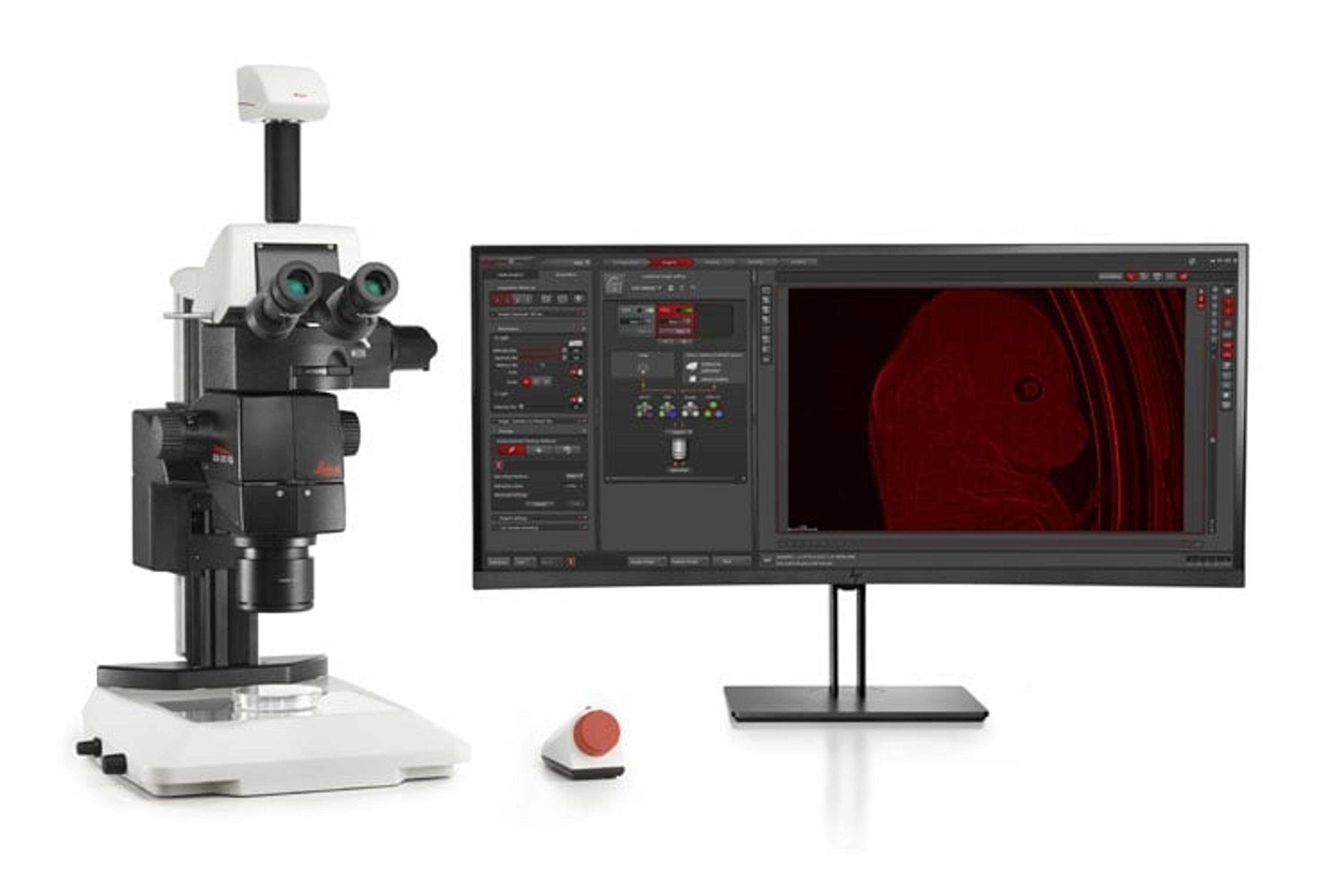
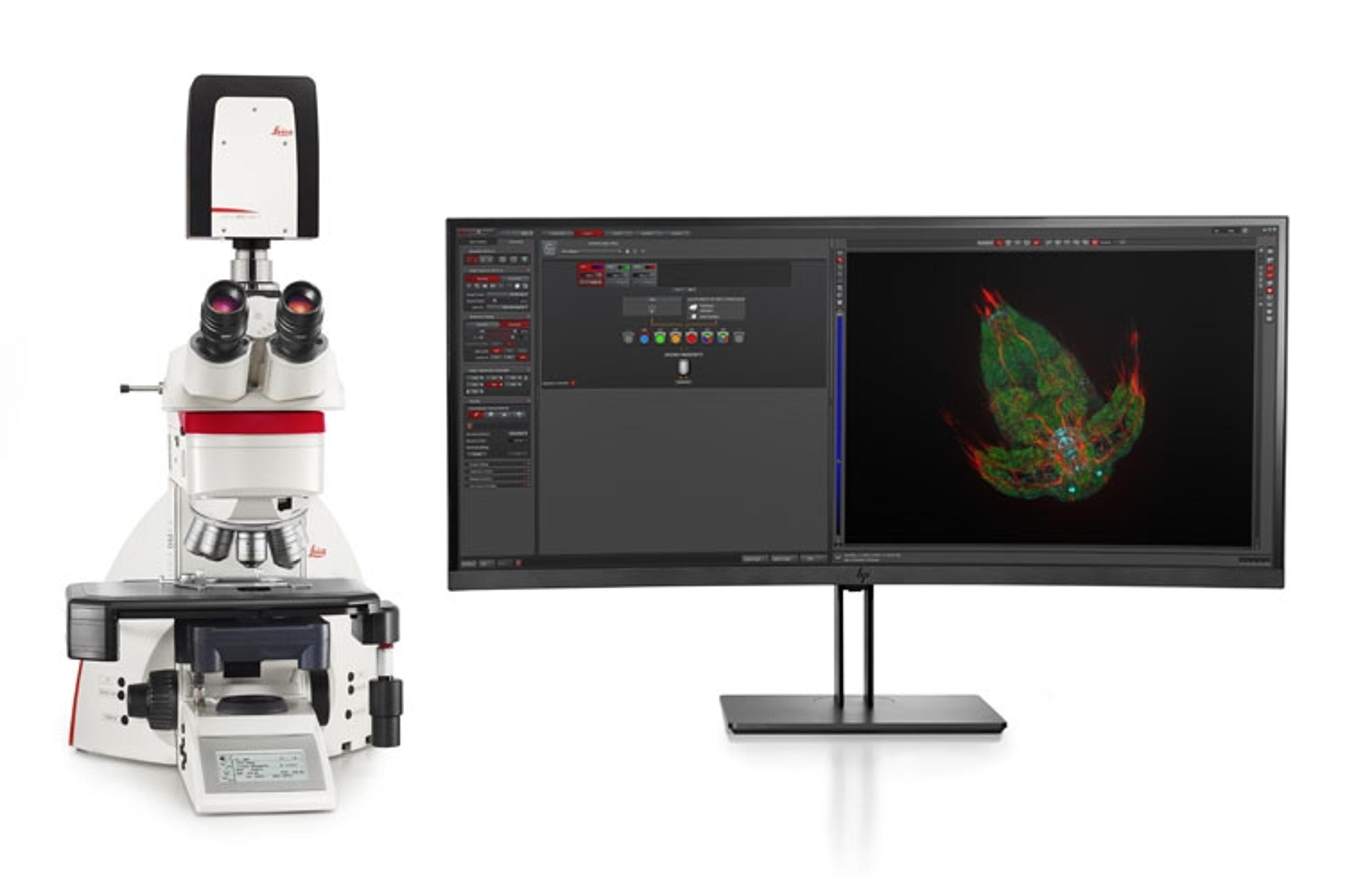
THUNDER Imager Model Organism
Fast and easy 3D exploration of whole organisms for developmental or molecular biology research
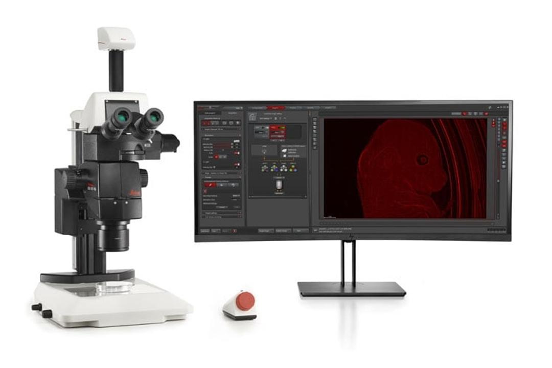
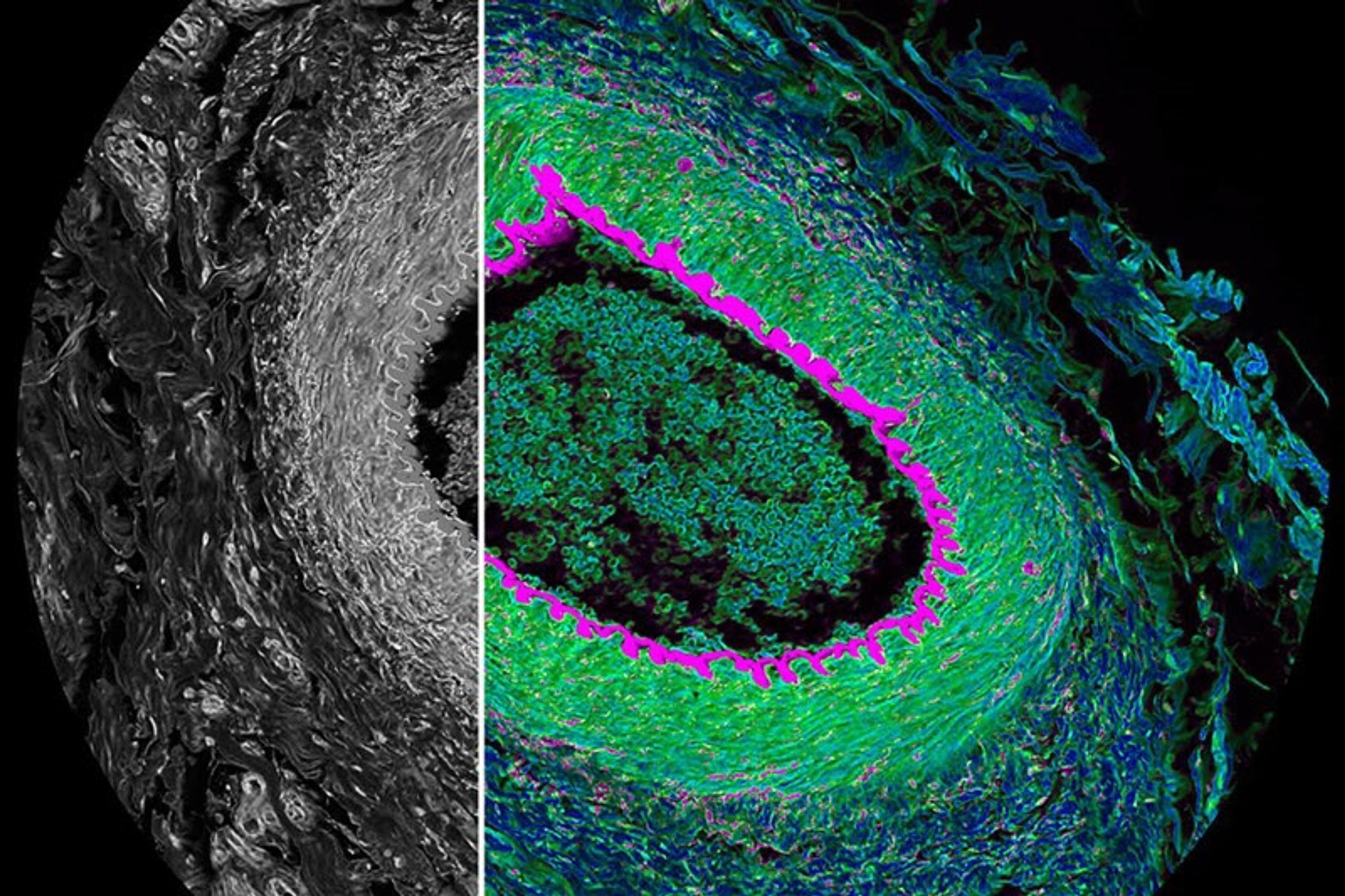
THUNDER Imager Model Organism – Mouse

Courtesy Yves Lutz, IGBMC (FR)


Receive your quote directly from the manufacturer.
The THUNDER Imager Model Organism allows fast and easy 3D exploration of whole organisms for developmental or molecular biology research. Thanks to Computational Clearing, your images reveal the finest structural details. No hassle with out-of-focus blur while maintaining the capabilities and ease-of-use typical for Leica stereo microscopes. A THUNDER Imager Model Organism is the optimal instrument for studying, e.g., Drosophila, C. elegans, zebrafish, plants, and mice. One device for screening, positioning, and imaging your specimen. Simplify your workflow and study model organisms from a large overview to the highest detail.
Advantages for your research are:
- Rapid acquisition of blur-free images showing fine details, even from deep within thick organisms
- Keep even large model organisms under excellent physiological conditions during imaging
- Simplify your organism handling for a more efficient imaging and analysis workflow
THUNDER Imagers feature the innovative Leica technology Computational Clearing. It efficiently removes out-of-focus blur in real time, enabling the meaningful use of 3D specimens with camera-based fluorescence microscopes. The high sensitivity of the system ensures low phototoxicity and photobleaching, i.e., higher throughput with optimal conditions.
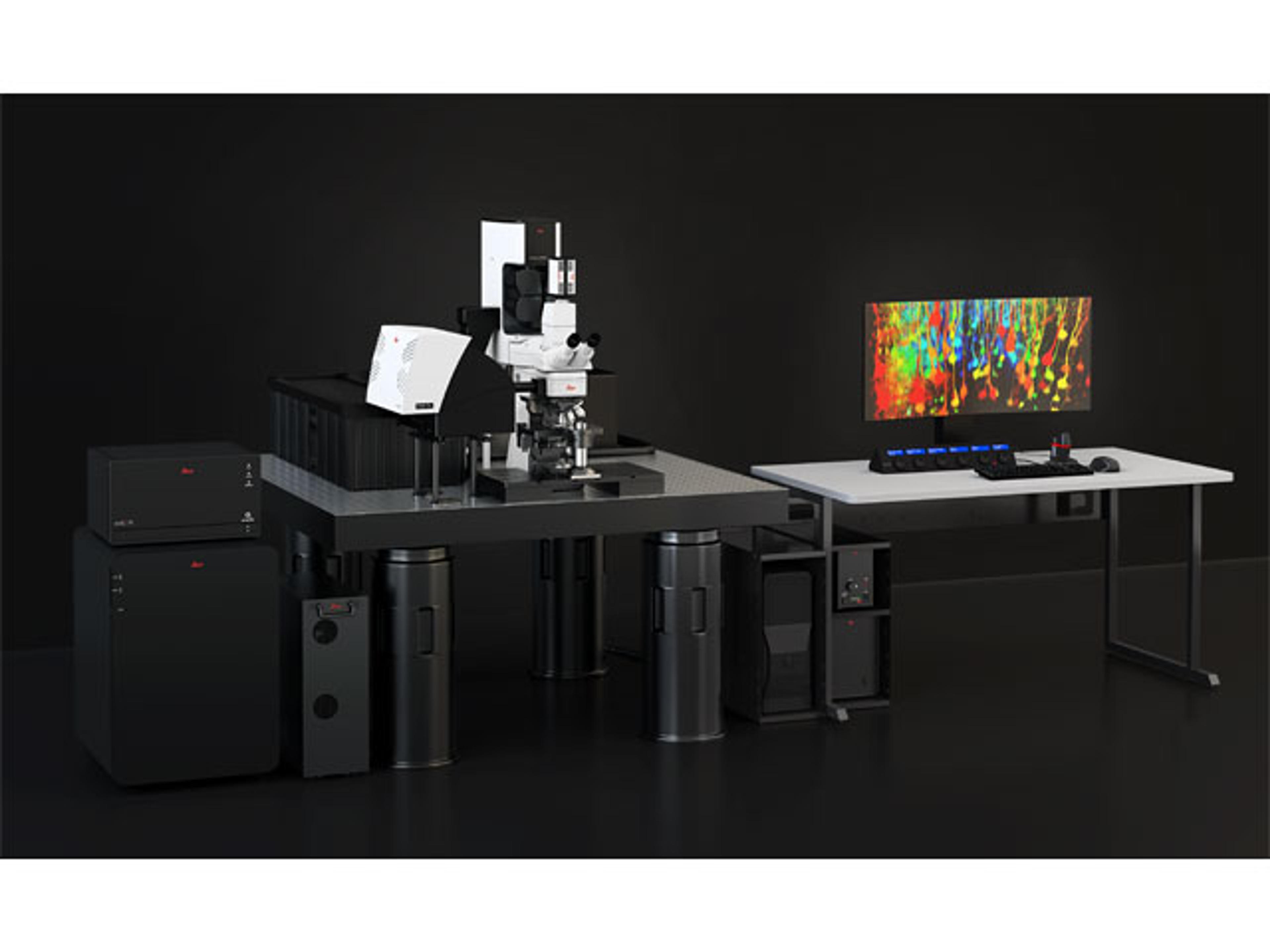
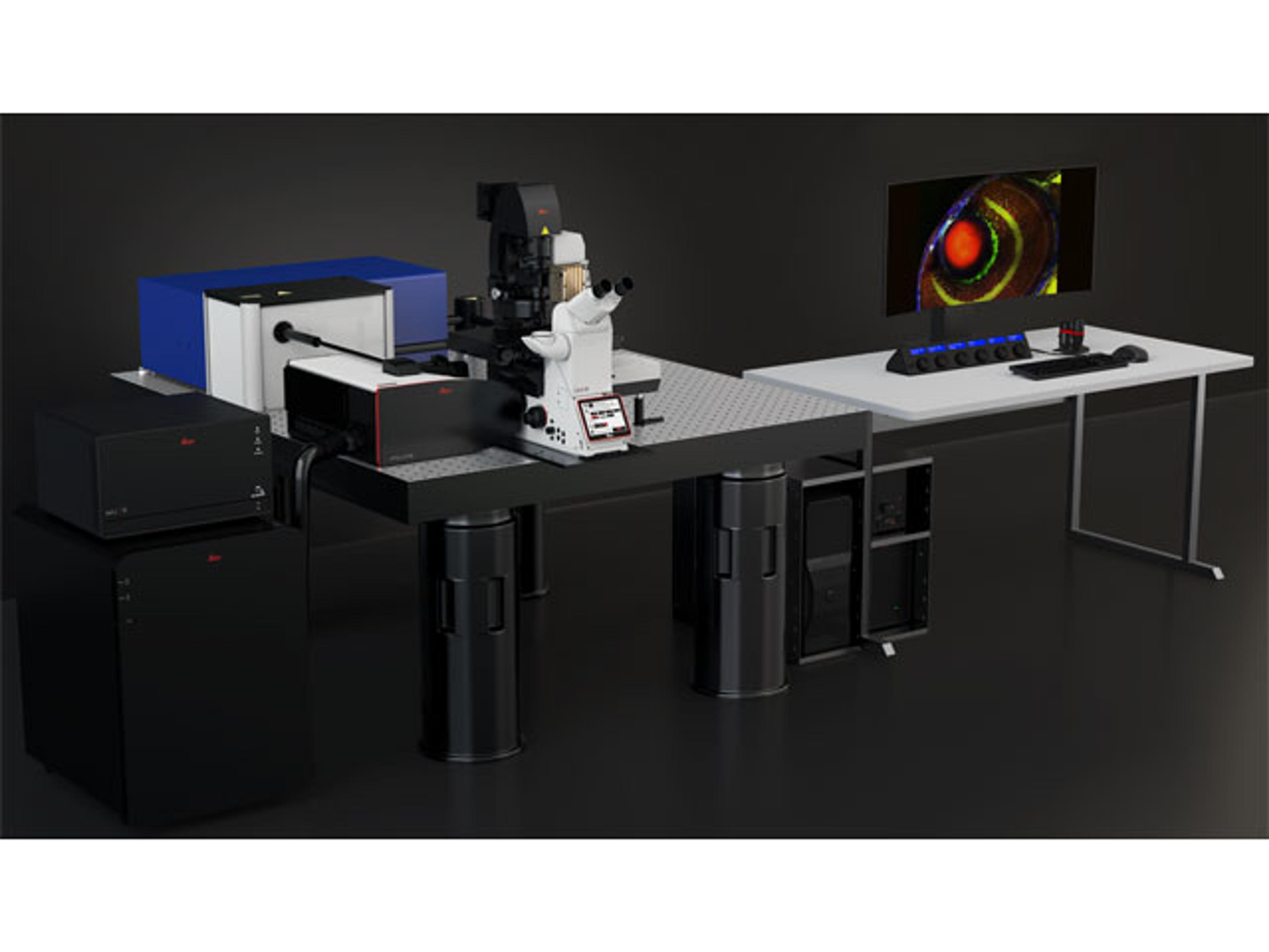
Find the THUNDER Imager that’s right for you
The THUNDER Imager Model Organism is part of the THUNDER family of imaging systems. Whether you are looking for a dedicated high-end imaging system that excels in a given application, or a versatile solution for a lab running different kinds of assays with various samples, we’ve got you covered.
Brochures

THUNDER Imaging Systems
THUNDER Imagers with Computational Clearing define a new class of instruments for high-speed, high-quality imaging of thick 3-dimensional specimens.
The power of pairing adaptive deconvolution with Computational Clearing
Deconvolution is a computational method used to restore the image of the object that is corrupted by the point spread function (PSF) along with sources of noise. In this technical brief, learn how the deconvolution algorithm offered by Leica Microsystems allows you to overcome losses in image resolution and contrast in widefield (WF) fluorescence microscopy due to the wave nature of light and the diffraction of light by optical elements. Explore the methods of user-controlled or automated deconvolution to see and resolve more structural detail.
An introduction to Computational Clearing - A new method to remove out-of-focus blur
Commonly in widefield (WF) microscopy, the imaging of objects in the object plane results in captured images that are compounded by image haze (referred to as background noise). The presence of background noise in an image obscures structural features from being resolved. Many software packages include background subtraction algorithms to enhance the contrast of features in the image by reducing background noise. The most common methods used to remove background noise from WF images are rolling ball and sliding paraboloid. Recently, Leica Microsystems introduced their own background subtraction method called Instant Computational Clearing (ICC), which is present in all Leica THUNDER WF imaging platforms. Find out more about how the technology works.
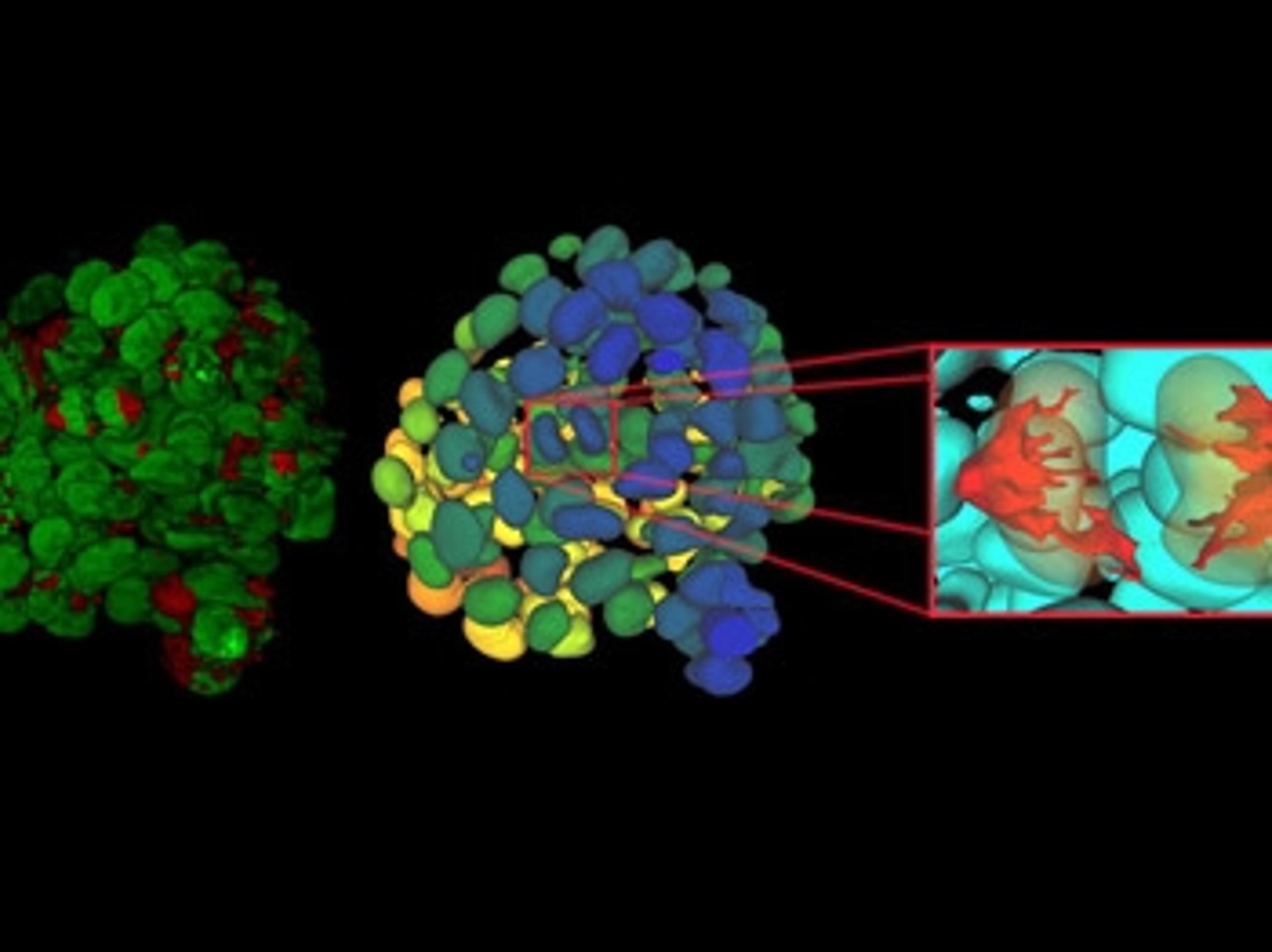
Computational Clearing with THUNDER Imagers
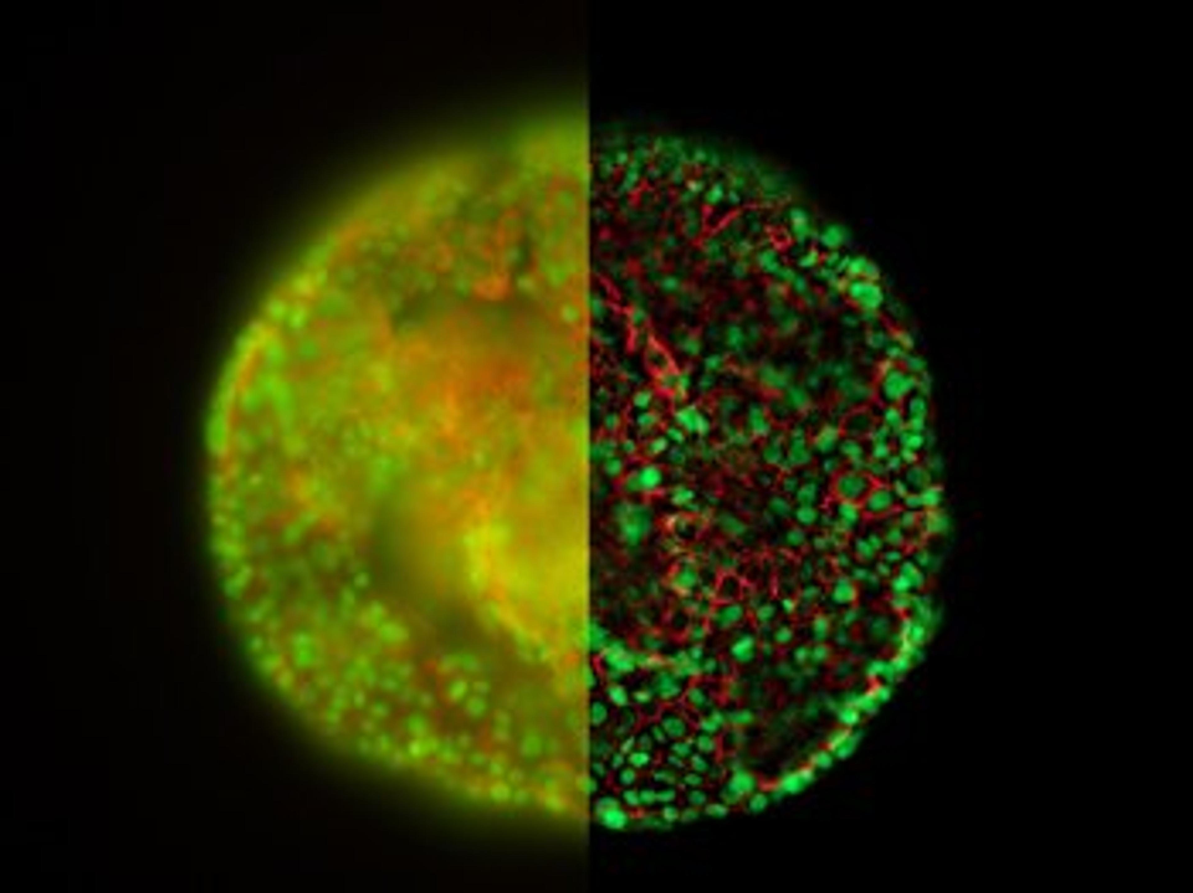
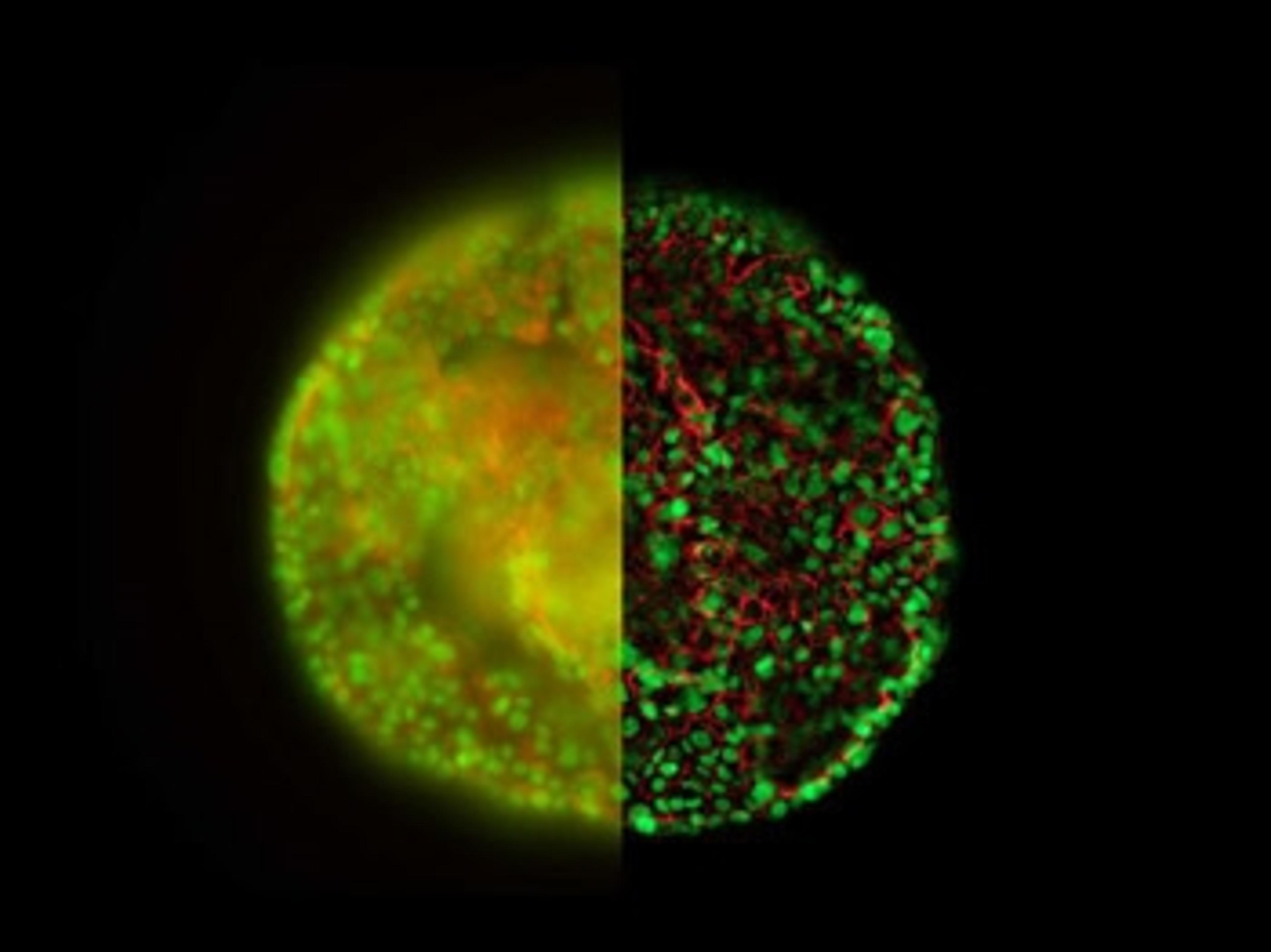
Computational Clearing efficiently differentiates between signal and background by taking the size of the targeted specimen features into account. This approach makes image details immediately visible which formerly were not revealed. Acquire one image and you have stunning results displayed instantly on the screen.
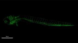
Zebrafish larvae (72 hours post fertilization). Blood vessels (green). Sample courtesy: Dr. Almary Guerra & Dr. Didier Stainier, Max Planck Institute for Heart and Lung Research, Bad Nauheim (Germany).

CrestOptics and Leica Microsystems collaborate to expand capabilities of spinning disk microscopy
The partnership will incorporate CrestOptics' CICERO spinning disk imaging unit within Leica Microsystems' THUNDER Imager Cell system
Cut Through the Haze: Can the Latest 3D Imaging Technology Help You Remove Out-of-Focus Blurring?
In this article, discover how Computational Clearing technology from Leica Microsystems quickly resolves the longstanding challenge of blurry contrast when imaging thick sections
New Leica imagers set to transform visualization of 3D samples
The THUNDER imager family enables users to decode 3D biology in real-time*