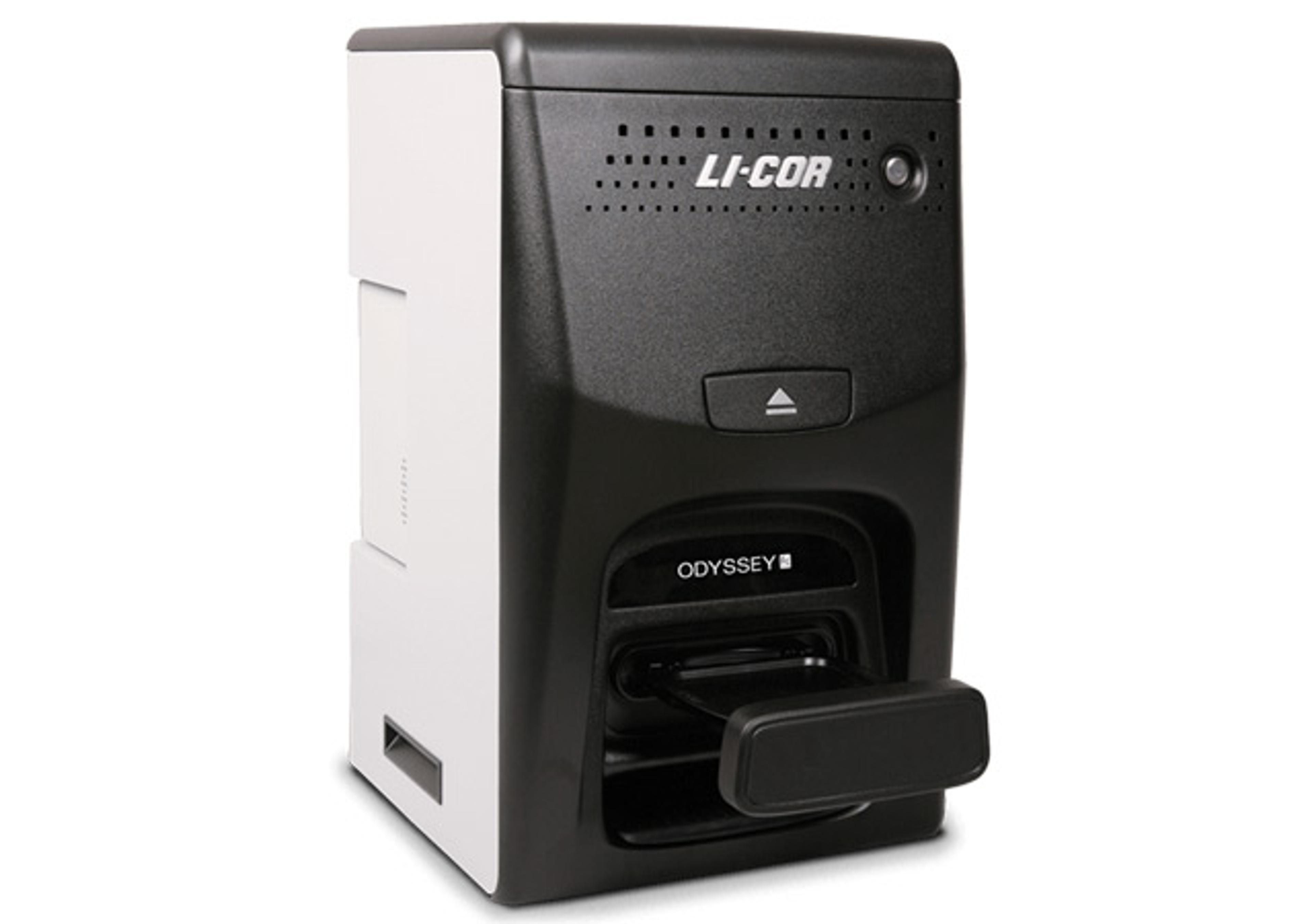
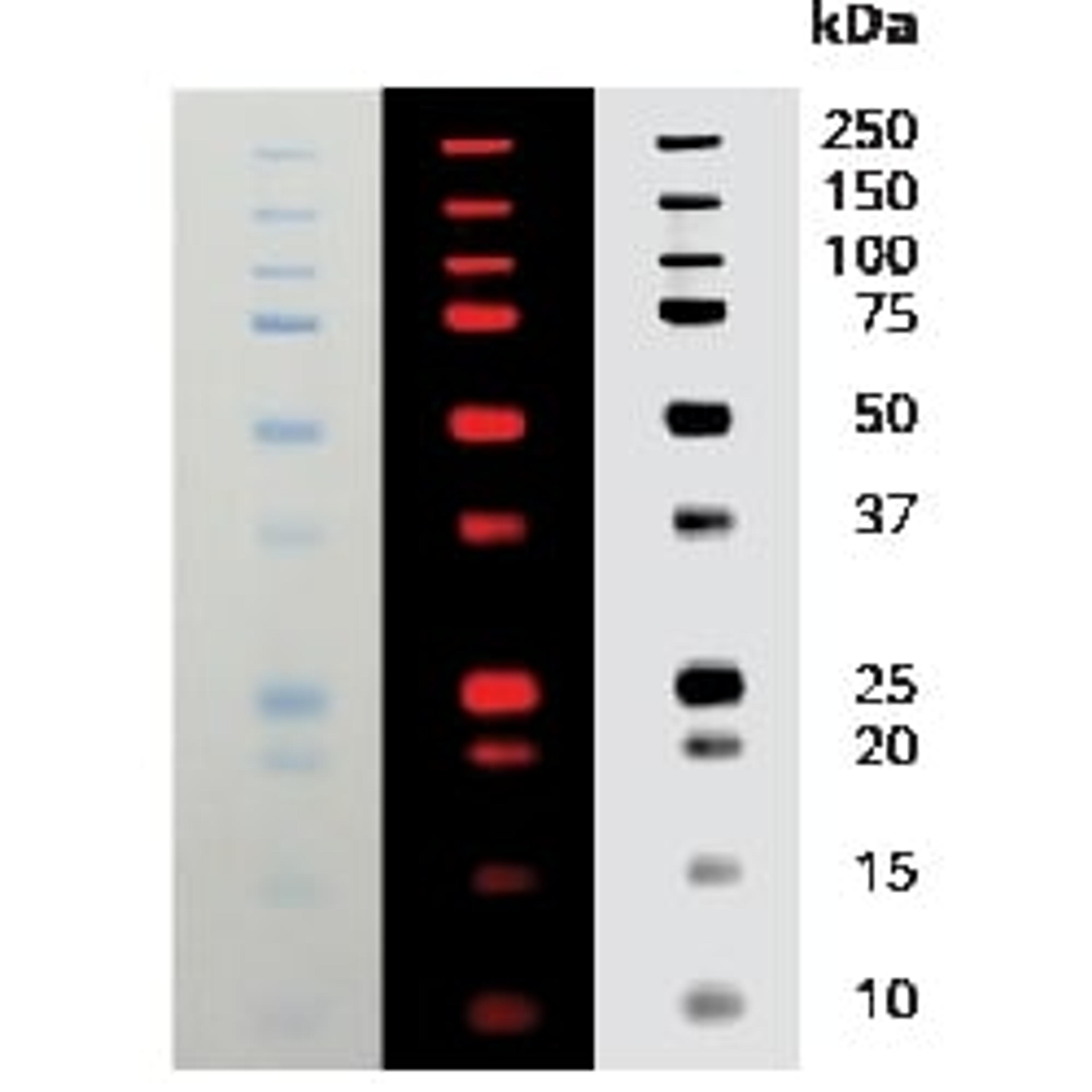
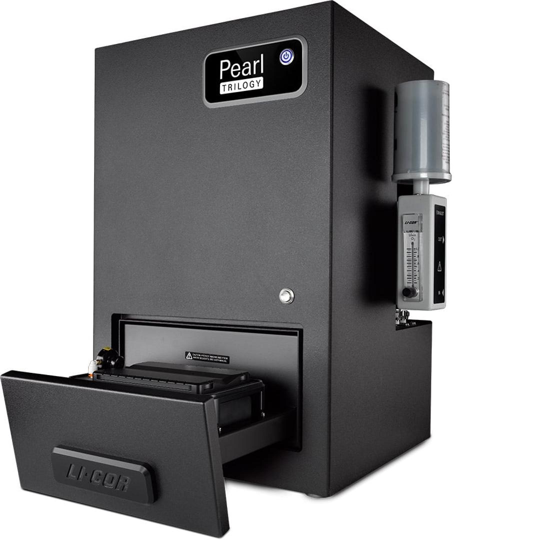
Pearl Trilogy Small Animal Imaging System
The Pearl Trilogy Small Animal Imaging System is a small animal in vivo imaging system that offers bioluminescence detection and dual-channel near-infrared fluorescent detection.
Receive your quote directly from the manufacturer.
The imager is very easy to use and very reliable.
animal imaging
The imager is small and compact. Very easy to use. The customer service we receive is great.
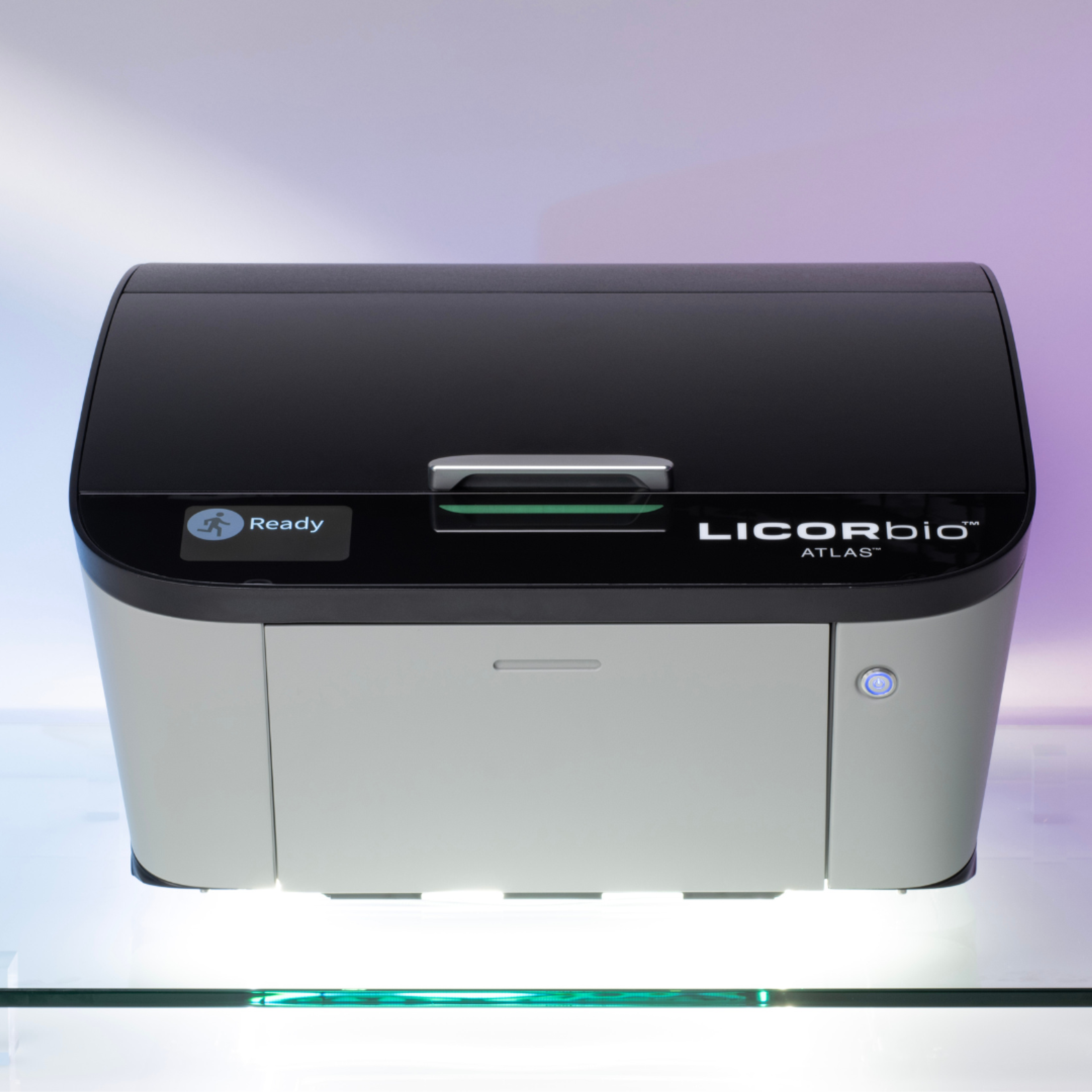
Review Date: 30 Nov 2015 | LICORbio
small animal imaging with Zenografts model
This machine is very simple to use. You get reliable results each time. Analysis is very straight forward
Review Date: 28 Sept 2015 | LICORbio
The Pearl Trilogy Small Animal Imaging System is a small animal in vivo imaging system that offers bioluminescence detection and dual-channel near-infrared fluorescent detection. The design simplicity minimizes user effort through one-button image acquisition. The revolutionary FieldBrite™ Xi2 optical technology results in exceptional sensitivity for NIR detection, with deeper tissue penetration and earlier target detection.
- Discover Optical Imaging in a New Light: Minimize tissue autofluorescence in your measurements. Get high signal-to-noise ratios and exceptional sensitivity for in vivo imaging with near-infrared (NIR) fluorescence detection.
- Acquire Consistent Data: Capture an accurate representation of in vivo molecular activity. With uniform illumination across the entire imaging area, you’ll get consistent data that does not vary spatially.
- Make Every Short Your Best Shot: No more adjusting camera settings before taking each image. Camera optics provide an optimal imaging setup for all your in vivo data acquisition needs, so you always get it right the first time.
Pearl Trilogy comes with Image Studio Software, an easy-to-use imaging software with an application-driven ribbon interface makes training for new users fast and simple. And, all LICORbio imaging systems and products give you access to our outstanding team of expert support scientists.

Near-Infrared Optical Imaging in Mouse Models of Parkinson's Disease, Alzheimer's Disease, and Contact Dermatitis
This scientific poster from LI-COR demonstrates that PSVue 794 is highly penetrable in the brain and may be used as an imaging probe in various nervous system models, and possibly other disease models. It is also shown that IRDye 800CW 2-DG can be used to visualize inflammatory environments.
<em>In Vivo</em> Breast Cancer Characterization Imaging using Two Monoclonal Antibodies Activatably Labeled with Near Infrared Fluorophores
Antibody-based immunohistochemistry (IHC) of tissue specimens is a common method of identifying expressed proteins in cancer cells. Molecular imaging is a potential method of performing similar IHC studies in vivo without the requirement for biopsy or tumor excision. This article discusses the use of optically activatable imaging agents, which are only fluorescent when bound to their cognate receptor, opening the possibility of doing in vivo multi-color IHC.
BrightSite™ Near-Infrared Fluorescent Targeting Agents for Optical Imaging of Disease
This application note by LI-COR Biosciences assesses the use of NIR dyes for clinical in vivo imaging. In the NIR wavelength range (700–900 nm) tissue autofluorescence is lower and light can penetrate more deeply than in visible light wavelengths. Here NIR fluorophores are used to image mice tumours and bone structure.