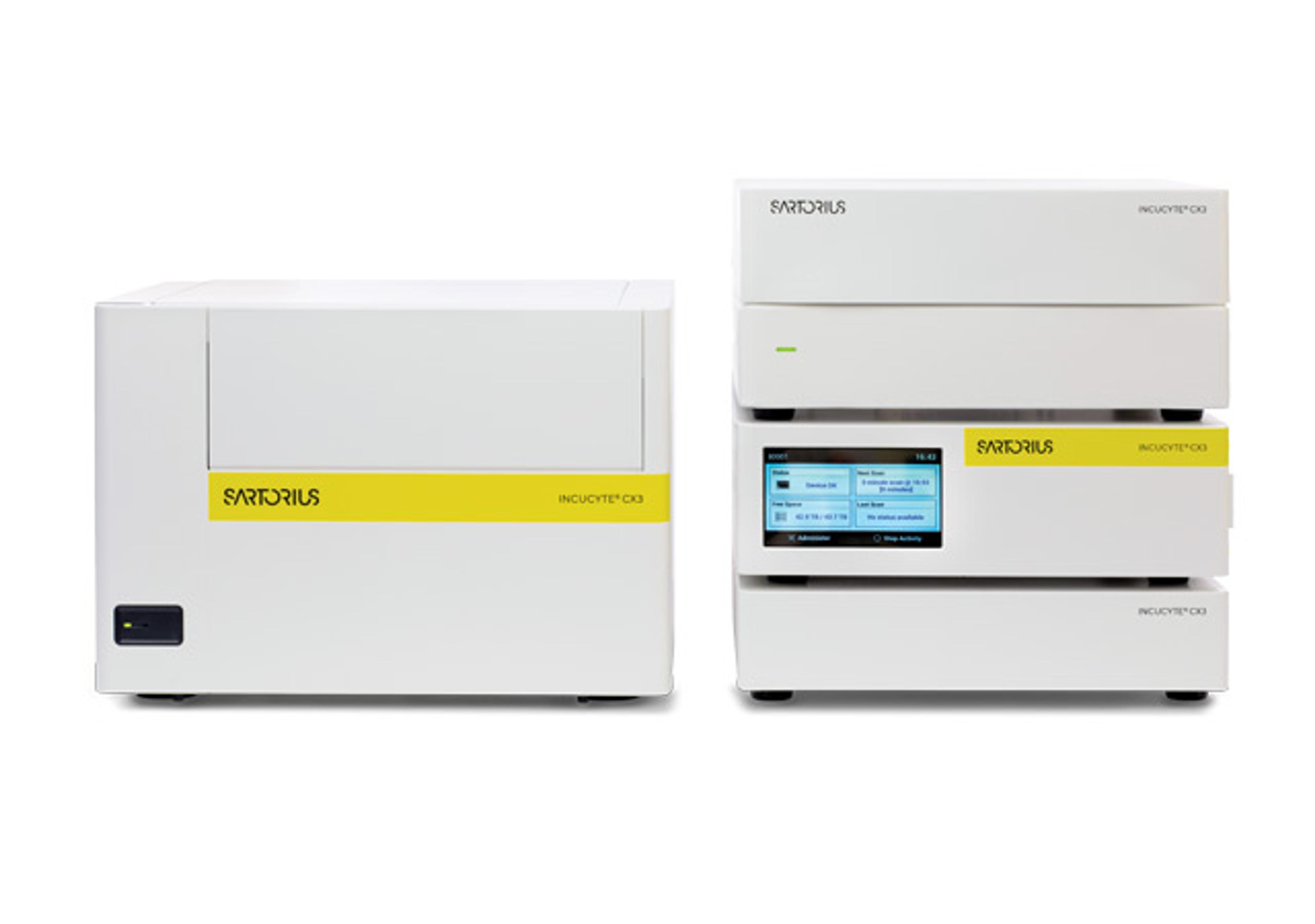
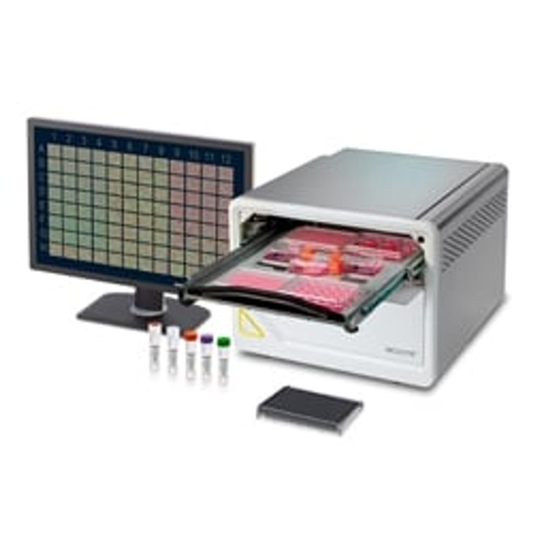
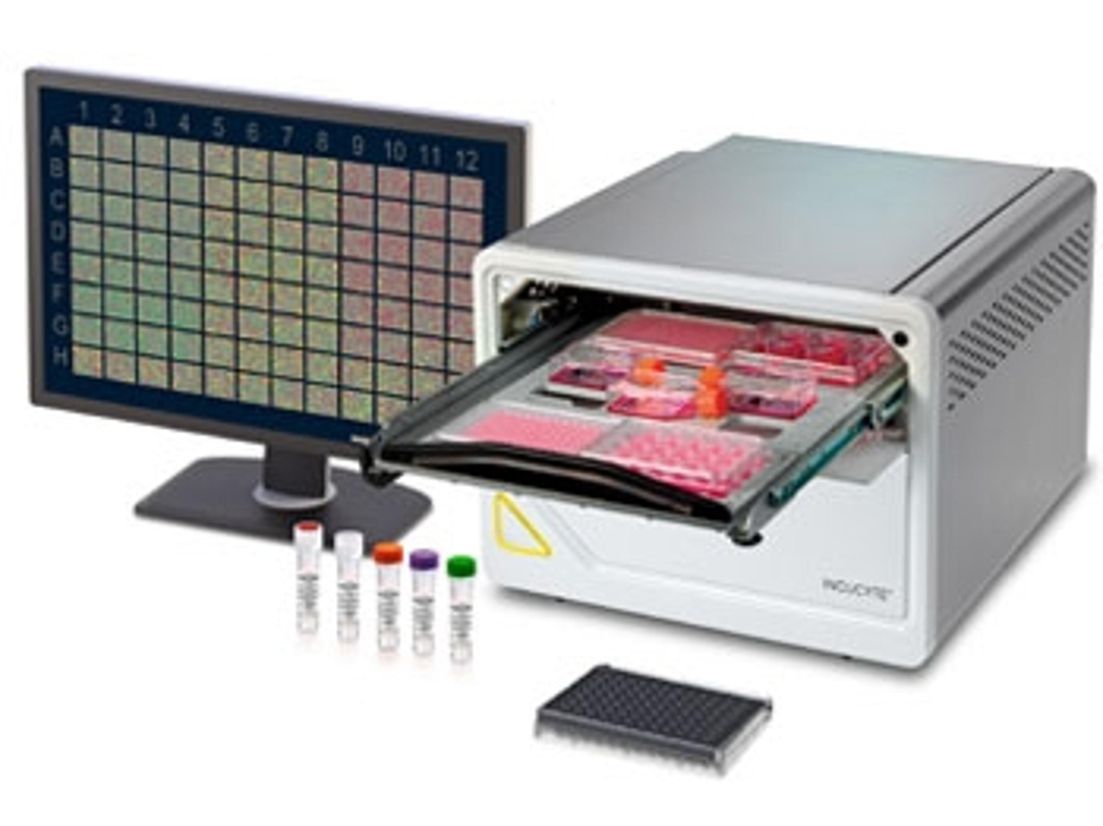
Incucyte® SX5 Live-Cell Analysis System
Leading the Way With Living Cells, our most advanced application offering.


Receive your quote directly from the manufacturer.
Great results with unbiassed reproducible and reliable data.
Life Sciences - Neuroscience, microbiology, cell biology
The product is really simple to set up and use. It's becoming a work horse for our lab. It gives great replicable data and is providing us with very insightful real time analysis of different treatment of live human primary cells in culture. This is enabling us to screen for various putative novel drugs and help us better understand signaling cascades downstream after addition of any biological agent.
Review Date: 3 Mar 2021 | Sartorius Group
Superior results - our scientists' favourite technology in the lab!
Live cell imaging and analysis
The Incucyte SX5 is an easy-to-use live-cell imaging technology, saving our scientists time in both experimental set-up and monitoring whilst providing exceptional, previously unobtainable readouts. Protocols for a wide range of lab-based assays are freely available and easy to follow. Many of our scientists have already switched many of their traditional lab-based assays to use the Incucyte technology instead. The ability to monitor your cells in real-time remotely is a hugely attractive feature to our scientists especially during this pandemic. The technology provides user-friendly built-in software to easily evaluate your experimental data offering a range of options to present your data graphically, via FACS plots or in movies – all of which are easily transferable to other software for presentation or publication purposes. Both the sales reps and application specialists were extremely helpful during our purchase and training and continue to provide support/advice to our scientists for their individual experimental needs. The fact that our Incucyte has been in non-stop use since our purchase is testament to our scientists' satisfaction with this technology and we would not hesitate in recommending this technology to other labs.
Review Date: 23 Feb 2021 | Sartorius Group
The Incucyte® SX5 is our most advanced application offering.
Study complex immune-tumor cell interactions, synaptic activity in neuronal co-cultures, metabolism in cancer cells, and much more — with a single platform.
See more information in every sample with the new Incucyte® SX5 featuring patent-pending optics. Do more with up to 5 different fluorescent channels specifically designed for live-cell analysis.
Enjoy more colors. More reagents. More purpose-built software for more applications. Derive deeper, physiologically relevant information about your cells.
Never miss powerful insights again.
Conventional approaches to cell analysis only capture a single time point, enabling only single-point and end-point measurements, and cells are perturbed or destroyed as part of the assay process.
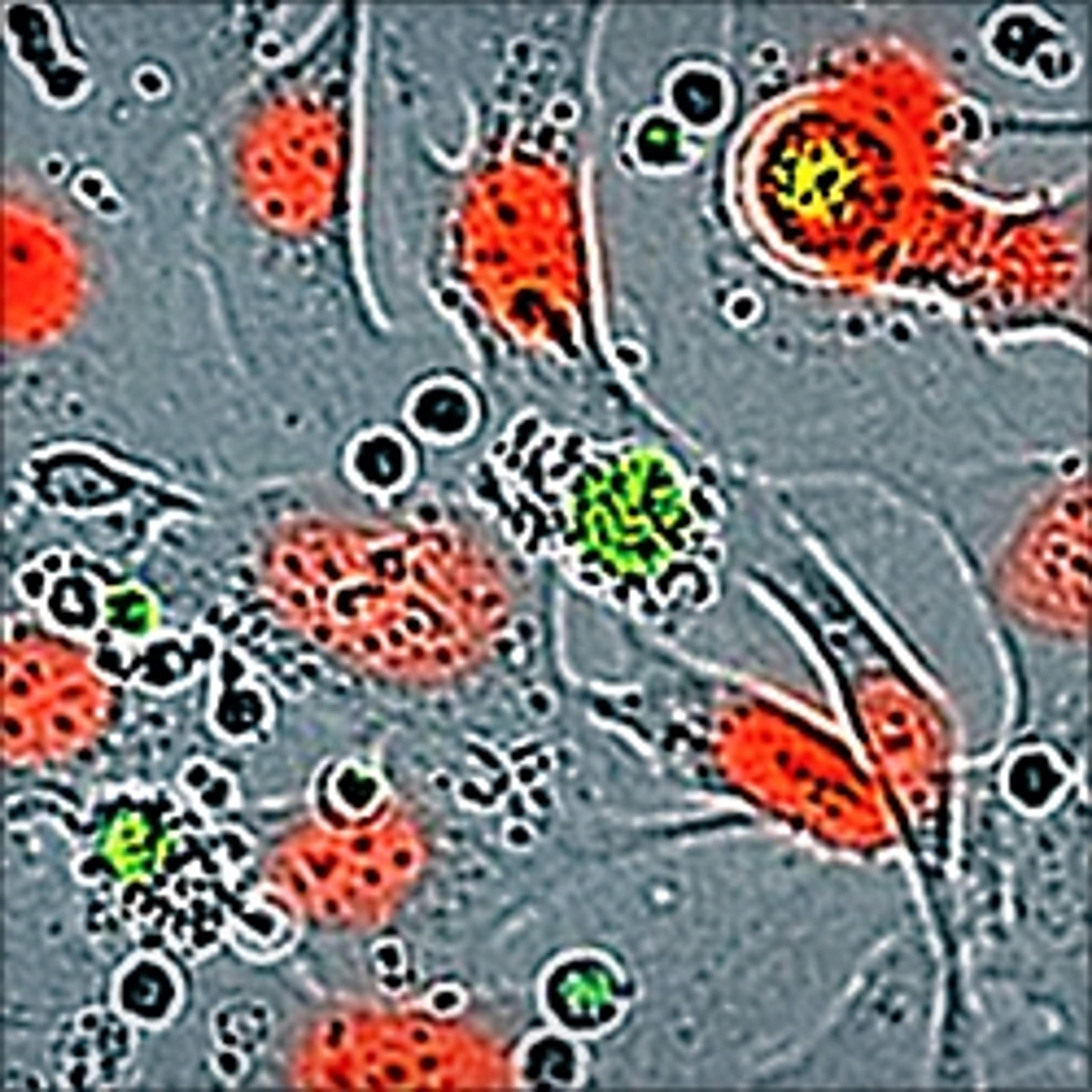
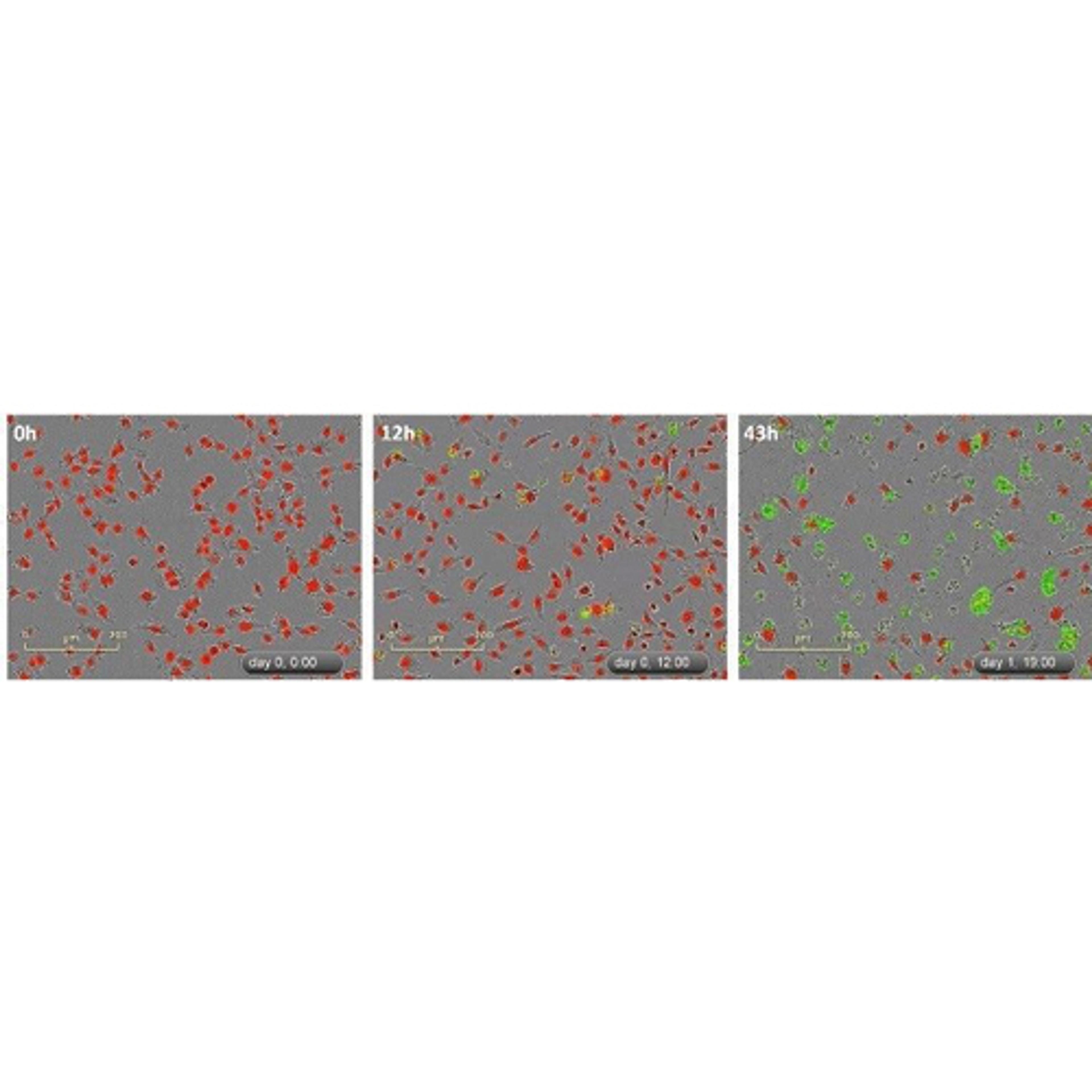
The Incucyte® SX5 Live-Cell Analysis System automatically acquires and analyzes images around the clock, enabling you to derive deeper and more physiologically relevant information about your cells, plus real-time kinetic data — without ever removing your cells from the incubator. Change can happen in an instant. See what your cells are doing and when they do it.
The Incucyte® SX5 instrument, proprietary assays and reagents provide you with the ability to gain new insights into biological processes via real-time, quantitative analysis of live cells.
Furthermore, the Incucyte® SX5 accommodates multiple users and applications seamlessly and combines information-rich, image-based analysis with the convenience and throughput of microplate assays. Incucyte® technology is featured in over 3,000 peer-reviewed publications in journals.
Key features
- Up to five different fluorescence channels, up to three at a time in a single experiment
- New 3-color optical module includes a long wavelength, low phototoxicity NIR channel and optimized reagents for turnkey applications
- Includes 4x, 10x, and 20x objectives on an automated turret
- Supports 3 interchangeable vessel trays and over 600 vessels, up to 6 microplates in parallel
- Seamless multi-user support via remote, networked access and unlimited, free licenses
Applications:
- Monitor metabolism in cancer cells
- Complex immune-tumor cell interactions
- Synaptic activity in neuronal co-cultures
- Proliferation (confluence and cell counts)
- Apoptosis (caspase 3/7 for live-cell imaging)
- Cytotoxicity
- Dilution cloning (whole-well imaging)
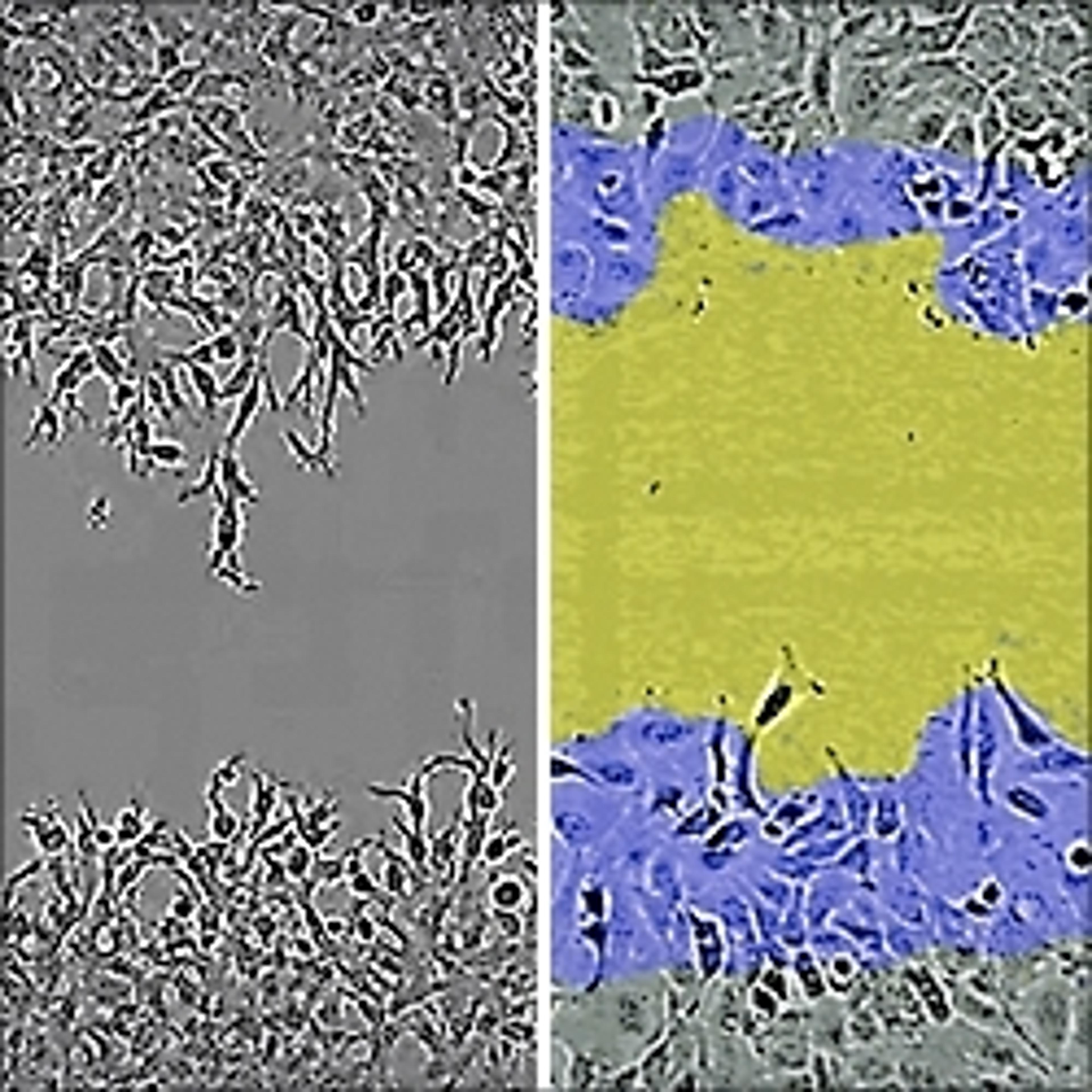
- Migration / Invasion
- Stem cell monitoring and reprogramming
- 3D-Spheroids Angiogenesis
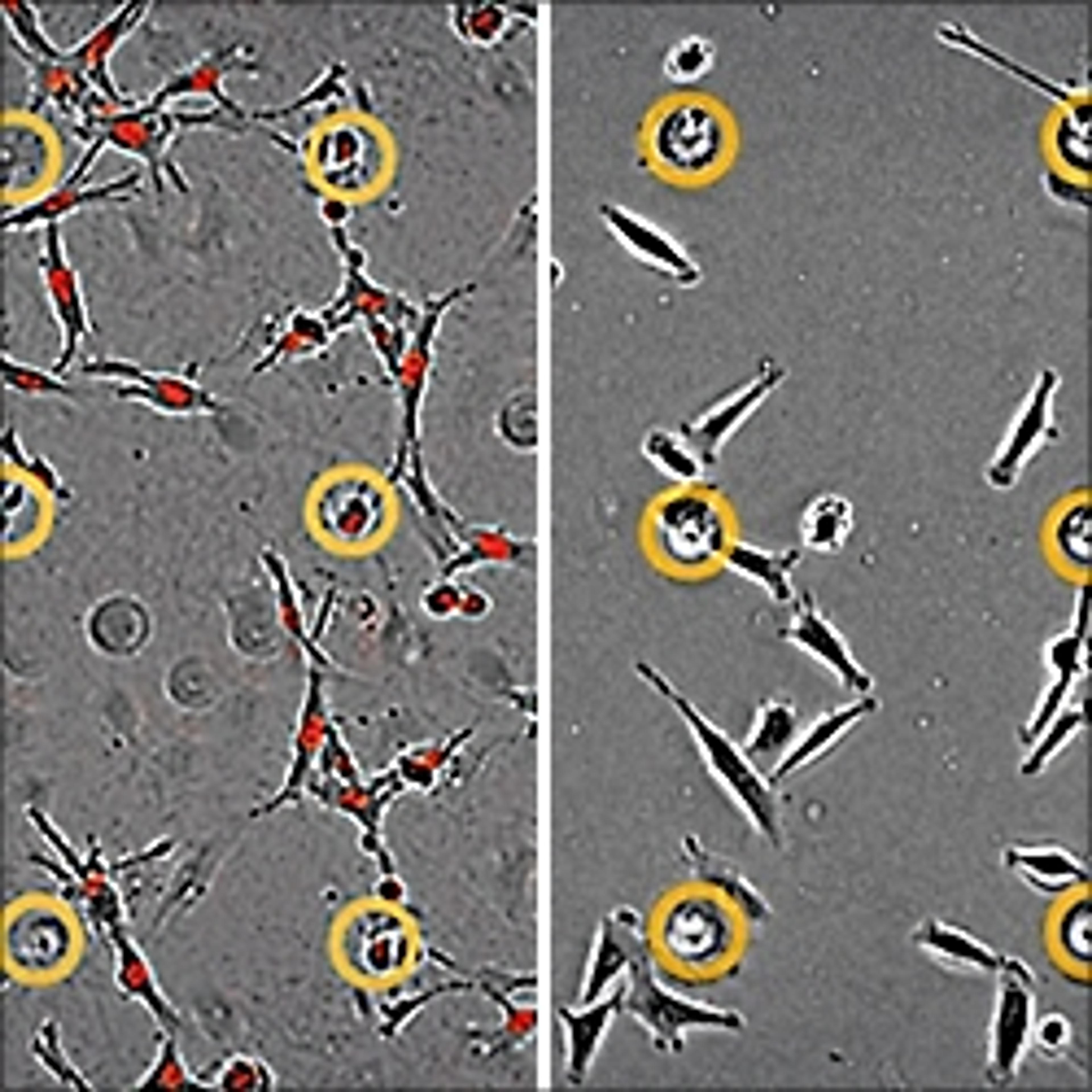
- Neurite outgrowth and dynamics
- Neuronal Activity
- Reporter gene expression
- Viral studies
- Immune response – Immune cell killing
- Antibody Internailization
- NETosis
- Phagocytosis
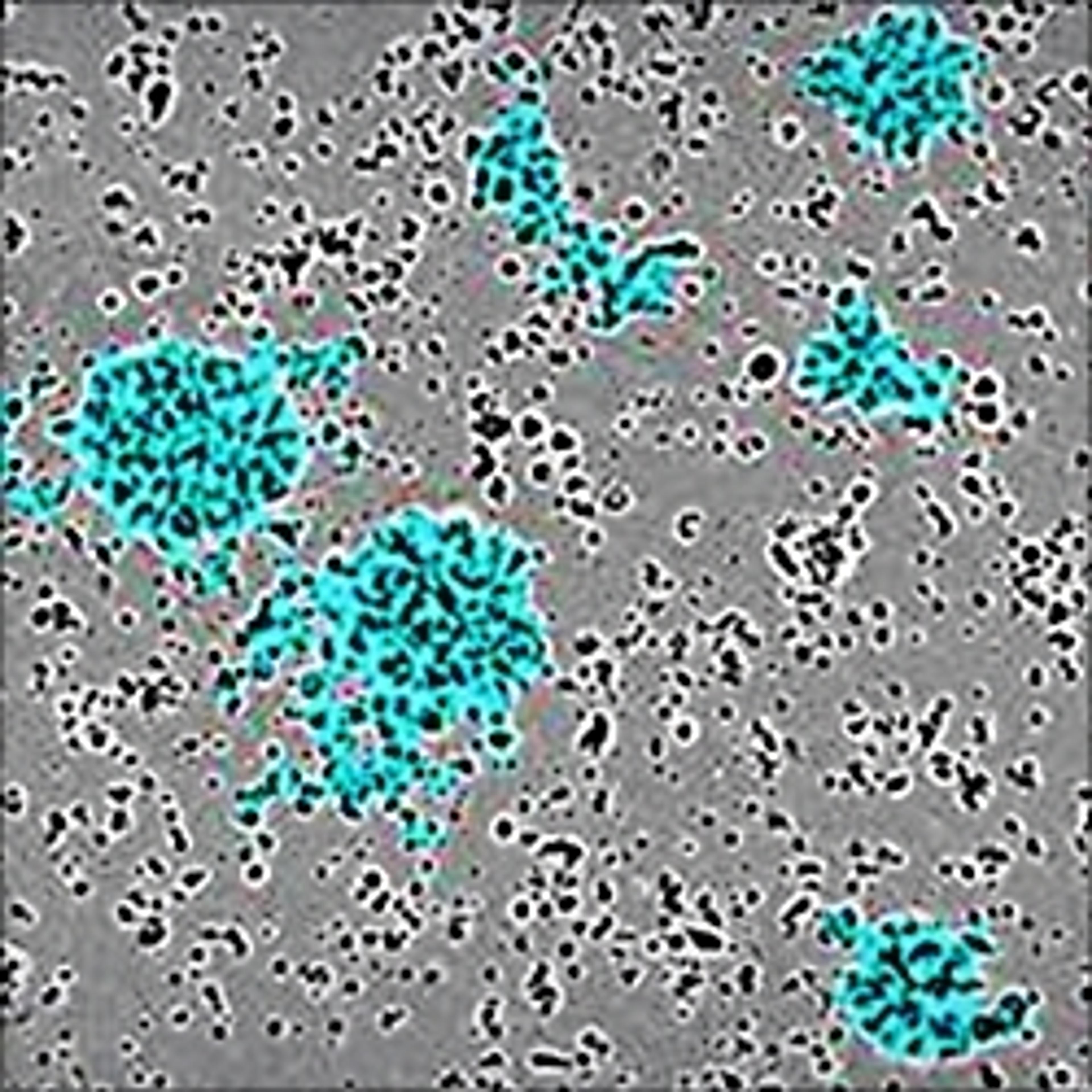
- Immune cell clustering
- Immunocytochmistry
- Cell-by-Cell Analysis
- Cell culture (& QC)
Brochures

Introducing the Incucyte SX5 Live-Cell Analysis System
The Incucyte SX5 Live-Cell Analysis System offers more channels, more reagents and more purpose-built software for more applications— allowing you to derive deeper, physiologically relevant information about your cells. Never miss powerful insights again, with the Incucyte SX5 Live-Cell Analysis System, software, reagents, and consumables.
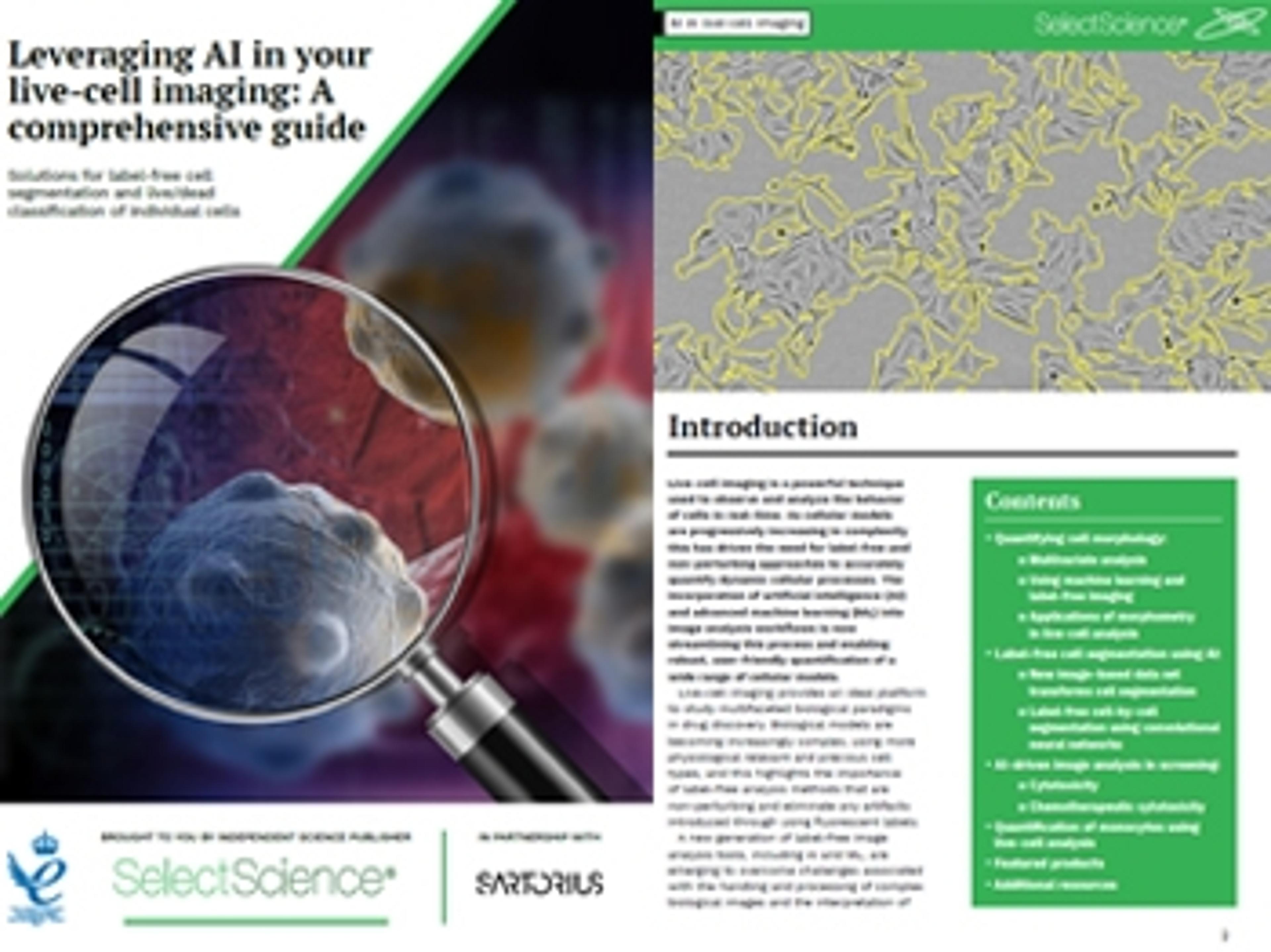
Artificial intelligence enhances label-free live-cell imaging
As cellular models are progressively increasing in complexity this has driven the need for label-free and non-perturbing approaches to accurately quantify dynamic cellular processes. The incorporation of artificial intelligence (AI) and advanced machine learning (ML) into image analysis workflows is now streamlining this process and enabling robust, user-friendly quantification of a wide range of cellular models.
This eBook provides an overview of recent label-free image analysis developments and focuses on how AI-driven image analysis is transforming live-cell imaging. This free guide will also highlight:
- How intuitive label-free analysis simplifies quantification of complex biological behavior
- The application of label-free analysis in the drug discovery process through providing high-throughput physiologically relevant insights into cell health and compound efficacy
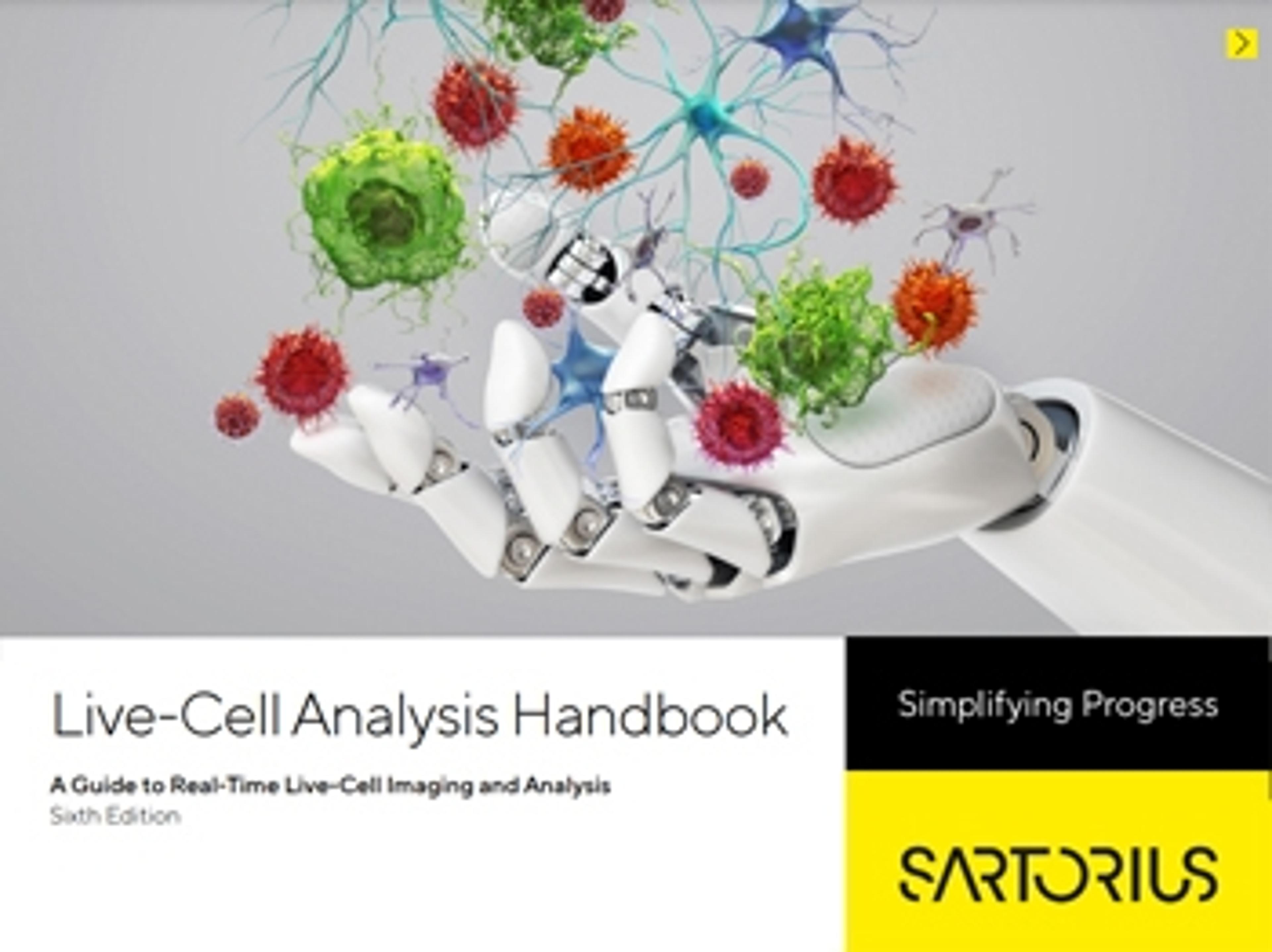
Live-cell imaging and analysis handbook
In this application note, Sartorius provides a comprehensive and interactive digital guide to real-time, live-cell imaging and analysis featuring new, AI-driven software and kinetic biosensor assays for analyzing cancer cells.
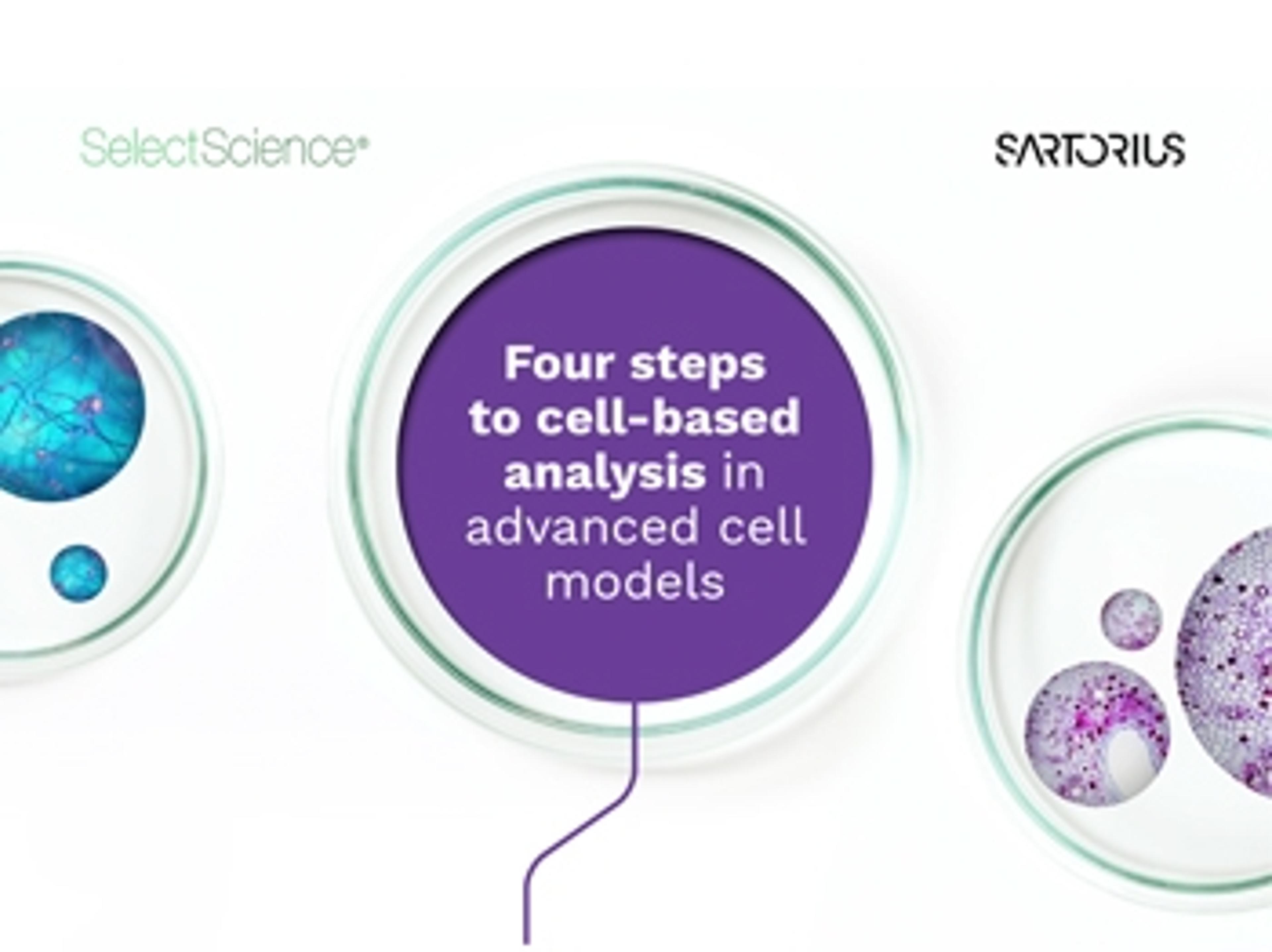
Four steps to cell-based analysis in advanced cell models
In this infographic, the challenges and opportunities presented by increasingly popular cell-based analysis in advanced cell models are considered, with live-cell monitoring via the Incucyte® from Sartorius offered as a solution to help explore and analyze these complex cell models. With precious samples requiring active management, real-time information supports a better understanding of the biological processes at play. This infographic presents the key advantages of live-cell analysis to optimize cultures, capture rare events and enable better decision-making in your experimental workflows.

Live-cell imaging for label-free toxicology analysis of hepatic organoids
Conventional 2D cultures of primary hepatocytes or established hepatic cell lines do not provide a true representation of the cellular mechanisms observed in vivo. In this webinar we will demonstrate an efficient and consistent protocol developed by STEMCELL Technologies to extract, grow, and differentiate liver cells in a complex 3D organoid culture system. These organoids more closely resemble the human liver and can be used in standard and high throughput assays to accommodate both academic and industry research needs.
This webinar will demonstrate the use of hepatic organoid cultures to validate a label-free live-cell toxicology screen using a powerful and flexible Incucyte® Live-Cell Analysis System.
Key learning objectives
- Organoid cultures – from tissue to mature 3D organoid culture and their downstream analysis
- Real-time kinetic analysis of organoids
- Label-free live-cell toxicology screening
- Key challenges of compound validations and data interpretation to improve the efficacy of drug discovery workflow
Who should attend?
Both academic and industry researchers using 3D in vitro models.
Certificate of attendance
All webinar participants can request a certificate of attendance, including a learning outcomes summary, for continuing education purposes.

Real-time visualization and quantification of Akt activity using live-cell imaging
Kinase signaling plays a key role in coupling extracellular stimuli with numerous downstream cellular functions including proliferation, survival, and migration. These pathways are highly interconnected, and their dysregulation has been implicated in several disease processes, including cancer initiation and metastasis, and chronic inflammation. Akt is a serine/threonine protein kinase that is upregulated across various diseases and has been extensively studied as a therapeutic target. Studying dynamic changes in kinase activity can be difficult, with standard approaches being limited to endpoint assays which cannot monitor the effects of treatment over time.
In this webinar, John Rauch, senior scientist at Sartorius, and Jasmine Trigg, scientist at Sartorius, will demonstrate the utility of the Incucyte® Kinase Akt Lentivirus Reagent, encoding a kinase translocation reporter based on a green fluorescent protein-tagged Akt substrate whose subcellular localization is phosphorylation-dependent, and a red fluorescent nuclear protein to denote the nuclear/cytoplasmic boundary. This biosensor enables Akt activity to be monitored in real-time, providing kinetic data on Akt activation and inhibition in living cells within a physiologically relevant environment.
Key learning objectives
- Gain an overview of validated assays combining the Incucyte® Kinase Akt Lentivirus Reagent and the Incucyte® Live-Cell Analysis System for image-based fluorescent readouts of Akt activity.
- Discover guidance on experimental set-up and how live-cell analysis can be built into your development workflow.
- Learn about case study data to support the use of live-cell analysis within the fields of oncology, inflammation and neuroscience.
Who should attend?
- Researchers involved in fundamental research and therapeutic development.
- Scientists interested in quantifying Akt activity, and its modulation, within the fields of oncology, inflammation, and neuroscience.
- Individuals who desire to implement live-cell imaging into their workflow.
Certificate of attendance
All webinar participants can request a certificate of attendance, including a learning outcomes summary, for continuing education purposes.

Classification of cell morphology with advanced multivariate analysis
Watch this on-demand webinar to discover a user-friendly workflow that yields quantitative analysis of a wide range of biological models
Sartorius launches the new Incucyte SX5 for live-cell analysis, offering new possibilities for live-cell analysis experiments
Up to five different fluorescence channels are designed to allow researchers to explore new applications, such as metabolism, with addition of new live-cell ATP and MMP assays