Incucyte® Scratch Wound Cell Invasion & Migration Software
Visualize and measure scratch wound migration and invasion in real time.
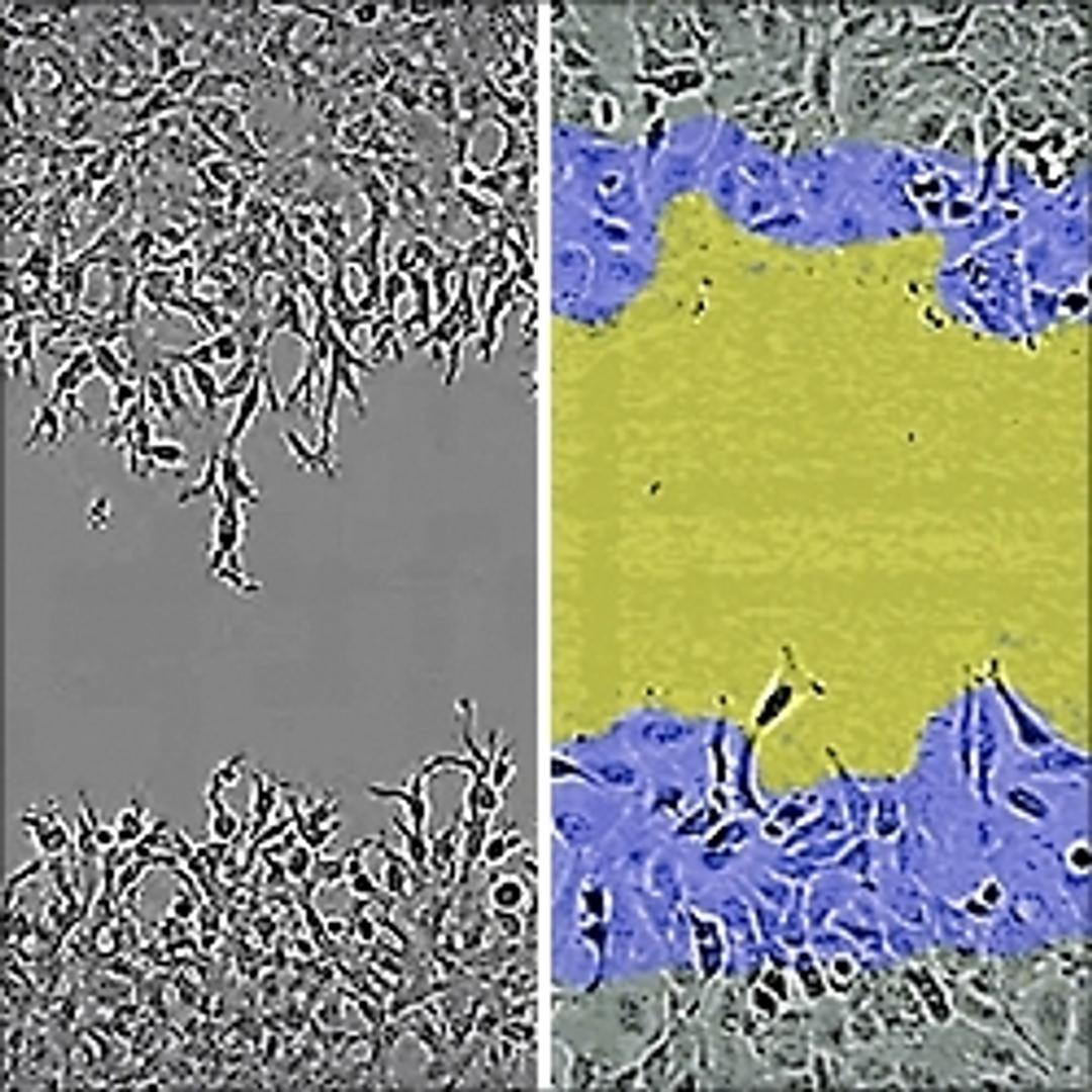
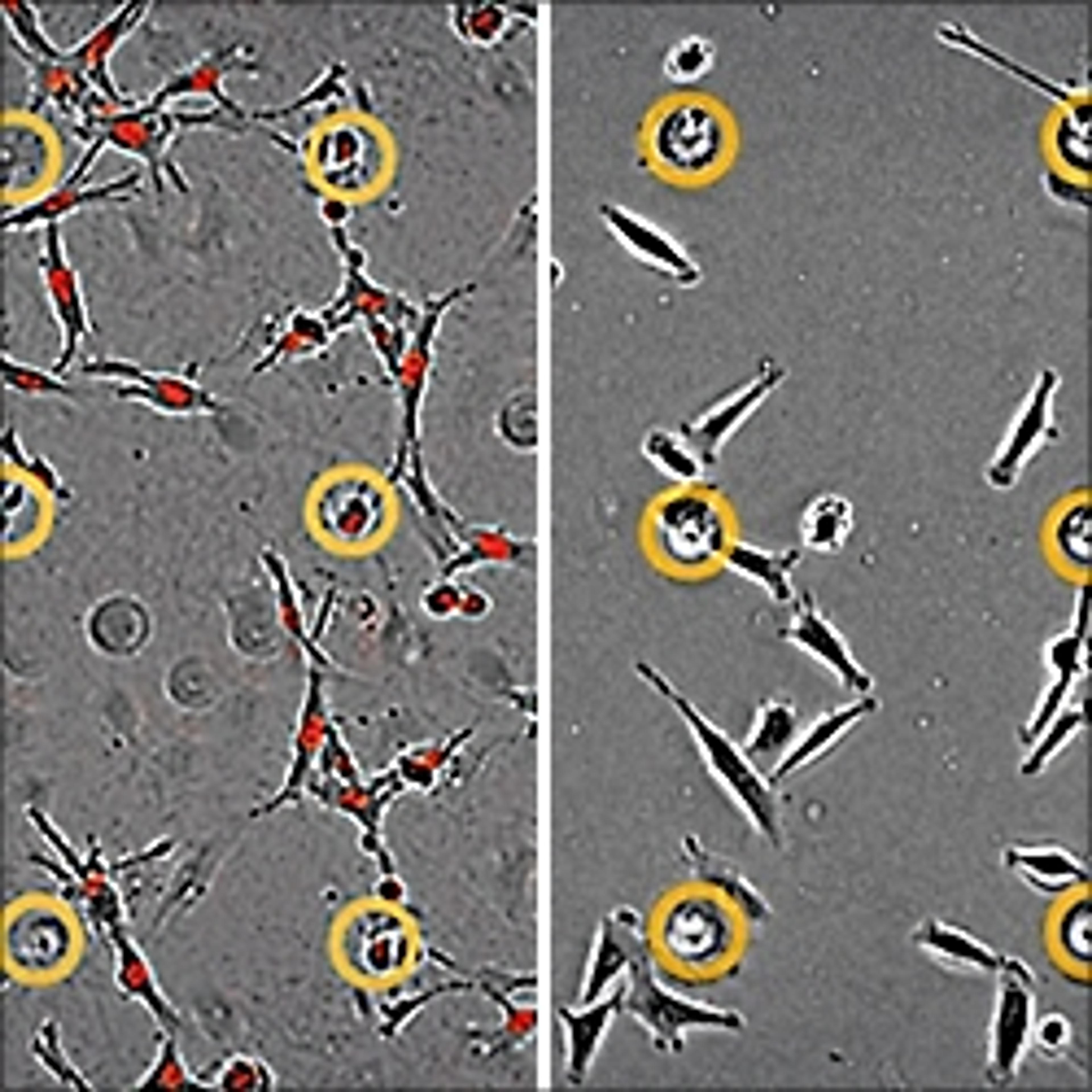
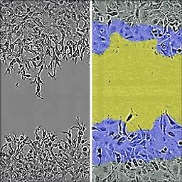
HT-1080 cells invade into the wounded area
IncuCyte® Scratch Wound cell invasion & migration assay
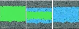
Visualize & analyze wound closure in real time

Receive your quote directly from the manufacturer.
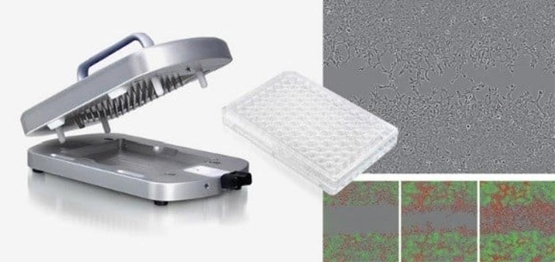
Use the Incucyte ZOOM® System to study cell movement without the need for chemotactic gradients.
- Wound healing/scratch wound assays for cell migration and invasion,
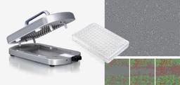
- Use the WoundMaker® to create a precise wound in each well of an ImageLock 96-well plate.
- Automatically image and quantify movement of label free, or labelled, cells into the wound area in real time.
- Cell movement can be either across a substrate (migration) or through a 3D gel matrix (invasion).

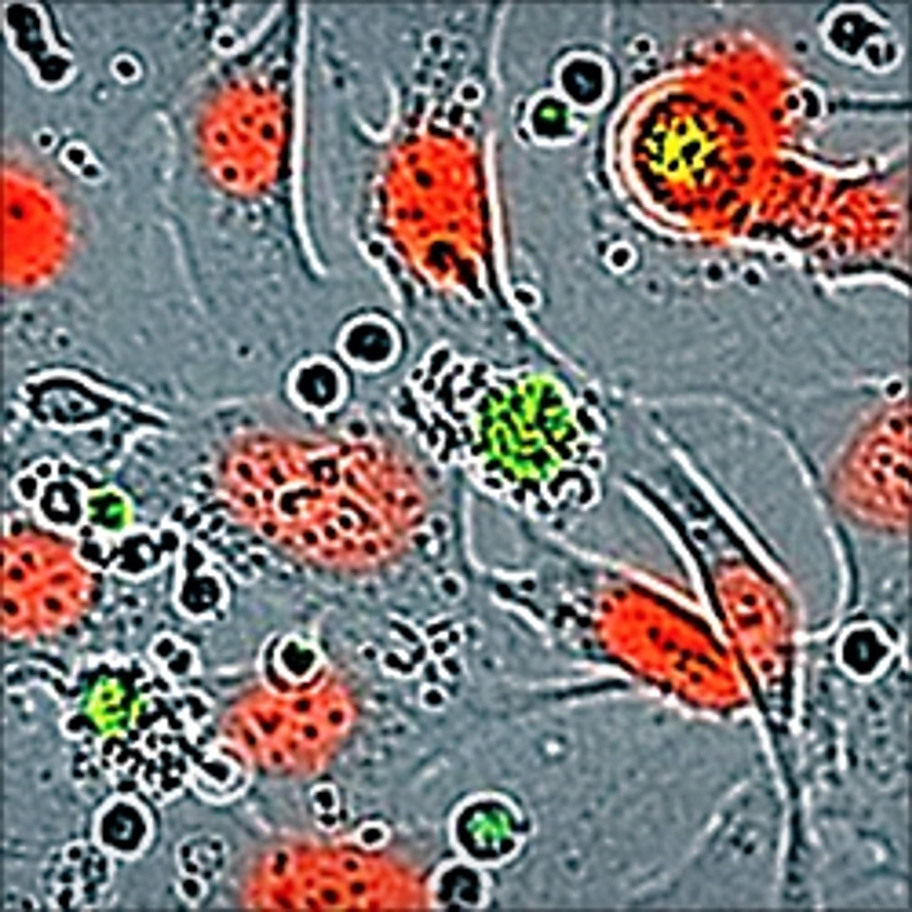
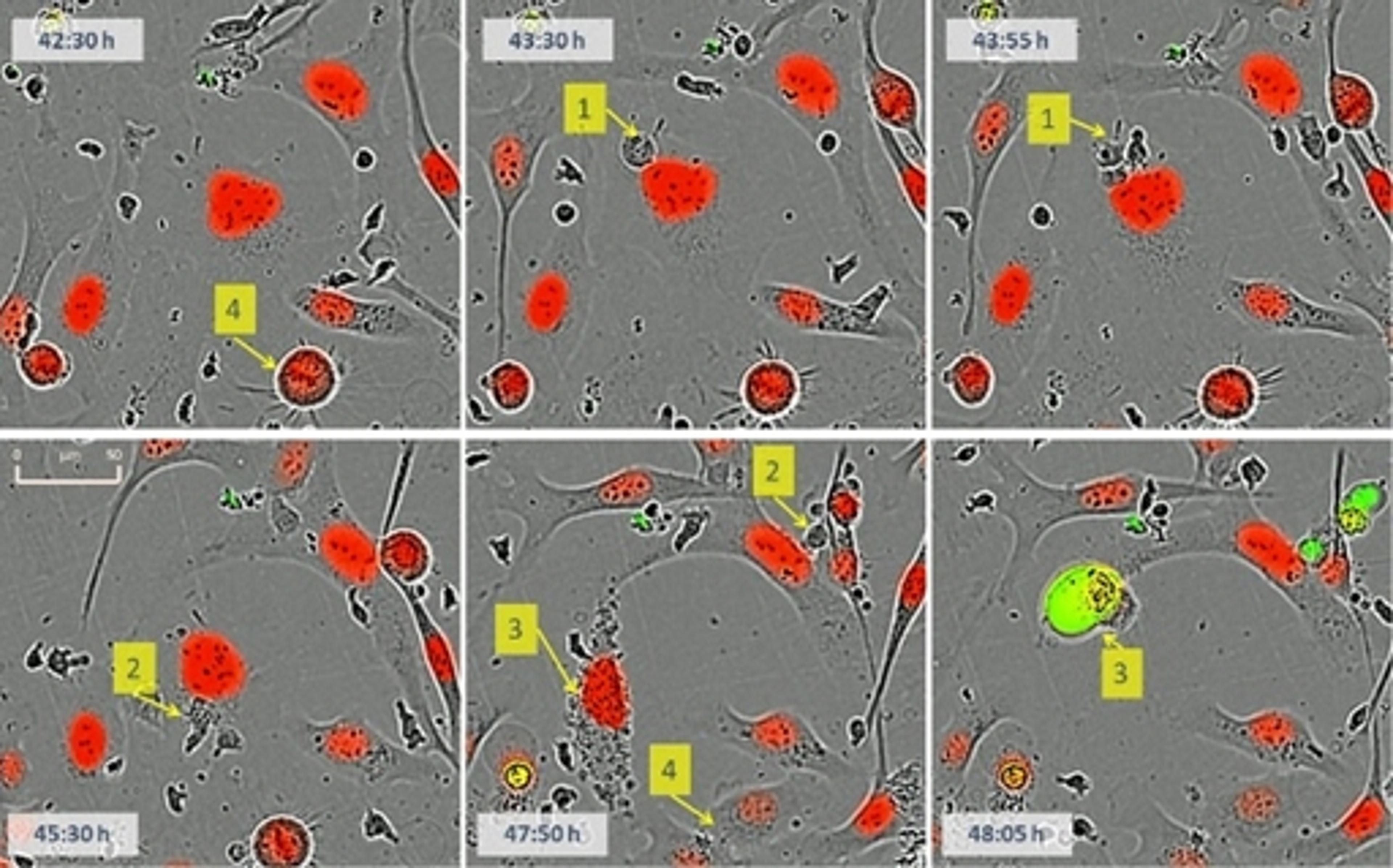
Analyzing Cell Subsets and Heterogeneity from Live Cells
Considerable heterogeneity of individual cells exists even in the simplest cell system. Such heterogeneity is mirrored by the diversity of pharmacological responses at the cellular level, where even seemingly identical cells may respond differently to drug treatments and perturbagens. Hence, analysis at the cell-by-cell level promises additional biological insights beyond which whole population measures may deliver. In this white paper, we illustrate techniques of analyzing living cells over time, and describe new, industrial-scale analysis solutions for quantifying the phenotypic biology of cell subsets in heterogeneous cultures.
IncuCyte™ 96-Well Real-Time Cell Migration and Invasion Assays
This application note demonstrates how the Migration Assay can be used to assess 2D cellular migration potential in the presence of experimental agents and as a starting point for creating a flexible, kinetic and quantitative 3D cell Invasion Assay.
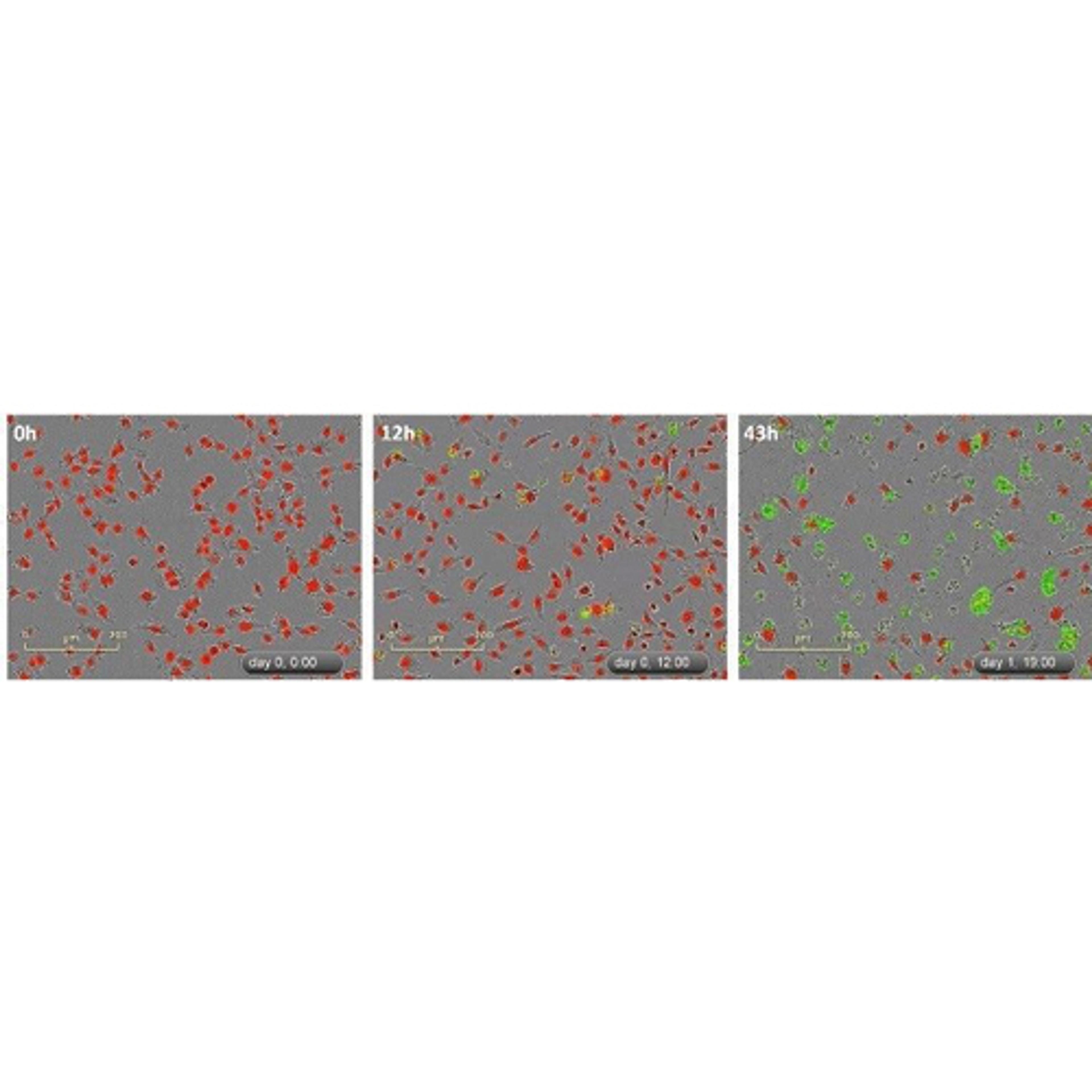
IncuCyte ZOOM™ Scratch Wound Processing Overview
This application note describes a study using the IncuCyte ZOOM Scratch Wound assay and WoundMaker-IncuCyte ZOOM-ImageLock Plate system to analyze both 2D-migration and 3D-invasion in label-free, live cells. This system allows analysis of genetic and pharmacological factors on cell migration.
Deciphering the Interplay Between Notch and Growth Factor Signaling Morphogenesis Signaling in the Complex Regulation of Vascular Morphogenesis
Angiogenesis is a multi-step, complex process regulated by growth factors, enzymes, and extracellular matrix molecules. In vivo, the angiogenic process involves multiple cell types acting in concert to cause endothelial cell proliferation, migration, differentiation, and, ultimately, micro-vascular arrays. Angiogenesis inhibitors are demonstrably effective in both preclinical models and clinical use, but their value can be transitory due to evasive and intrinsic resistance. The discovery of new classes of anti-angiogenic drugs has proven difficult, as very few in vitro systems adequately model the entire process. This poster investigates the relationship between two major regulatory pathways, Notch and VEGF, using a kinetic co-culture model of angiogenesis with a compact fluorescent imaging instrument
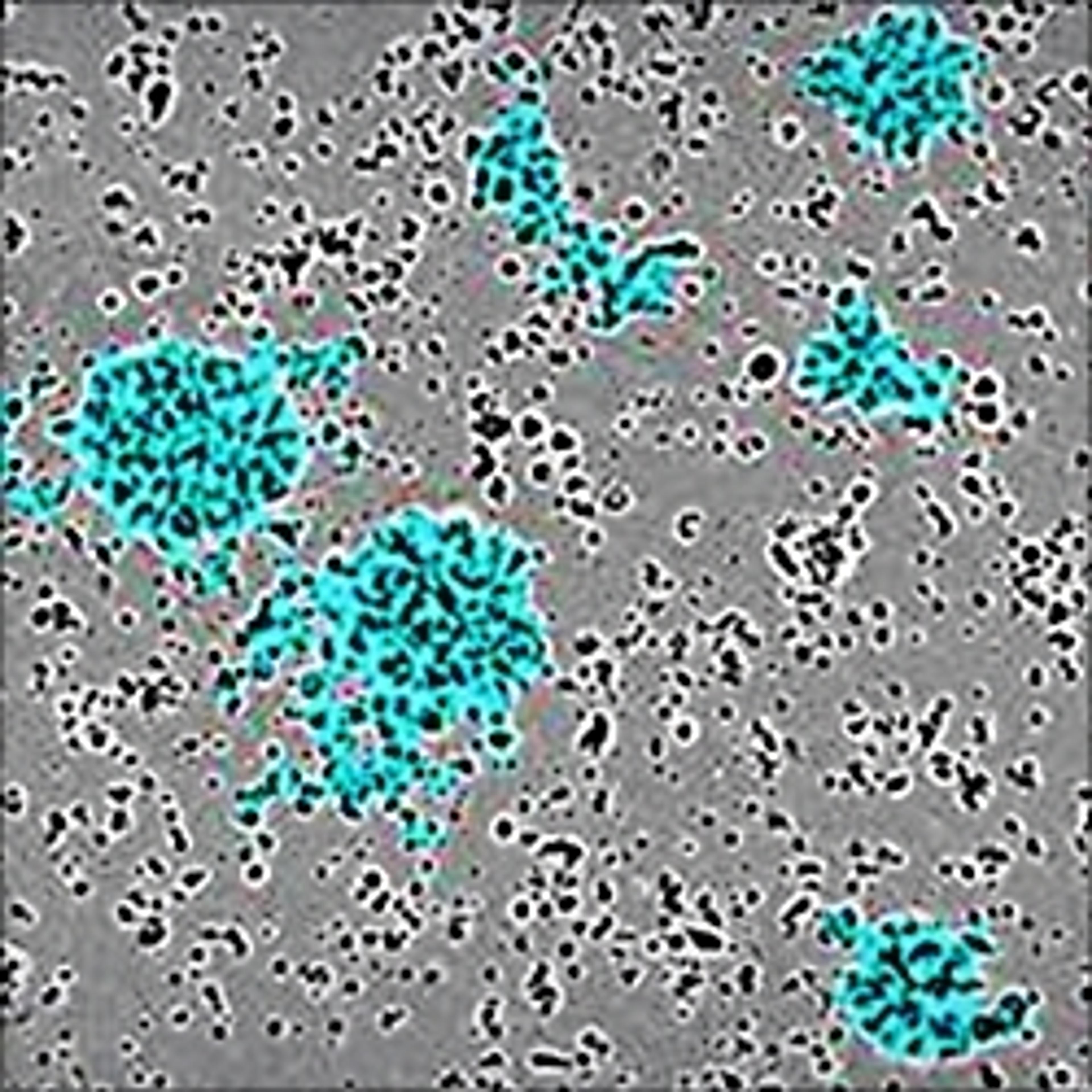
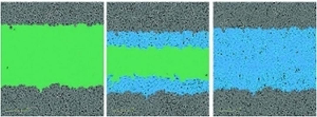
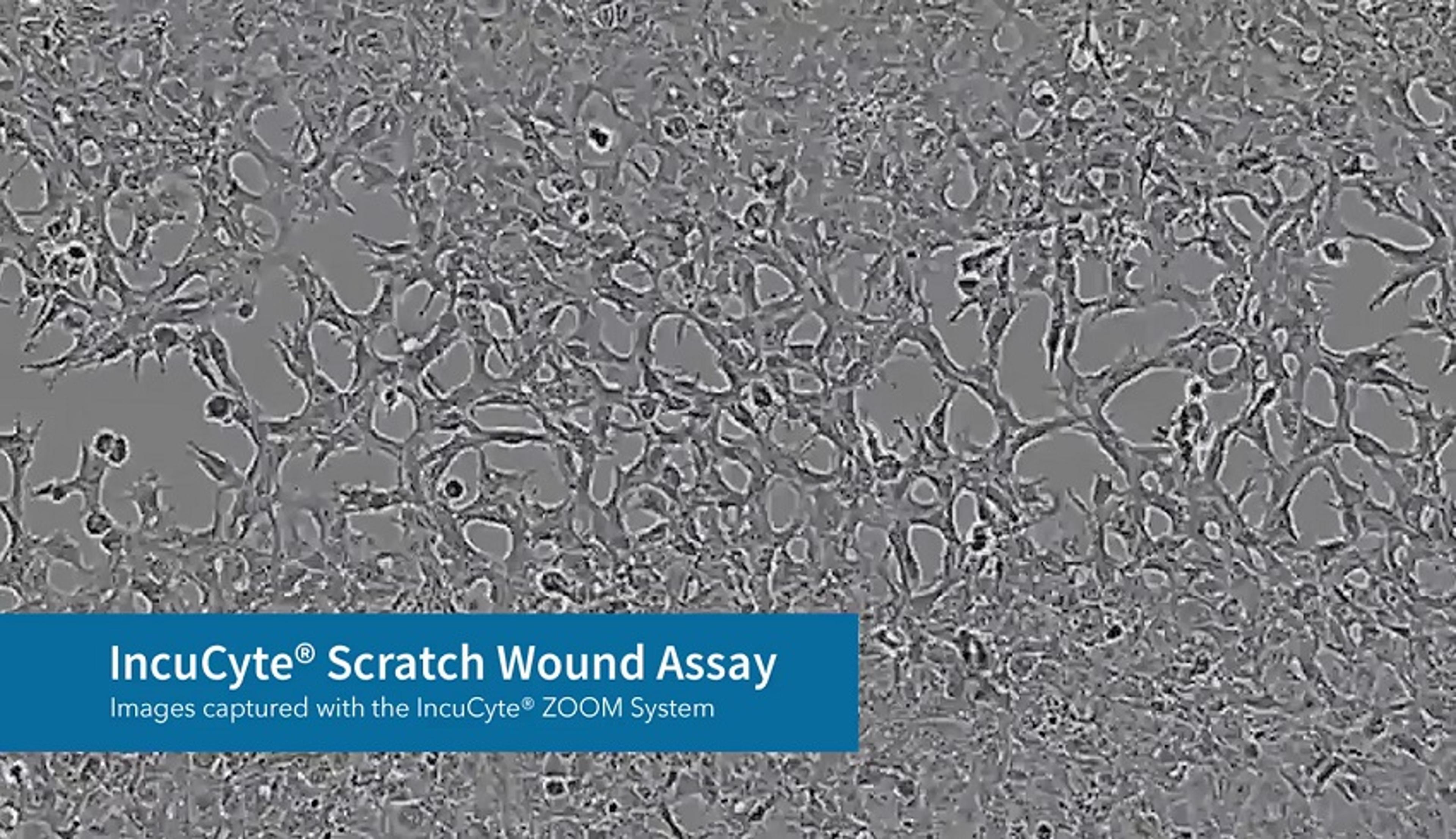
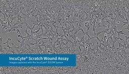
IncuCyte® Scratch Wound Assay
This video shows images captured with the IncuCyte® ZOOM System of an IncuCyte® Scratch Wound Assay.
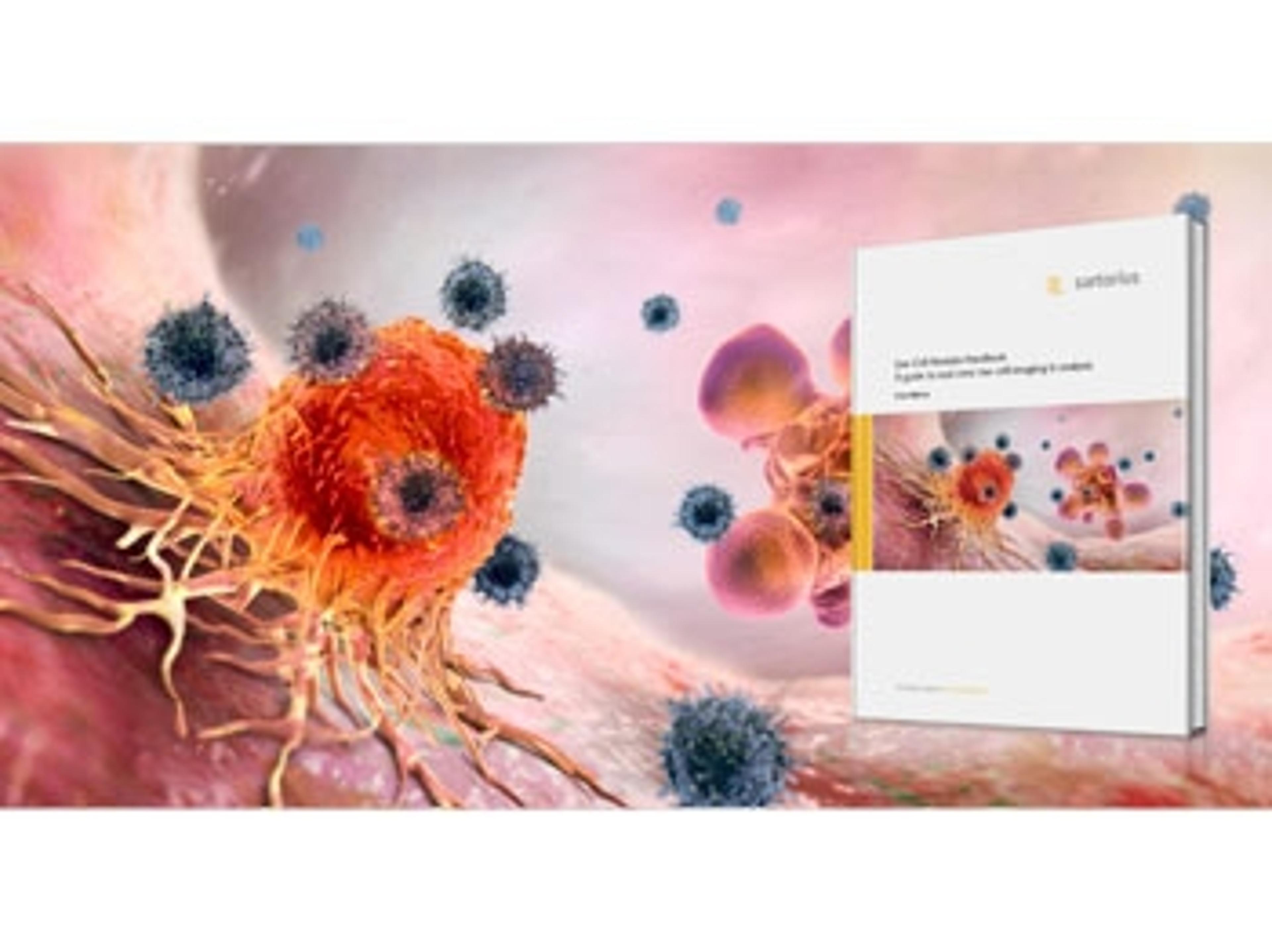
The third edition of the Live-Cell Analysis Handbook published
Latest edition includes two new chapters for scientists who perform cell biology assays

How to Use Live-Cell Analysis for Neuroscience Applications
Learn how live-cell analysis techniques can benefit your neuroscience research, in this expert on-demand masterclass