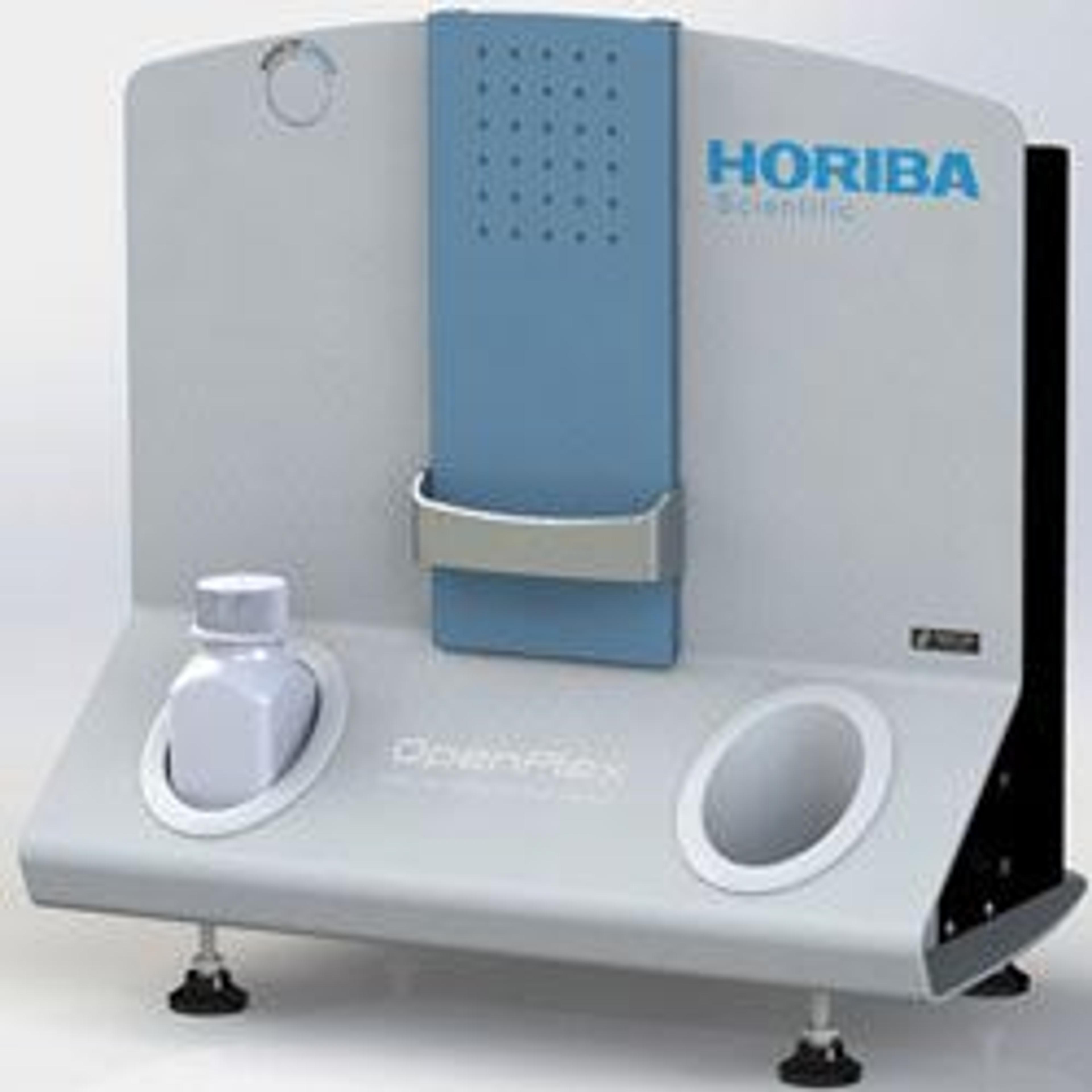
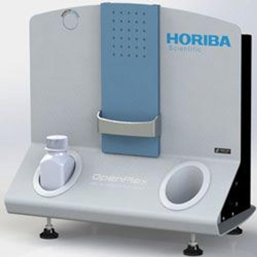
OpenPlex - Flexible Surface Plasmon Resonance Imaging System
Designed to meet the demands of biologists, biochemists and biophysicists, the OpenPlex is a flexible surface plasmon resonance imaging system. OpenPlex is your companion for the development of label-free and multiplexed bio-assays and molecule detection. It is a robust and compact system designed for simple use and high versatility. Its open format, dedicated sensor chips and manual operation enable numerous types of experime…


The supplier does not provide quotations for this product through SelectScience. You can search for similar products in our Product Directory.
Designed to meet the demands of biologists, biochemists and biophysicists, the OpenPlex is a flexible surface plasmon resonance imaging system.
OpenPlex is your companion for the development of label-free and multiplexed bio-assays and molecule detection. It is a robust and compact system designed for simple use and high versatility. Its open format, dedicated sensor chips and manual operation enable numerous types of experiments to be explored without compromise, covering chemistry, biochemistry, physico-chemistry and biomolecular interactions.
Open your research areas with OpenPlex! Choose between three different flow cell configurations without concession:
• Normal flow cell for standard surface plasmon resonance imaging experiments
• Window flow cell, compatible with fluorescence measurements
• Cuvette cell, compatible with electrochemistry measurements

Label-Free Ligand Fishing in Human Plasma Using Surface Plasmon Resonance and Mass Spectrometry Imaging
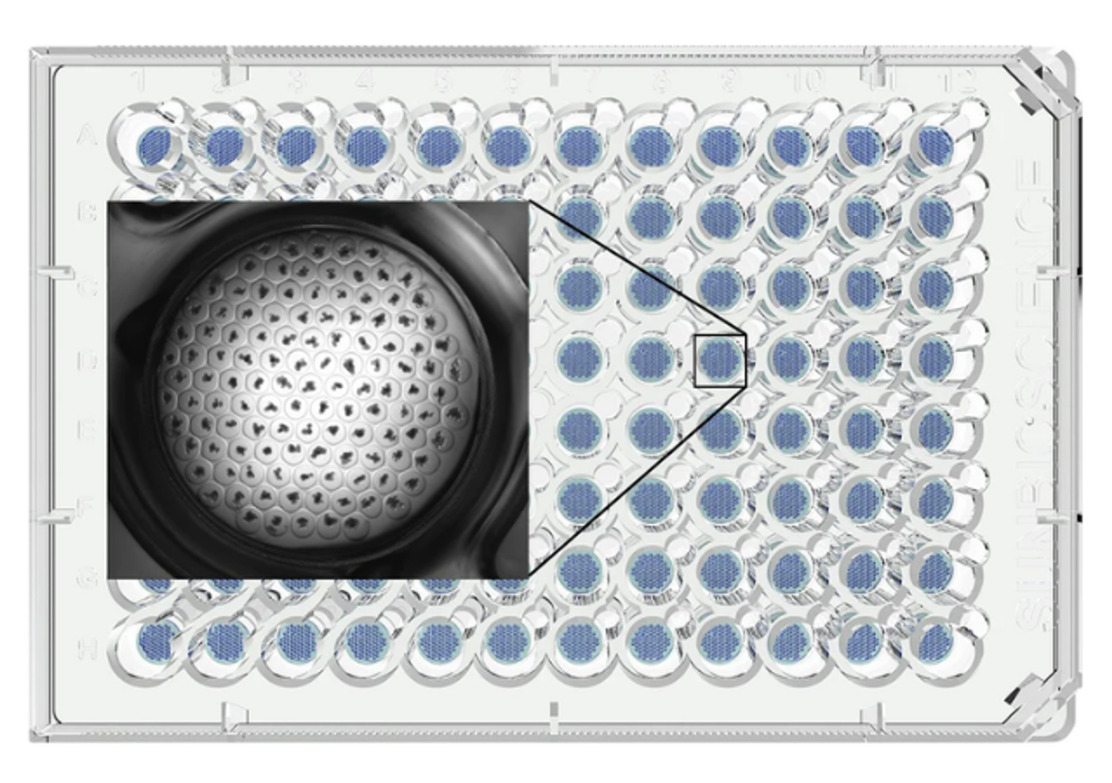
The Surface Plasmon Resonance imaging (SPRi) system enables visualization of biomolecular events occurring across an entire biochip surface, down to the nanomolar scale. In this application note, Horiba discusses how this technology can be coupled with Matrix Assisted Laser Desorption Ionization Mass Spectrometry (MALDI-TOF), to enable detailed structural characterization of proteins by molecular weight and peptide sequence. This technology permits high throughput analysis of patient samples for disease biomarkers, and may allow for accelerated identification of novel biomarker proteins in the future.
How to start a SPRi experiment
In the following application note, Horiba addresses several questions to consider before conducting a surface plasmon resonance imaging experiment. For example, multiple biochip sensors can be used and a number of functionalized surface chemistries are available to directly immobilize ligands of interest. Controls are another important consideration as refractive index changes and non-specific interactions are subtracted from signals. Running buffers can improve specific interactions and samples must be carefully prepared to avoid refractive index changes. Furthermore, the capacity for ligand regeneration is discussed, as well as how to evaluate the data produced by SPRi software.
Validation of the Activity of G-Protein-Coupled Receptors (GPCRs) Using SPRi
G-Protein coupled receptors (GPCRs) are a large family of membrane proteins with integral functions in most physiological processes. There are several challenges, however, associated with finding suitable GPCR expression systems and measuring their interactions with ligands at binding sites. Here, Horiba discusses surface plasmon resonance imaging (SPRi) as a technology that facilitates the study of GPCR-ligand interactions and the binding site itself, by covalently immobilizing proteoliposomes on a SPRi-BiochipTM.
Peptide-Antibody Interaction Specificity Using SPRi
This application note demonstrates the relevance of using SPRi technology for research into peptide-antibody interactions. First, streptavidin grafting is performed by using GenOptics pyrrole electro-polymerization protocol. A mix of pyrrole and pyrrolated streptavidin is co-polymerized on the gold layer of a glass prism. Biotinylated peptides are then injected in the detection cell and create a homogenous peptide layer, due to the strong affinity between streptavidin and biotin. Use of SPRi technology allows real-time studies of peptide-antibody interactions on a large number of spots analyzed in parallel.
Biomarker Discovery using Surface Plasmon Resonance Imaging
Surface Plasmon Resonance (SPR) is an optical technique used to follow molecular interactions (binding) in real time without labeling. It provides information on kinetic processes (association and dissociation rates), binding affinity, analyte concentration and real time molecule detection. This article describes the principle of SPR imaging for high-throughput measurements. An example of a clinical application for the capture and characterization of a potential biomarker of breast cancer is illustrated.

HORIBA presents 2020 Young Fluorescence Investigator Award
Dr. Jelle Hendrix, Assistant Professor at the Biomedical Research Institute of Hasselt University in Belgium, was presented with this honor