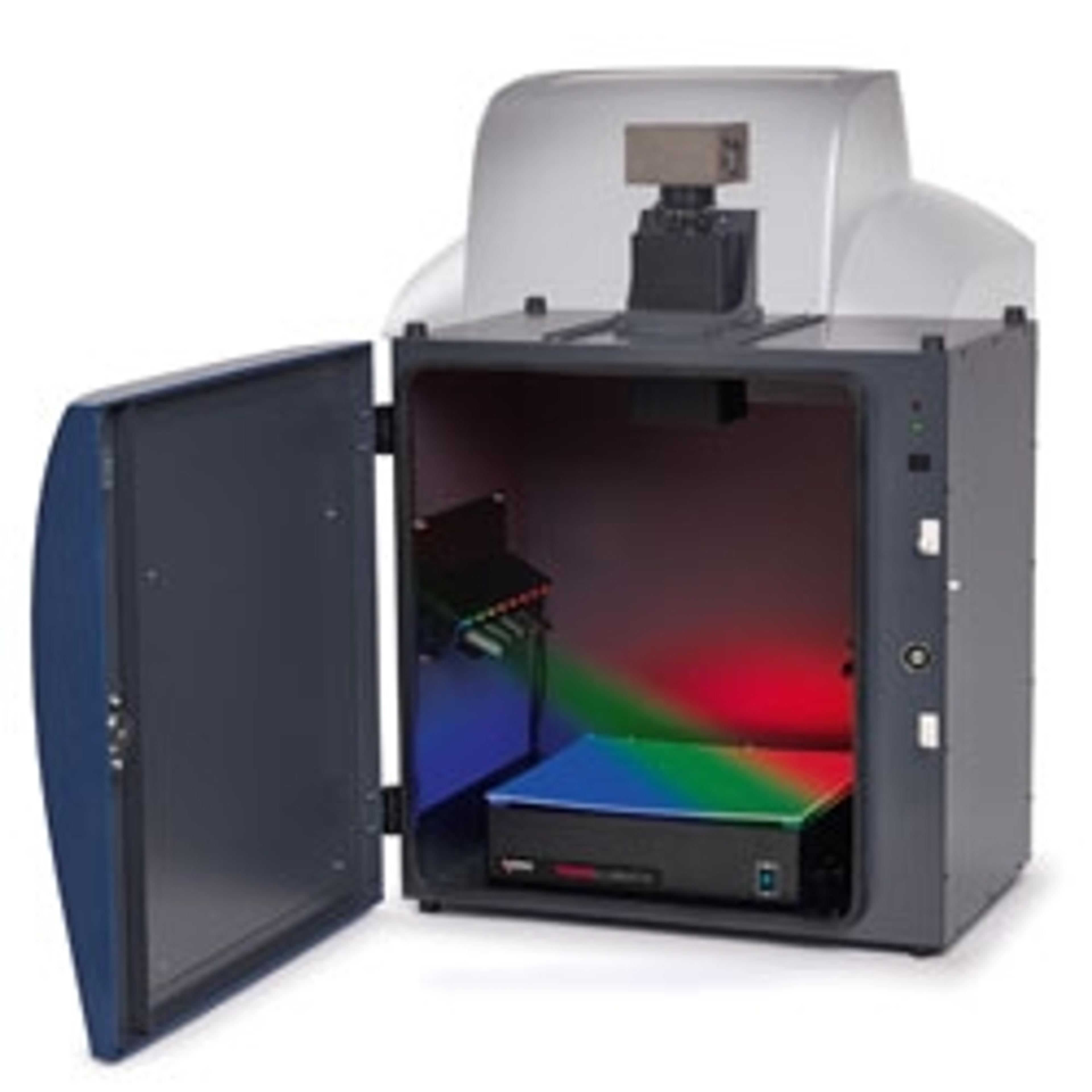
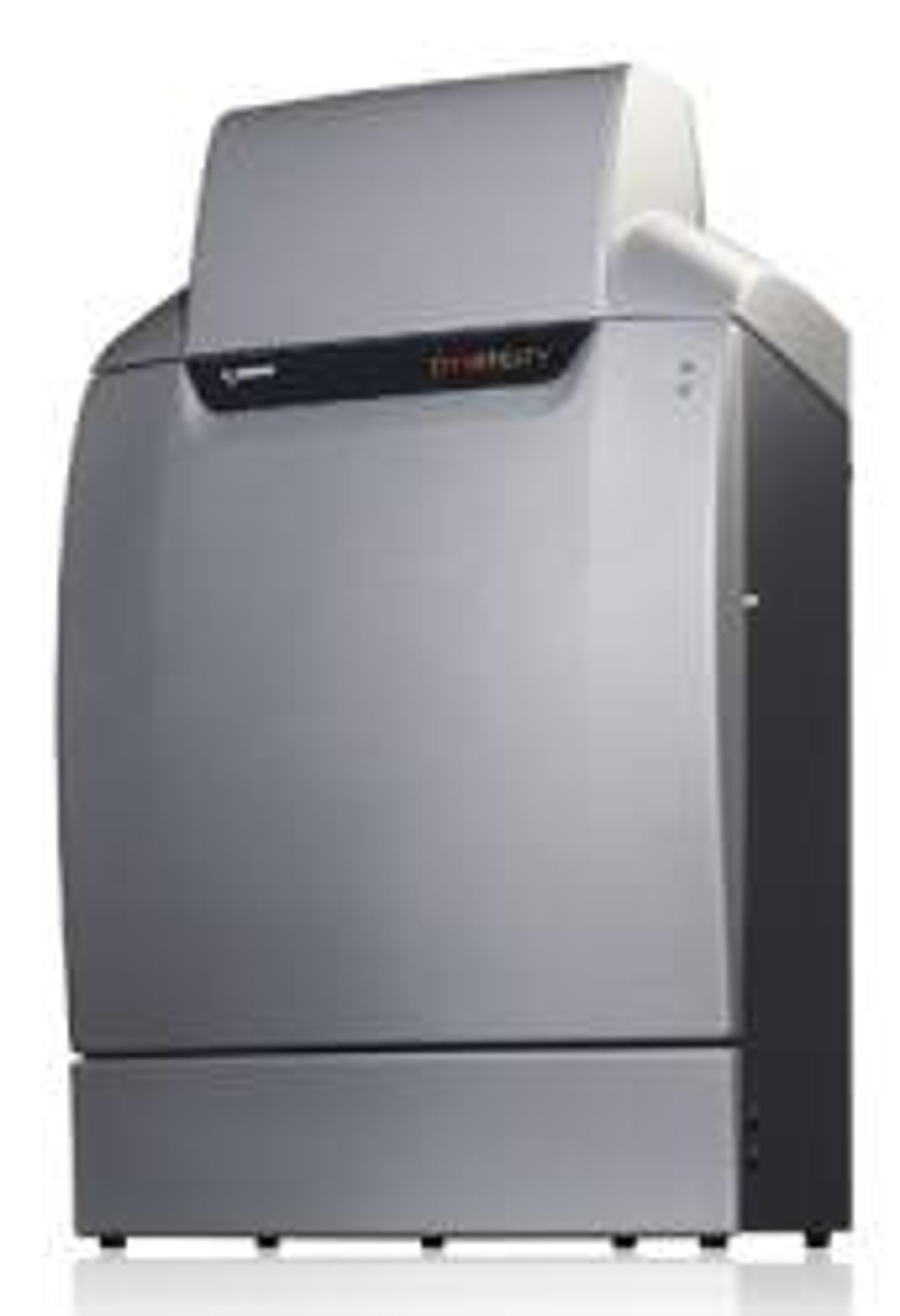
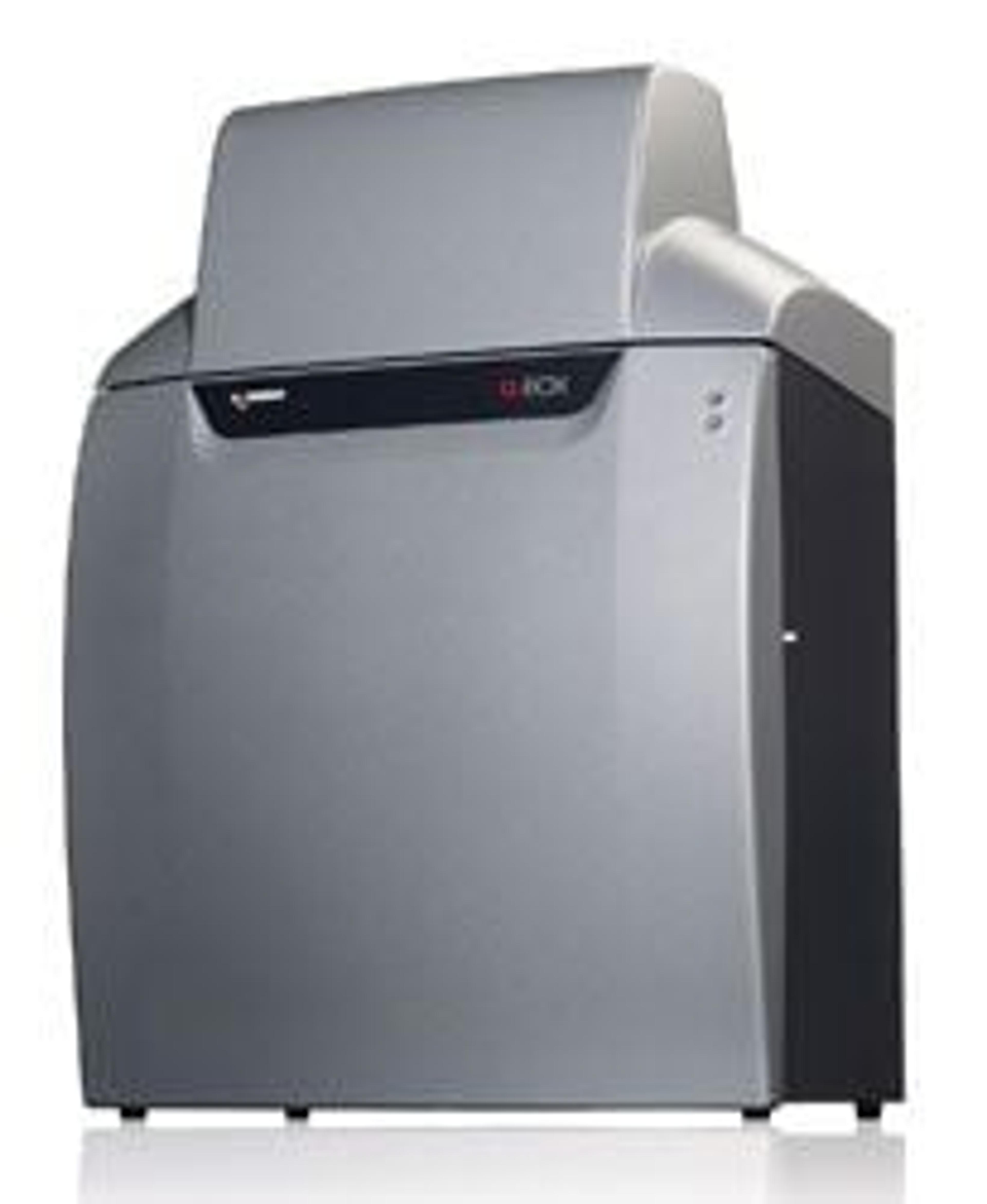
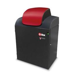
G:BOX F3
An entry level system for imaging fluorescence and visible applications, the G:BOX F3 gel doc is the ideal solution for your laboratory.

The supplier does not provide quotations for this product through SelectScience. You can search for similar products in our Product Directory.
Gel imager
This system had several issues in the beginning with the camera. we got it fixed and since then its working pretty well.
Review Date: 16 Nov 2015 | Syngene
Excellent performance for gel imaging. Also does other gel stains, including fluorescence.
Review Date: 23 Sept 2015 | Syngene
Very convenient for quantification.
Review Date: 29 Jul 2011 | Syngene
An entry level system for imaging fluorescence and visible applications, the G:BOX F3 gel doc is the ideal solution for your laboratory.
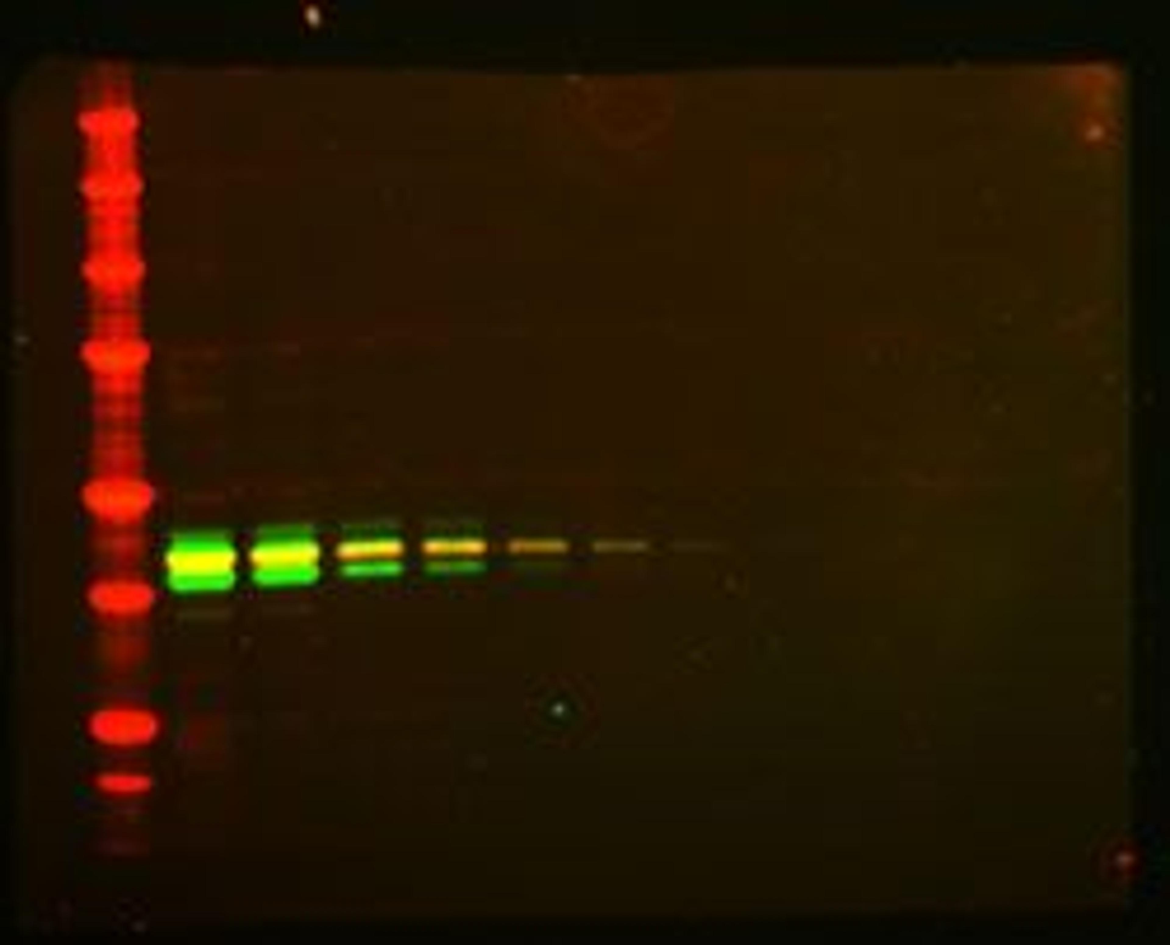
Using your choice of dyes or stains, the G:BOX F3 automatically selects the right lighting and filters to detect close bands on both small and large gels. Choosing white light options for Coomassie Blue, UV and blue lighting options, ethidium bromide or SYBR® Safe gels, this system is the ultimate in gel doc flexibility.
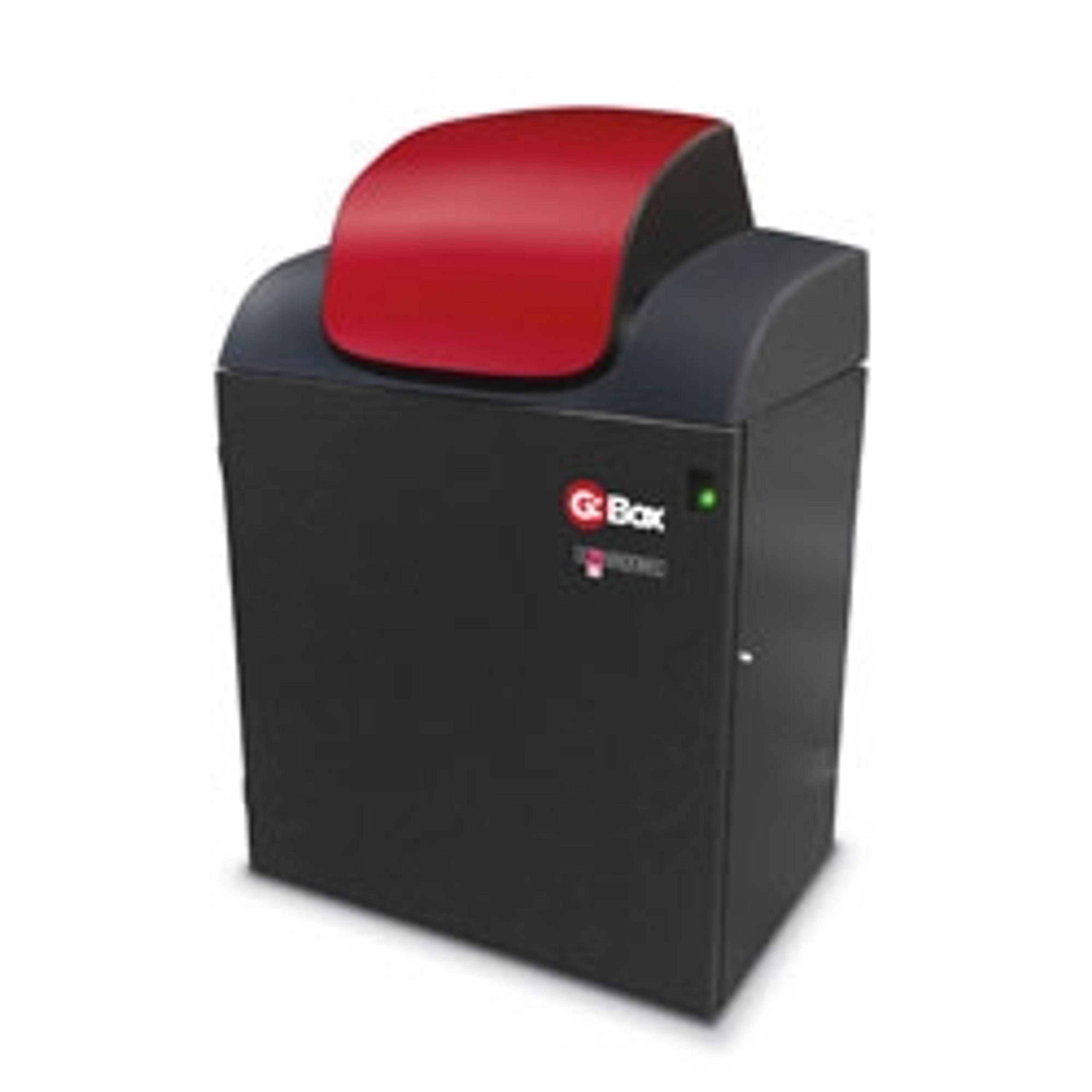
The stylish and modular designed G:BOX F3 gel doc system includes a high resolution 5m pixel camera which is capable of giving outstanding images with incredible spatial resolution as well as a motor driven zoom lens and a motor driven filter wheel as standard. G:BOX F3 also comes with a Lens Feedback option allowing you to optimise your image capture using user protocols.
The system is controlled by GeneSys application driven image capture software and comes complete with unlimited copies of GeneTools analysis software.
Features:
- Spacious darkroom
- Image gels up to 32cm x 24cm
- 5m pixel camera
- Resolves close bands and detects nanogram amounts
- Motor driven zoom lens and filter wheel
- One click set up for easy image capture
- Optional Lens Feedback available
- Optimise image capture by using user protocols
- White UV and blue lighting options
- Flexibility to image fluorescent and visible stained gels
- GeneSys application driven image capture software
- Contains extensive database of dyes and imaging protocols. All you need to know is the type of gel you’re using and GeneSys automatically selects the optimal lighting and filters to produce the perfect image
- GeneTools analysis software (unlimited copies)
- Analyse data at your own computer

Comparing Criterion TGX Stain-Free gels to standard Coomassie staining procedures
In this application note, Syngene outlines how users can successfully image Criterion TGX Stain-Free gels using any Syngene G:BOX imaging system by using the recommended lighting and filter combination, 302nm UV transilluminator and UV filter. The stain-free method of visualizing cells has comparable sensitivity to that of more traditional techniques such as Coomassie Blue Safe staining and also has the potential to significantly reduce the lengthy SDS-PAGE workflow.
Overview of Ethidium Bromide Dyes
This application note provides an overview of ethidium bromide dyes, commonly used in agarose gel electrophoresis to detect double-stranded DNA (dsDNA) and single-stranded DNA (ss) or RNA.
An Overview of Alexa Fluor 647 Dyes
This application note provides an overview of Alexa Fluor dyes, a series of fluorescent dyes produced by Molecular Probes that span the visible spectrum.
Dynamic Fielding for Syngene Image Capture Systems
Some manufacturers use a Flat Field Correction method in order to address uneven illumination of light sources. This involves subtracting the image information from an empty field of view or ‘perfectly flat’ fluorescent reference sample from that of the same field of view with the gel added. As this process involves the subtraction of one image from another, the integrity of the raw data of the initial captured image is compromised. Such manipulation is incompatible with Good Laboratory Practice (GLP). Syngene uses the Dynamic Fielding Correction method to address uneven light illumination whilst maintaining GLP compliance.
G:BOX iChemi Image Analyzer and KPL Chemiluminescent Substrates make Detecting Proteins in Picogram and Femtogram Range Quick and Easy
This application note demonstrates the level of flexibility and sensitivity that this innovative image analyzer can achieve and makes the G:BOX iChemi XR chemiluminescent imaging system an excellent tool for detecting poorly expressed protein.