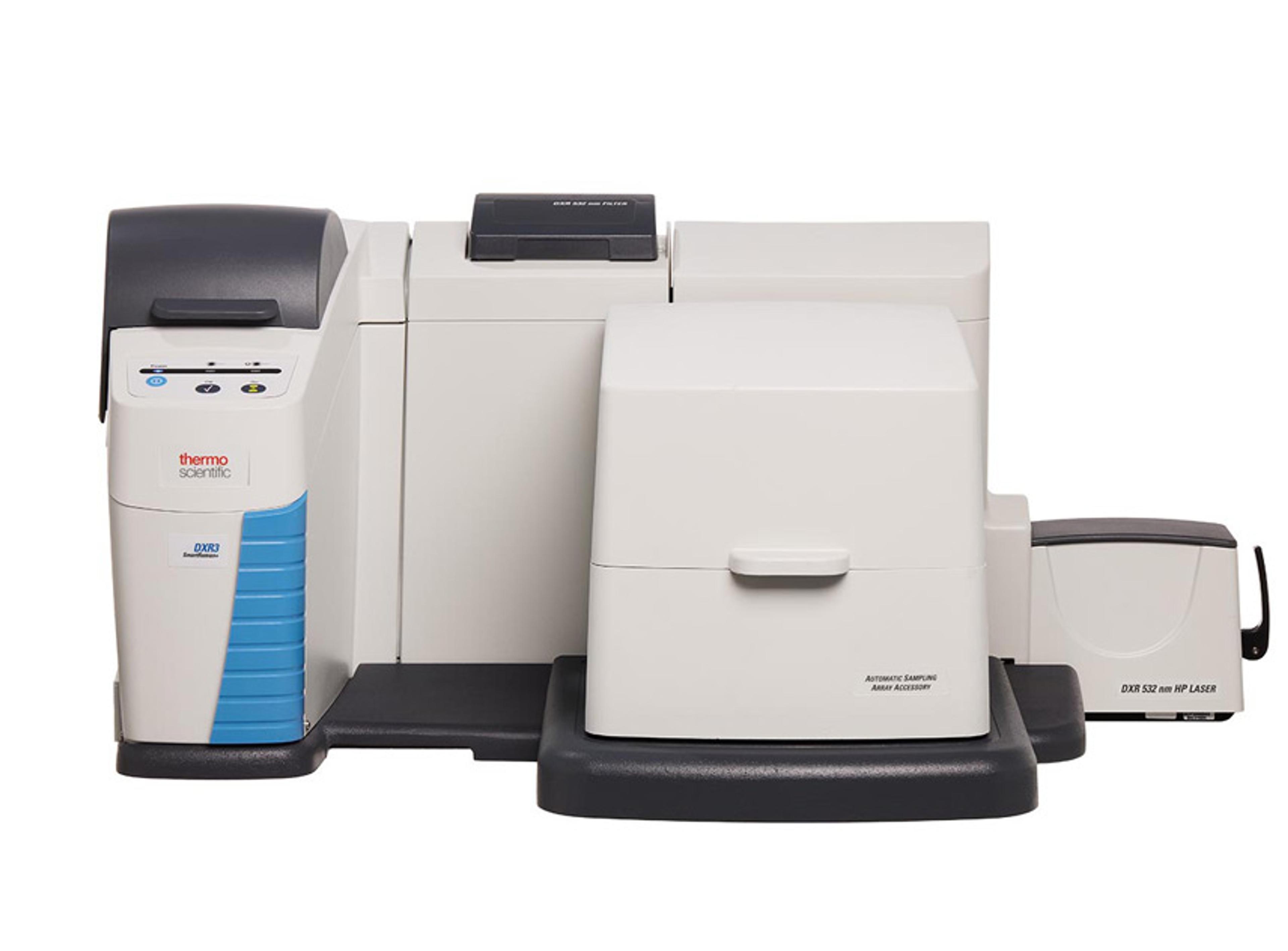
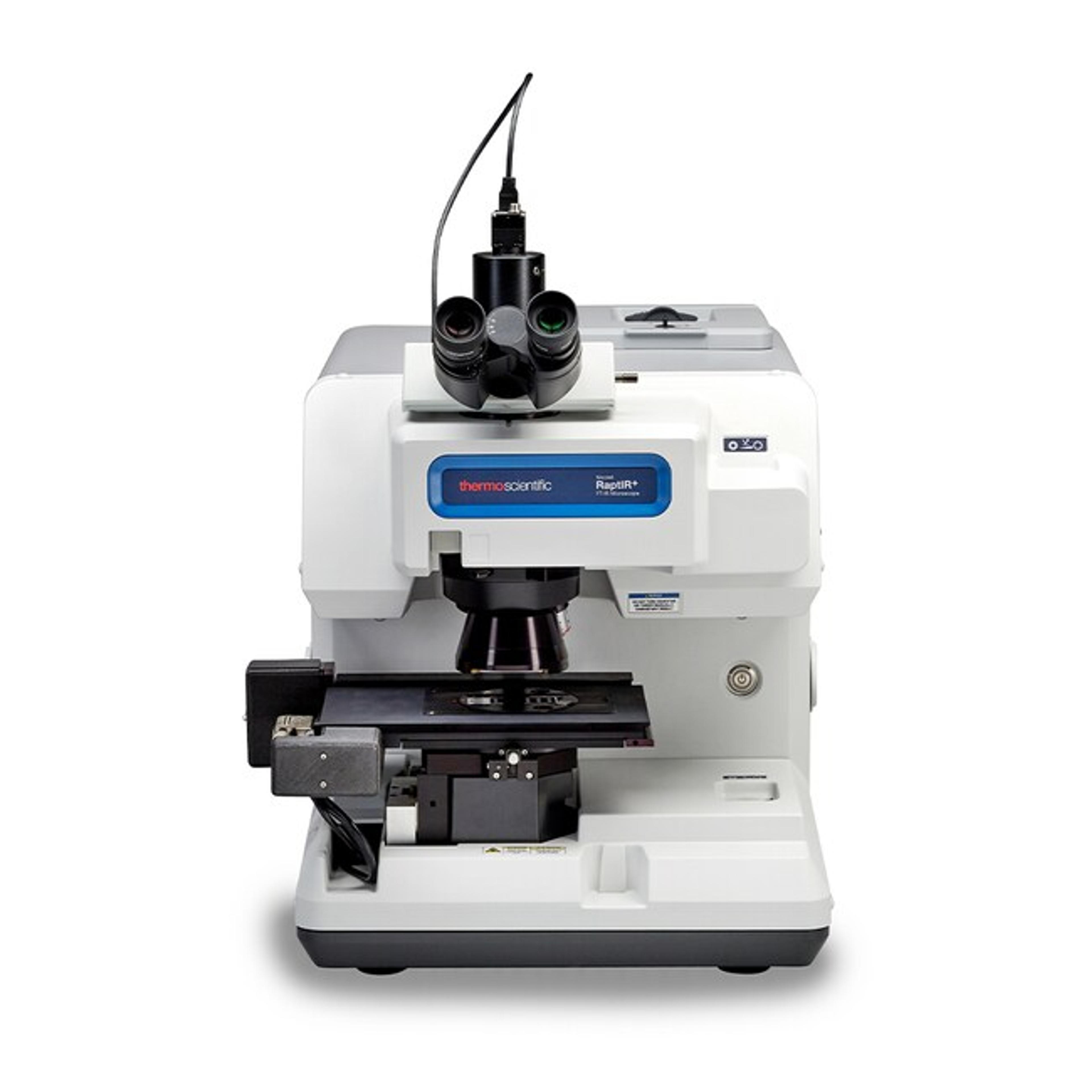
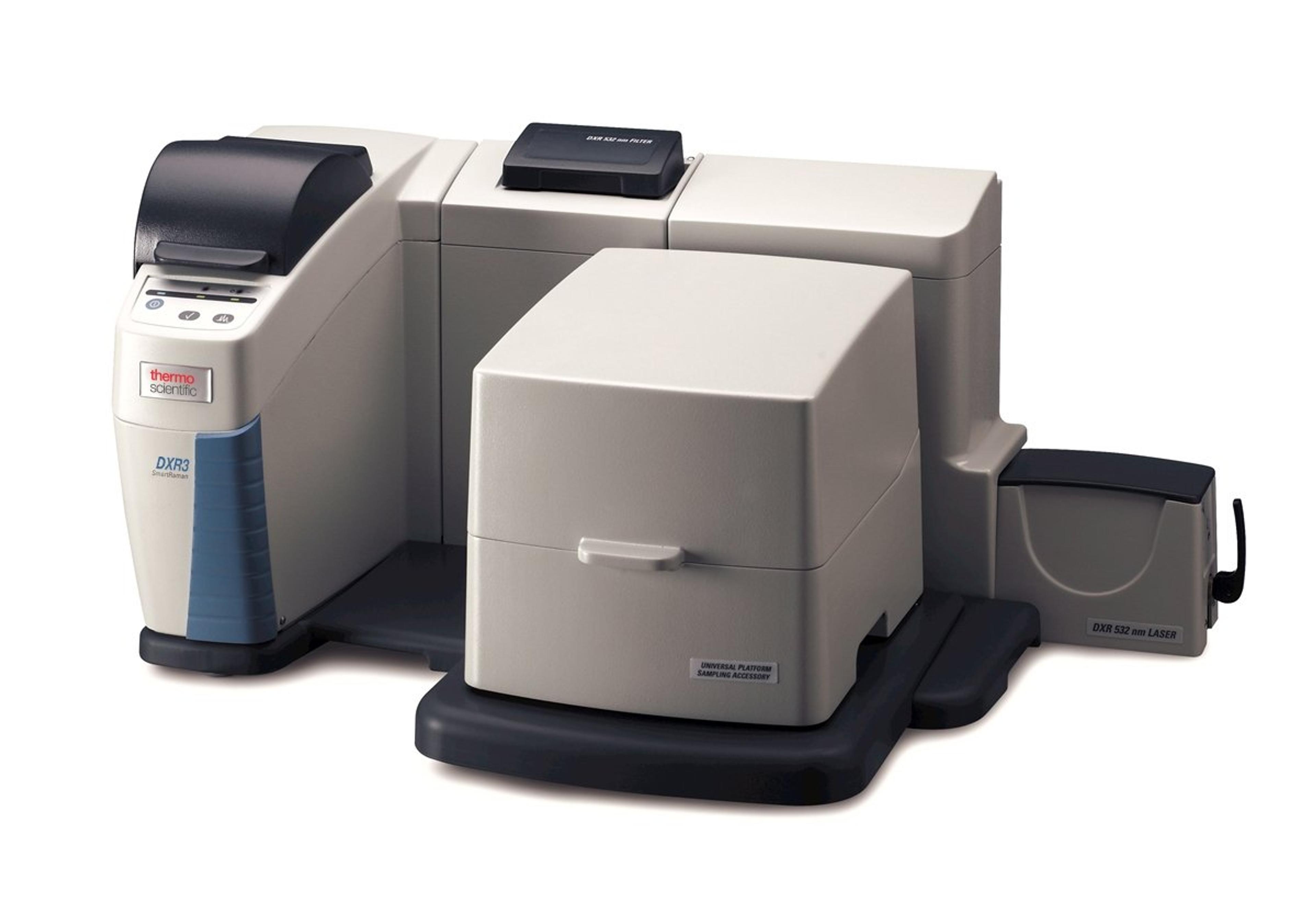
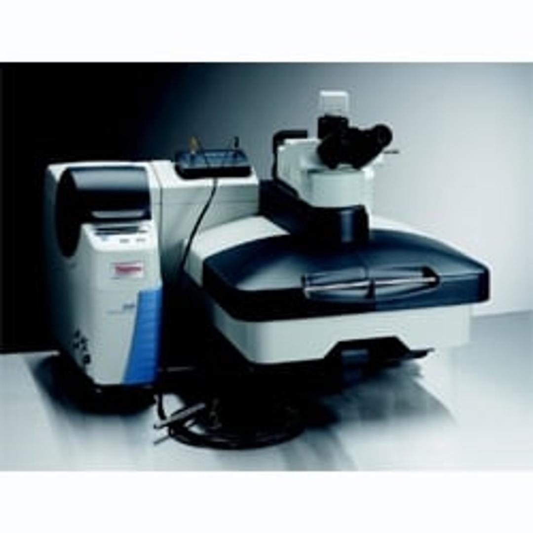
Thermo Scientific™ DXR3 Raman Microscope
Achieve exceptional sensitivity and spatial resolution for working applications with the Thermo Scientific DXR3 Raman microscope.


Receive your quote directly from the manufacturer.
The technique allows us to compare the chemical composition of diverse materials.
Polymers, biomaterials
As a beginner, the use of this equipment is really easy. It is a good option because it is not necessary to modify or adapt the samples for the analysis. Besides, the determination of chemical composition and as an alternative test brings a lot of information and background for future applications. For material science, the research area let the combination of chemical group determination and structural behavior analysis.
Review Date: 4 Apr 2022 | Thermo Fisher Scientific
Easy to use , an efficient equipment with reproducible results
Pharmaceutical technology
We used RAMAN spectroscopy in our lab to map API in final dosage form. The equipment is very easy to use and its results are reproducible.
Review Date: 16 Nov 2021 | Thermo Fisher Scientific
Good results, really useful for an academic institution.
Materials characterisation
This is an easy-to-use benchtop micro Raman, that can be effectively used by undergraduates. Both postgrads and undergraduates can be trained within a 3-hour session to work with the instrument. The ease of switching between lasers is really useful and the capability to measure laser intensity delivered to the sample is particularly useful for peer-reviewed papers. The software is the same platform as the ATR-FT-IR which on the one hand is a good thing. On the other hand, ease of deconvolution is missing, and to get a proper deconvolution different software must be used.
Review Date: 16 Jul 2021 | Thermo Fisher Scientific
Great for a variety of samples, on various surfaces to get rapid results.
Explosive materials
Good help from specialized engineers post- purchase. Very easy to use interface and the mosaic imaging system and real-time Raman is very handy!
Review Date: 3 Mar 2020 | Thermo Fisher Scientific
Great instrument with good value.
Ambient aerosol particles
The instrument is very easy to use. They offer a good library to search spectra. Good after sell service is available.
Review Date: 23 Mar 2019 | Thermo Fisher Scientific
Not necessary to carry out sample preparation for analyzing with this instrument.
Identification of minerals and compounds
Confocal Raman is very useful for identifying unknown minerals and compounds in samples. This instrument is also easy to use. Sample preparation is not necessary for analyzing with this instrument.
Review Date: 18 Feb 2019 | Thermo Fisher Scientific
Great results with image.
Analyze mineralization in skeleton and tissue sample
It is very easy to use with no sample preparation. It gives very accurate results.
Review Date: 29 Jul 2016 | Thermo Fisher Scientific
Protein quantification
This is a very nice, compact piece of equipment. It was very easy to use - I was using it confidently myself after only a few minutes. It is very user friendly, both the instrument itself and the operating/analysis software. The best feature is the ease of changing the laser wavelength - literally less than a minute!
Review Date: 6 Sept 2012 | Thermo Fisher Scientific
Access Raman data faster than ever with this easy-to-use point-and-shoot Raman Microscope. Advanced imaging capabilities make the Thermo Scientific™ DXR3 Raman Microscope faster and more reliable than ever. Users can more confidently use their Raman data thanks to Automatic X-axis Calibration which improves reliability and stability. With speed and flexibility from new lasers and gratings the DXR3 Raman Microscope is ideal for applications like pharmaceutical, academic, advanced polymers, microplastics and many more.
Capabilities:
- New, Particle Analysis for quickly identifying and analyzing microparticles
- High performance confocal Raman microscopy in a robust, integrated design
- Autoalignment and calibration ensures scientifically accurate measurements, without tools or manual procedures
- Real-time preview, automated fluorescence correction, autoexposure, and cosmic ray rejection
- System status indicator shows the user at a glance that the system is optimized and ready to collect data
- Three-path fine beam autoalignment maintains peak performance and sampling integrity
- Laser power regulation ensures consistent sample excitation over the lifetime of the laser
- Advanced spectrograph design with no moving parts simplifies use and make the detection system and calibration robust
- Pre-aligned and lock-in-place components use automatic recognition and stored alignment, allowing any user to reconfigure an instrument in seconds
- Lasers and other components can be interchanged and shared with every instrument in the DXR3 Raman family
- Optional, automated polarized Raman capabilities provide structural information that complements chemical information
- Add new wavelengths without tools or service engineer visit
Laser Safety
- Microscope independently certified as class-1 laser safe. (Note: Optional fiber-optic accessory and some other optional accessories are class IIIb laser devices requiring laser safety precautions and laser safety eyewear.)
- Laser is physically blocked from visual viewing path to prevent exposure to the eyes while viewing.
Validation
- Complete package for DQ/IQ/OQ/PQ validation is available including extensive documentation and automated software protocols
- Omnic D/S software offers CFR 21 part 11 compliance
Exceptional Performance
- Spatial resolution— as fine as 540 nm resolution on ideal sample
- Confocal depth resolution— as fine as 1.7 μm on ideal sample
- Full spectral range of 3500-50 cm-1 captured with a single exposure of the CCD avoids stitching artifacts. Extended spectral range to 6,000 cm-1 available for 532 nm laser
- Supports multiple excitation lasers. 455 nm, 532 nm high brightness, 532 nm high power, 633 nm high brightness, 633 nm high power, 785 nm high brightness, and 785 nm high power
- Automatic fluorescence correction available with all excitation lasers
- Research-quality Olympus viewing optics
- Automatic intensity correction generates spectra that are comparable between instruments and different excitation lasers
- Multidimensional wavelength calibration provides accurate wavelength calibration across entire spectral range
- Proprietary Triplet spectrograph provides exceptional peak shape and maintains focus at all wavelengths
- Software controlled switching between confocal and high-energy collection modes
- Proprietary algorithm for automated cosmic ray rejection and high-quality laser line filters ensure artifact-free spectra
- Lasers depolarized to avoid confusing sample orientation effects
- Laser Power Regulator provides active monitoring of laser power and ensures reproducible laser excitation power over the lifetime of lasers and even after replacing lasers
- Optimized gratings for each excitation laser avoid performance sacrifices of systems that share gratings for multiple excitation lasers
- Proprietary autoalignment ensures system can be maintained in like-new condition
- Proprietary Smart-backgrounds automatically remove CCD dark energy profile
Recommended for:
- Forensics – trace evidence and illicit drug identification
- Pharmaceutical – polymorphs, particulate contaminants, and diffusion studies
- Art Restoration/Conservation – identify and characterize pigments, resins, glazes, and inks
- Archaeology – characterize horn, shell, bone, and ceramic artifacts
- Solar – Silicon crystallinity; characterization of photovoltaic materials
- Polymers – inclusions and gel defects, weathering effects, tie layers in laminates, and crystallinity
- Failure analysis – characterizing particulates and small features on surfaces
- Gemology – rapid ID of colored stones, distinguishing natural and synthetic diamonds, characterizing inclusions and adulterants
- Nanotechnology – characterize graphene, CNTs, DLC coatings and other nanostructures
- Academic research – useful in material science, biological studies, and many applied research fields
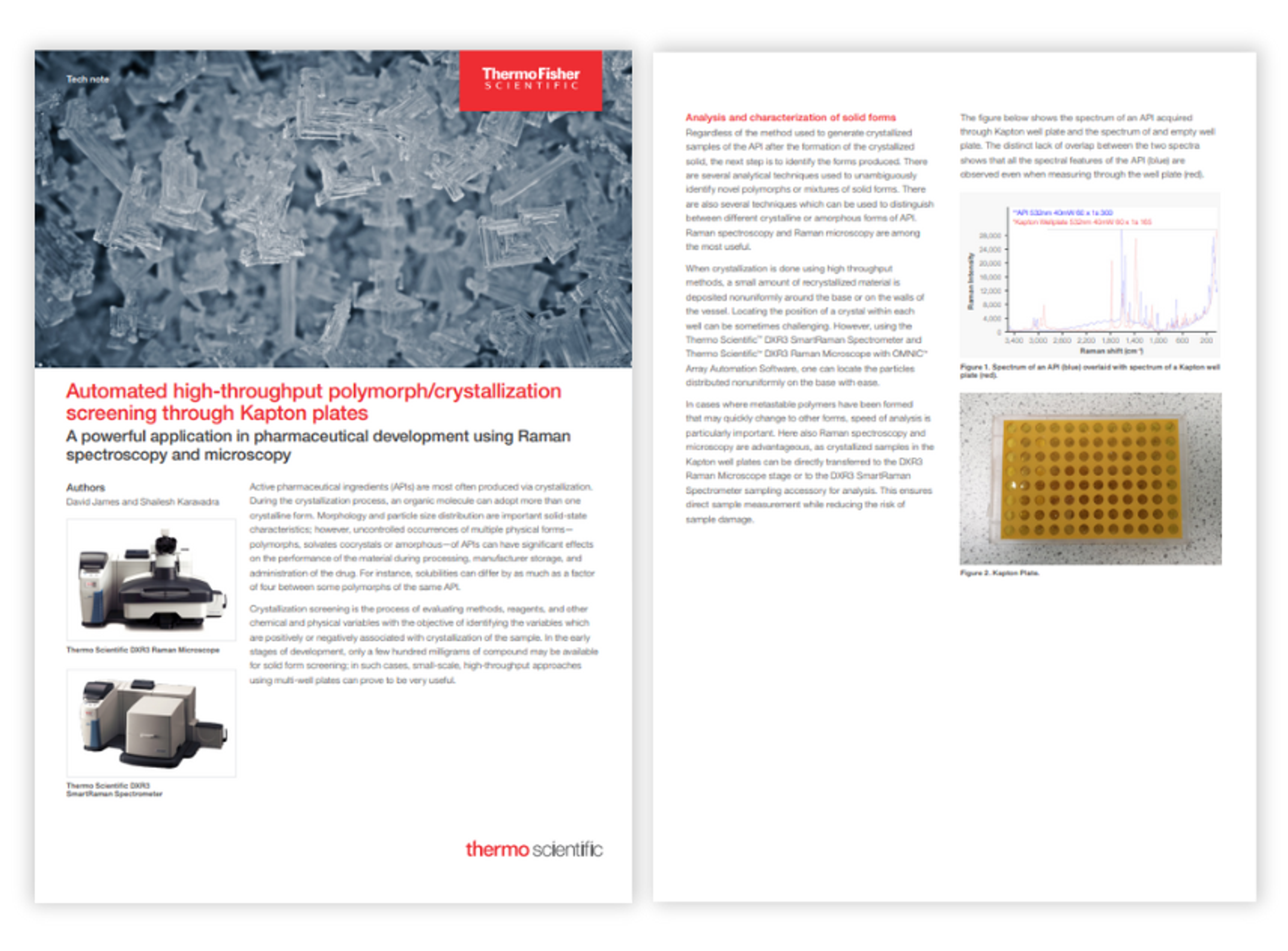
Automated high-throughput polymorph/crystallization screening through Kapton plates
During the crystallization process, an organic molecule can adopt more than one crystalline form. Morphology and particle size distribution are important solid-state characteristics; however, uncontrolled occurrences of multiple physical forms of APIs can have significant effects on the performance of the material during processing, manufacturer storage, and administration of the drug.
In this application note, Thermo Fisher Scientific demonstrates a workflow for high-throughput polymorph screening using its DXR3 SmartRaman Spectrometer.

Characterization of Amorphous and Microcrystalline Silicon Using Raman Spectroscopy
Silicon deposited on glass or silicon carbide is widely used in manufacturing photovoltaic cells. Raman spectroscopy is an ideal technique for this application. By mapping an area of deposited silicon the uniformity of the distribution of the two silicon forms can be monitored. This application note shows typical results and discusses some of the practical aspects and potential pitfalls of using Raman spectroscopy for measuring amorphous and crystalline silicon.
Measuring Diamond-like Carbon Films by Dispersive Raman Spectroscopy
Raman spectroscopy is a powerful technique for monitoring the physical properties of diamond-like carbon (DLC) films. In this application note, Raman spectroscopy exhibited the ability to monitor DLC films that resulted from varying the plasma conditions. It is known that deposition conditions have a strong influence on the atomic structure of the resulting films. This work suggested that harder, more inert films result from higher plasma current and higher partial gas pressures.
Raman Mapping of Single-Walled Carbon Nanotube Distribution on Phase Separated Polystyrene and Polymethylmethacrylate
In this application note, phase separated polystyrene and polymethylmethacrylate polymers were vapor deposited onto silicon. To ensure that these materials were indeed phase separated, dispersive Raman mapping was used to show the distribution of the polymers. The size of the structures varied from 10-50 microns in width. Single-walled carbon nanotubes were then vacuum-deposited onto the surface.
Characterizing Carbon Materials with Raman Spectroscopy
This application note describes how Raman Spectroscopy is a very powerful and valuable technique for the characterization of carbon nanomaterials. Raman is particularly well suited to detect small changes in structural morphology of carbon nanomaterials making it an indispensable tool for many material scientists working with carbon nanostructures. Raman instruments are very fast and provide a great deal of flexibility in samples that can be accommodated. Every lab that is characterizing carbon nanomaterials will benefit from having access to Raman instrumentation.
Characterizing Graphene with Raman Spectroscopy
Raman spectroscopy is a great tool for the characterization of graphene. Few techniques will provide as much information about the structure of graphene samples as Raman spectroscopy and any lab doing graphene characterization without Raman would be at a significant disadvantage. In this application note, the Thermo Scientific DXR Raman microscope is used for graphene characterization to provide a high level of stability, control and sensitivity needed to produce confident results.
Evaluating Active Ingredients in Pharmaceutical Hot Melt Extrusion Products with Raman Imaging
Hot Melt Extrusion (HME) is a highly beneficial alternative to traditional methods for the manufacture of pharmaceutical products. In this application note, discover how Raman imaging can verify the distribution and integrity of the various components used in HME as well as identify contaminants throughout the process in order to assure a reliable and reproducible product.