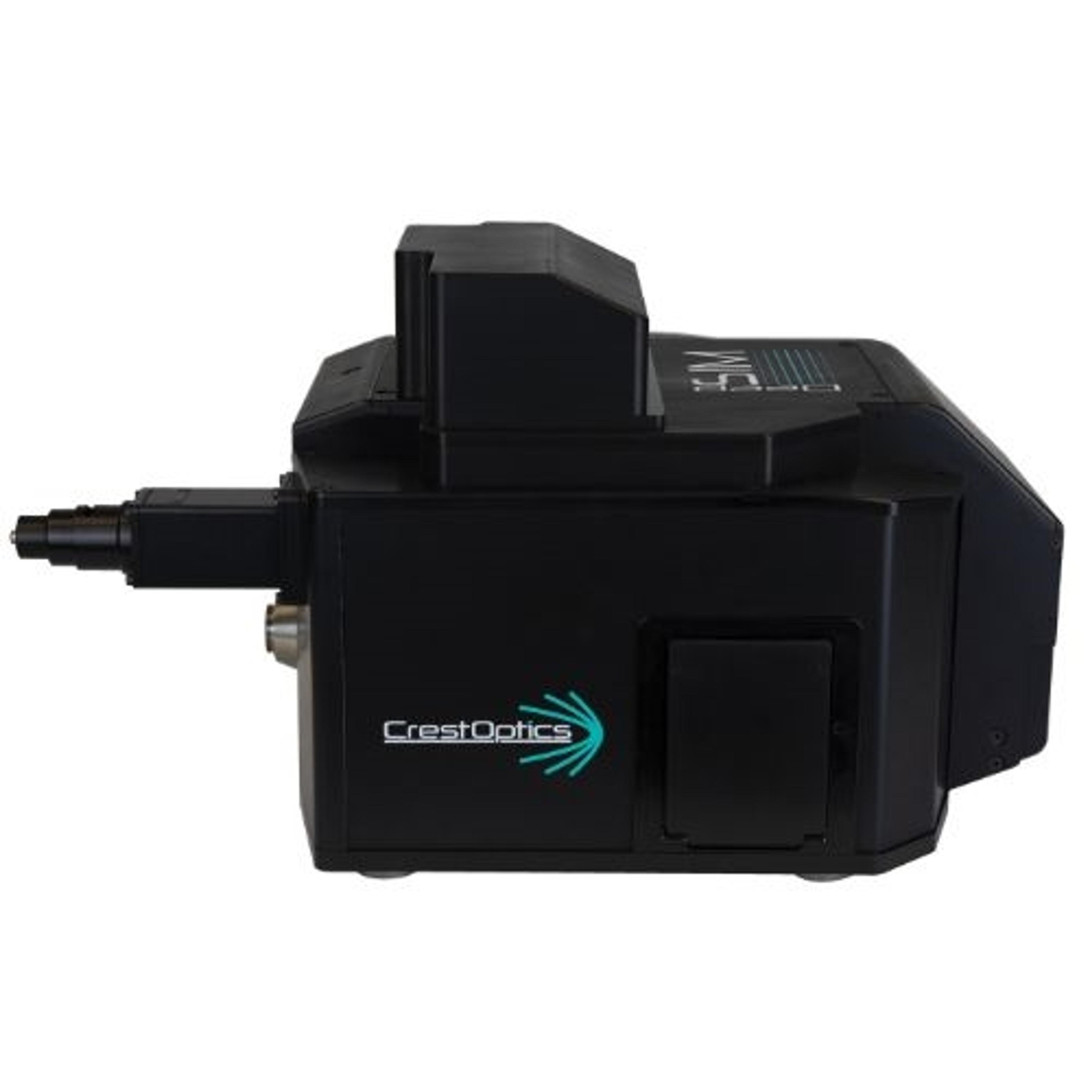
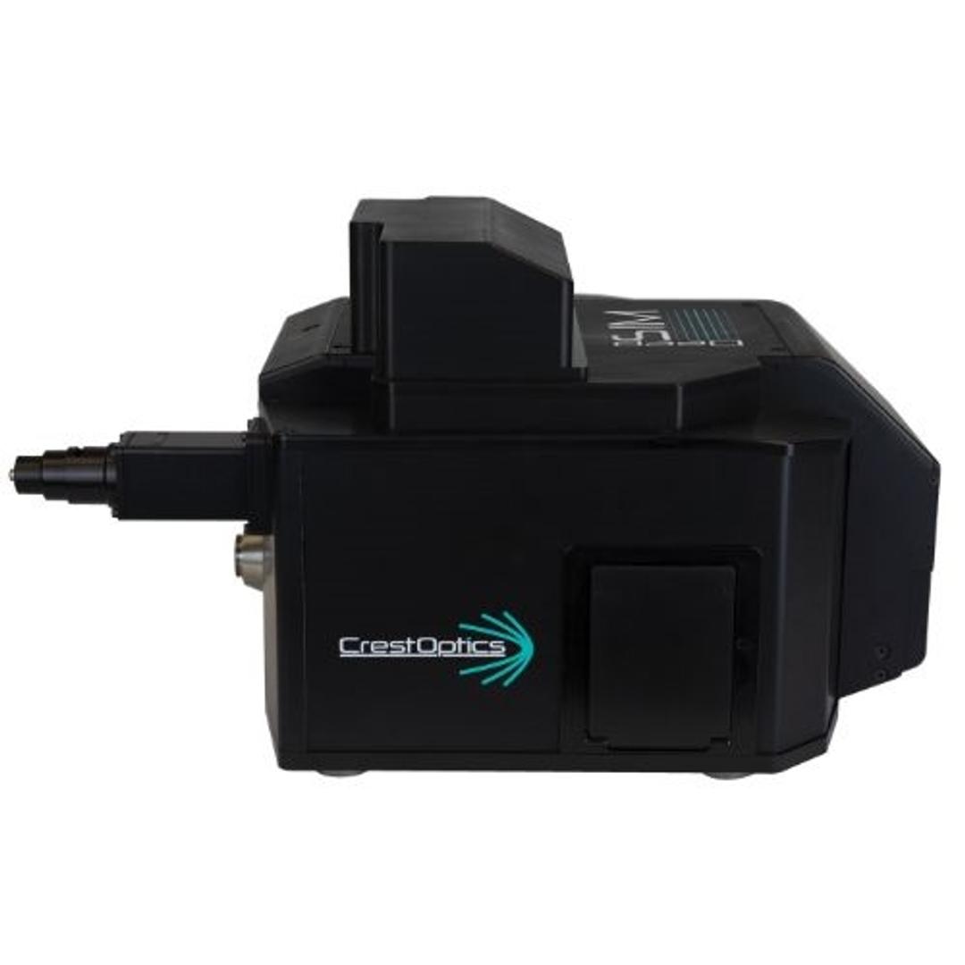
DeepSIM
"A Seamless Evolution of Your Microscopy". DeepSIM super-resolution technology is based on multi-spot lattice structured illumination and provides a reliable and affordable solution to study subcellular structures with an XY resolution of 100 nm. The DeepSIM super-resolution system is easy-to-use and enables scientists to access deep data from their biological samples without requiring any special sample preparation protocol…


The supplier does not provide quotations for this product through SelectScience. You can search for similar products in our Product Directory.
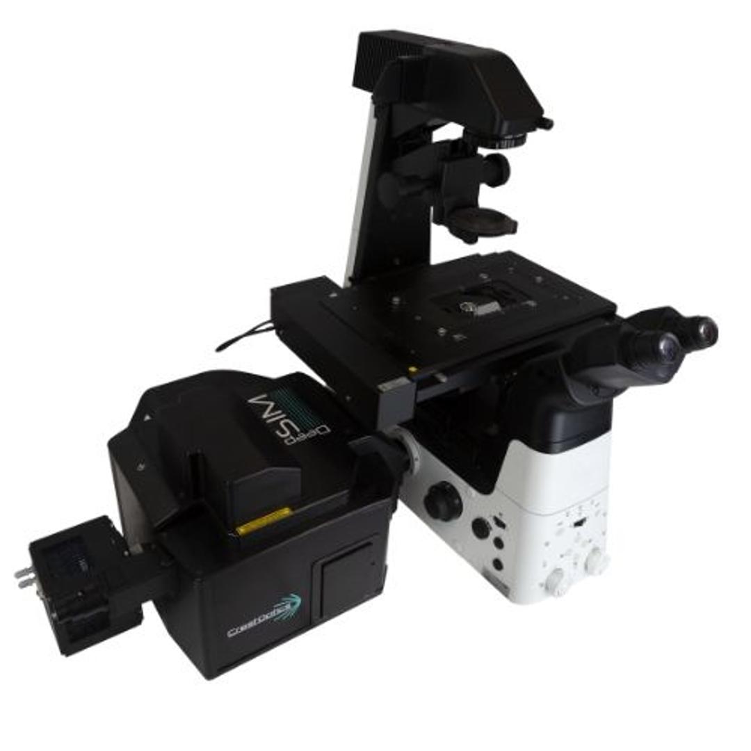
At CrestOptics, we believe that super-resolution should be accessible for all scientists to progress their research. This is the reason behind the development of the DeepSIM, the first Lattice SIM super-resolution module compatible with any existing upright or inverted microscope with a camera port. The DeepSIM constitutes the seamless evolution of any diffraction-limited microscopy approach.
It is as easy to use as a widefield system and reaches the same Z-depth penetration of a confocal microscope, enabling scientists to access super-resolved data through conventionally prepared, deep, thick specimens, even in challenging sampling conditions. DeepSIM does not require any special probes and researchers can maintain their standard sample preparation protocols. The system is characterized by robust system calibration, pre-optimized acquisition modalities, and automated data processing to break down the learning curve. Aware that establishing Super Resolution - SIM as a common tool for routine life science research applications, DeepSIM has been developed to be a truly enabling technology, combining performance with flexibility and advanced application integration.
KEY FEATURES:
1) Imaging Across Scale: Multiple Lattice SIM Illumination Patterns
The 2D Lattice SIM technology behind DeepSIM enhances the illumination efficiency and homogeneity with minimal phototoxicity in live cell imaging experiments. Besides, the 2D Lattice SIM Illumination delivers a better contrast that significantly improves in-depth Z acquisitions and the image reconstruction robustness. More importantly, the user can choose among 3 Lattice SIM Illumination Patterns for approaching specimens of increasing thickness and morphological complexity. The well-matched sampling conditions significantly mitigate the risk of artifacts in image reconstruction.
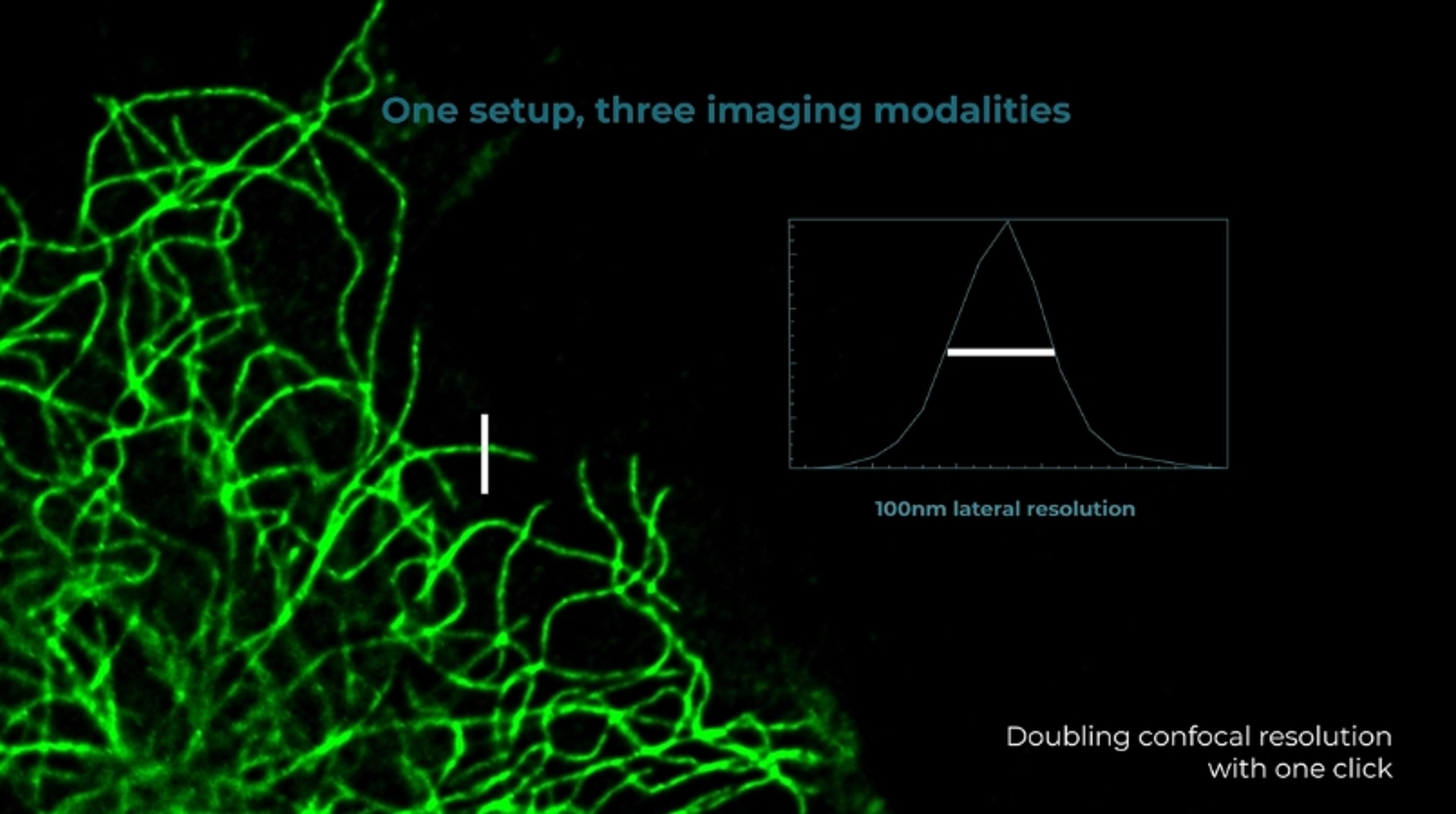
2) Down to 100 nm lateral resolution
Featuring the structured lattice illumination with the computationally super-resolved data reconstruction, DeepSIM improves the spatial resolution in all three dimensions reaching 100 nm laterally and 300 nm axially.
3) Deep imaging through thick specimens
Deep super-resolved data across conventionally prepared thick samples. The DeepSIM is designed to work with samples of thicknesses comparable to those used with Confocal microscopy, giving super-resolved data over 50 μm Z-depth. Z-stacks take full advantage of the entire objective working distance and meaningful data can be obtained from a native heterogeneous and intricate sample, providing researchers with a complete understanding of its structure and composition.
4) Suitable with standard confocal staining up to Near-Infrared 750 nm Excitation
Multiplexed imaging is an emerging way to gather information from multiple cellular markers simultaneously. DeepSIM performs optimally with the standard staining markers for confocal microscopy. It is the only SIM technology covering the full spectrum from 405 nm to NIR 750 nm excitation wavelength. DeepSIM offers the maximum flexibility in fluorophore choice and reduces the spectral overlap in multicolor experiments.
5) Compatible with Low Mag objectives and with any kind of Immersion Media
The DeepSIM was conceived to maintain high-performance standards over a large set of objectives from Low Magnification 20x up to High Magnification 100x. The compatibility with dry lenses and any immersion media expands the range of suitable applications over a large cohort of sample and support types. DeepSIM offers the best trade-off between FOV and resolution to catch fine details in extra-large objects.
6) Resilient resolving power across specimen scale: from 3D organoids to small animal models
The DeepSIM resolving power is preserved across a wide spatial scale, including 3D organoids, tissue slices, and small animal models (Zebrafish, C. elegans). The extended flexibility suggests its use with Spinning Disk technology in a correlative bioimaging approach.
7) Gentle Lattice Structured illumination suitable for live-cell imaging
Thanks to its temporal resolution of over 10 fps, the DeepSIM allows the capture of meaningful data at high resolution with minimized light exposure and photo-toxicity risk. Delicate and live specimens can be explored in real time to monitor cellular and subcellular changes.
8) Evolution through compatibility
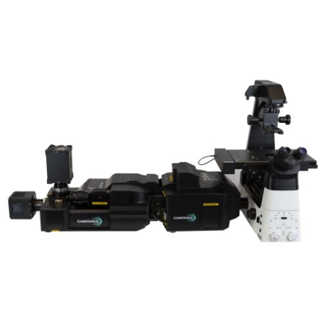
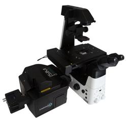
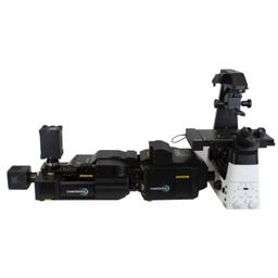
By choosing DeepSIM technology, you will be able to create a modular, expandable, and highly performant system, resulting in the creation of a truly enabling technology. The DeepSIM can be used both with CrestOptics’ X-Light V3 confocal system as well as independently as a stand-alone system for any microscope that has a camera port.
CONCLUSION
DeepSIM is designed to be a truly enabling technology where performance is combined with the flexibility of a modular, expandable system. DeepSIM can seamlessly upgrade any existing frame with a camera port, be it upright or inverted, thus leading to a significant cost-saving setup.
Brochures

DeepSIM brochure
In this product brochure, CrestOptics introduces DeepSIM, a super-resolution microscopy system that addresses deep biological questions with ease. The goal of CrestOptics is to make super-resolution accessible to all scientists to advance their research. For this reason, it developed DeepSIM, the first super-resolution module that is compatible with any existing upright or inverted microscope and can be used like a confocal microscope to facilitate access to super-resolved deep data of biological samples.
Discovering cellular organelles with illumination microscopy
An organelle is a subcellular structure that contributes to a variety of cellular functions through its molecular composition and environmental interactions. CrestOptics discusses the significance of organelles in cellular functions and the limitations of conventional fluorescence microscopy in studying subcellular structures smaller than 200 nm. DeepSIM, a SIM super-resolution module that is compatible with any existing upright or inverted microscope with a camera port, is presented as a reliable, easy-to-use, and affordable solution to study sub-cellular structures with an XY resolution of 100 nm without requiring any special sample preparation protocol. DeepSIM's capability in visualizing various intracellular structures, such as endosomes, actin, mitochondria, tubulin, endoplasmic reticulum (ER), and lysosomes, at the subcellular level is also demonstrated.
Increasing amplicon spots detection efficiency
The field of spatial transcriptomics aims to map the RNA transcriptome of single cells within tissues while preserving spatial information. CrestOptics addresses challenges faced in imaging-based spatial biology, particularly optical crowding when using epifluorescence microscopy. A study is presented that explores the impact of structured illumination microscopy (SIM), specifically the DeepSIM super-resolution (SR) system, on single-gene transcript detection. CrestOptics demonstrates that DeepSIM enhances RNA spot detection compared to widefield (WF) and confocal (CF) modes, improving contrast, resolution, and spot detection performance.
Introducing the DeepSIM super-resolution system, a seamless evolution of your microscopy
CrestOptics believes that super-resolved microscopy data should be accessible to all scientists to progress their research. This is the reason why it has developed DeepSIM, a super-resolution module based on a lattice multi-spot structured illumination. In this application note, CrestOptics presents the DeepSIM. The DeepSIM relies on a 2D lattice multi-spot SIM technique, reaching 100 nm XY resolution and 300 nm Z resolution (100X, 1.45 NA Plan Apochromat objective). The set of three dedicated micro-lens masks and the compatibility with low and high magnification objectives expand the application of the DeepSIM to a variety of biological samples, from cell monolayers to 3D cell cultures and cleared tissues over 100 µm thick with Z-depth penetration comparable to a confocal microscope. Featuring high photon-efficiency in illumination with a temporal resolution over 10 fps, the DeepSIM is also well suited for live-cell imaging with multiple colours and conventional fluorophores within the 405-750 nm excitation spectral ranges.
Deep structured illumination microscopy of neurobiological samples
Widefield (WF) and confocal (CF) microscopy might be sufficient to study cellular and tissue morphology and dynamics but are often not enough to accurately answer all biological questions. Getting a further level of information could allow new discoveries and, in this regard, super-resolution (SR) microscopy has become an increasingly popular and robust tool across life sciences to study fine cellular structures and sub-cellular components, as well as their dynamic processes. CrestOptics are working to make SR accessible to all researchers and to advance the potential of their scientific discoveries, moving them to a next level. Its newest product, the DeepSIM, was developed with this purpose and it is the first SR module based on structured illumination compatible with any existing upright or inverted microscope with a camera port, it is as easy to use as a CF microscope, and it enables scientists to access deep data about their biological samples. The DeepSIM is designed to work with samples of thickness comparable to those used in CF microscopy and in this application note, CrestOptics focuses on super-resolved deep imaging of different neurobiological samples at high magnification.
Structured illumination microscopy with low magnification objectives
In this application note, CrestOptics demonstrates that two-fold enhanced spatial resolution can be obtained with a magnification objective as low as 20X, making the super resolution (SR) module a reliable, simple to use, and affordable solution to study sub-cellular details.
DeepSIM super-resolution acquisition
One of the aims of modern microscopy is to overcome the barrier due to the light diffraction limit in order to observe biological structures at the nanometer scale. Furthermore, because of light scattering caused by the inhomogeneity of tissues, achieving this goal is even more difficult within thick specimens where scattering limits the light penetration depth. To demonstrate the DeepSIM optimal performance in terms of penetration depth and light throughput at different sample depths, CrestOptics highlights SR images in complex and intricate biological structures.
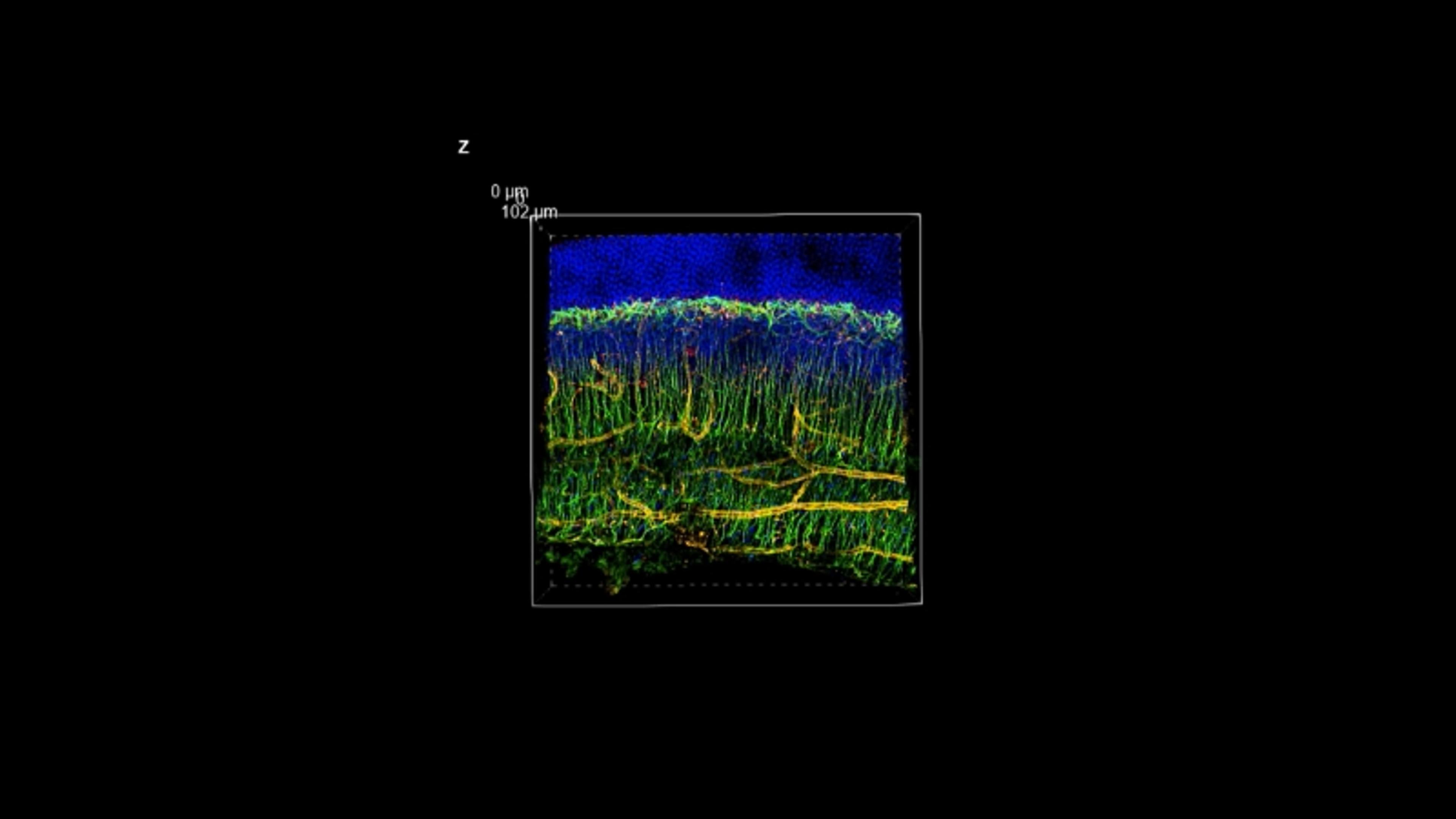
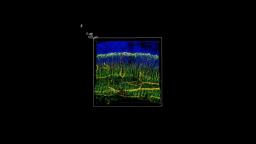
DeepSIM mouse retina section
The DeepSIM was conceived to maintain high standards of performance over a large set of objectives from Low Magnification 20x up to High Magnification 100x. The compatibility with dry lenses and any immersion media expands the range of suitable applications over a large cohort of sample and support types. DeepSIM offers a trade-off between FOV and resolution to catch fine details in extra-large objects. CrestOptics presents an example of DeepSIM capture from a mouse retina section.
CrestOptics DeepSIM super-resolution system
CrestOptics showcases its DeepSIM system, a Lattice SIM super-resolution module that is compatible with any existing upright or inverted microscope with a camera port. It allows for one setup with three imaging possibilities. Featuring structured lattice illumination with computationally super-resolved data reconstruction, DeepSIM promises to improve the spatial resolution in all three dimensions reaching 100 nm laterally and 300 nm axially.