Discovering cellular organelles with illumination microscopy

17 Apr 2024

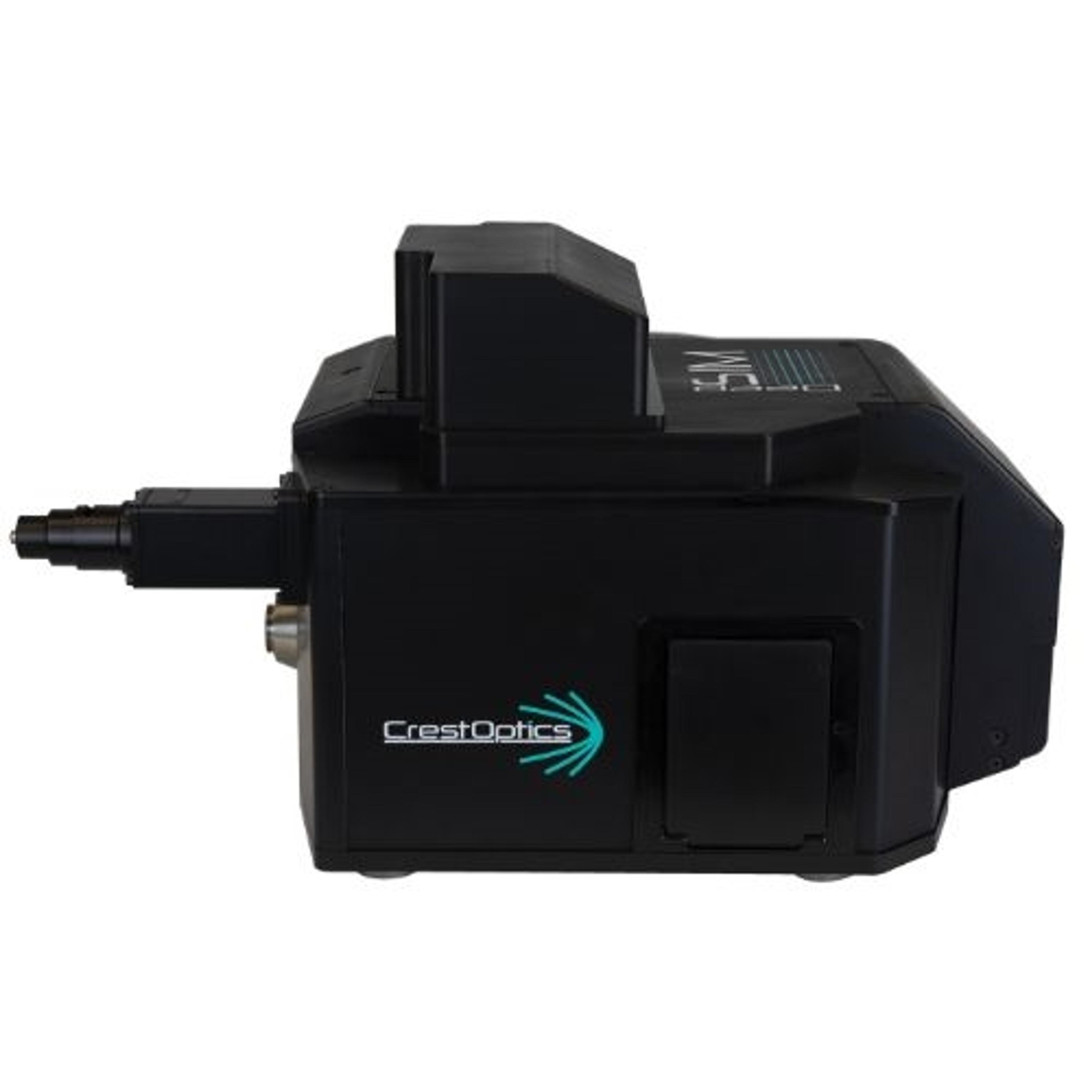
An organelle is a subcellular structure that contributes to a variety of cellular functions through its molecular composition and environmental interactions. CrestOptics discusses the significance of organelles in cellular functions and the limitations of conventional fluorescence microscopy in studying subcellular structures smaller than 200 nm. DeepSIM, a SIM super-resolution module that is compatible with any existing upright or inverted microscope with a camera port, is presented as a reliable, easy-to-use, and affordable solution to study sub-cellular structures with an XY resolution of 100 nm without requiring any special sample preparation protocol. DeepSIM's capability in visualizing various intracellular structures, such as endosomes, actin, mitochondria, tubulin, endoplasmic reticulum (ER), and lysosomes, at the subcellular level is also demonstrated.

DeepSIM

CrestOptics S.p.A

"A Seamless Evolution of Your Microscopy". DeepSIM super-resolution technology is based on multi-spot lattice structured illumination and provides a reliable and affordable solution to study subcellular structures with an XY resolution of 100 nm.  The DeepSIM super-resolution system is easy-to-use and enables scientists to access deep data from their biological samples without requiring any special sample preparation protocol.  

(0)

Links

Tags

Light MicroscopyLight microscopes or optical microscopes are used to visualize microscale objects under magnification, including cells, clinical specimens and materials. Lab equipment for light microscopy includes confocal microscopes, fluorescence microscopes, zoom and stereo microscopes. Microscope slides and imaging reagents are available for visualizing samples, as well as various microscope stages and incubators for large or temperature-sensitive samples. Find the best light microscopes in our peer-reviewed product directory: compare products, check customer reviews and receive pricing direct from manufacturers.Electron MicroscopyElectron microscopes (EM) are used to create high-resolution images of samples at the nanoscale by means of an accelerated beam of electrons as a source of illumination. Types of electron microscope include scanning electron microscopes (SEM), transmission electron microscopes (TEM), scanning transmission electron microscopes (STEM) and cryo-electron microscopes. Focused ion beam (FIB) microscopes are useful for modifying or milling a sample surface with nanometer precision, as well as imaging. Find the best electron microscopes in our peer-reviewed product directory: compare products, check customer reviews and receive pricing direct from manufacturers.Fluorescence MicroscopyFluorescence microscopy has become an essential tool in biology, as well as in materials science. The application of many fluorochromes has made it possible to identify cells and sub-microscopic cellular components with a high degree of specificity. Using multiple fluorescence labels, different probes can simultaneously identify several target molecules.MitochondriaMicroscopyMicroscopy is a technique used to observe small objects in detail, from cells to materials, using light or electron microscopes. It enables researchers to examine structures with high resolution, aiding in fields such as biology, medicine, and materials science. With advanced microscopy techniques, scientists can gain insights into cellular processes, tissue structures, and material properties. Explore the best microscopy solutions in our peer-reviewed product directory, compare products, read customer reviews, and get pricing directly from manufacturers.Super Resolution Microscopy