Live cell imaging made easy: The Celloger Pro
13 Aug 2023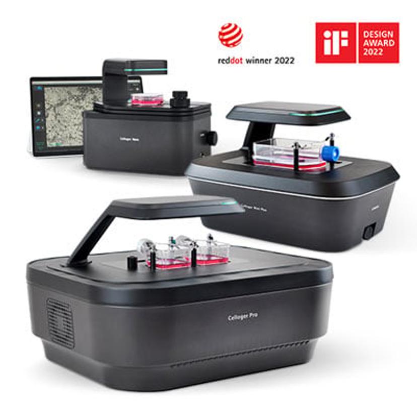
Although microscopes were discovered in the 16th century, live-cell imaging was not established until the 21st century. Despite its relatively recent discovery, it has made great technological and academic advances in the field of cellular biology. Because the implications of live cell imaging are endless, it has already become a vital tool to the study of life science; it can be used to address unanswered biological questions and provide information on the dynamics of cells. Universities, research labs, hospitals and other companies utilize live cell imaging for a myriad of uses, including cancer research, cell biology, stem cell research, drug discovery, pharmaceuticals, and more. The limitless possibilities of live cell imaging have opened a new market in the biotechnological field, which, according to Strategic Market Research, is currently worth about $2.29 billion and is projected to increase to $3.96 billion within the next seven years.
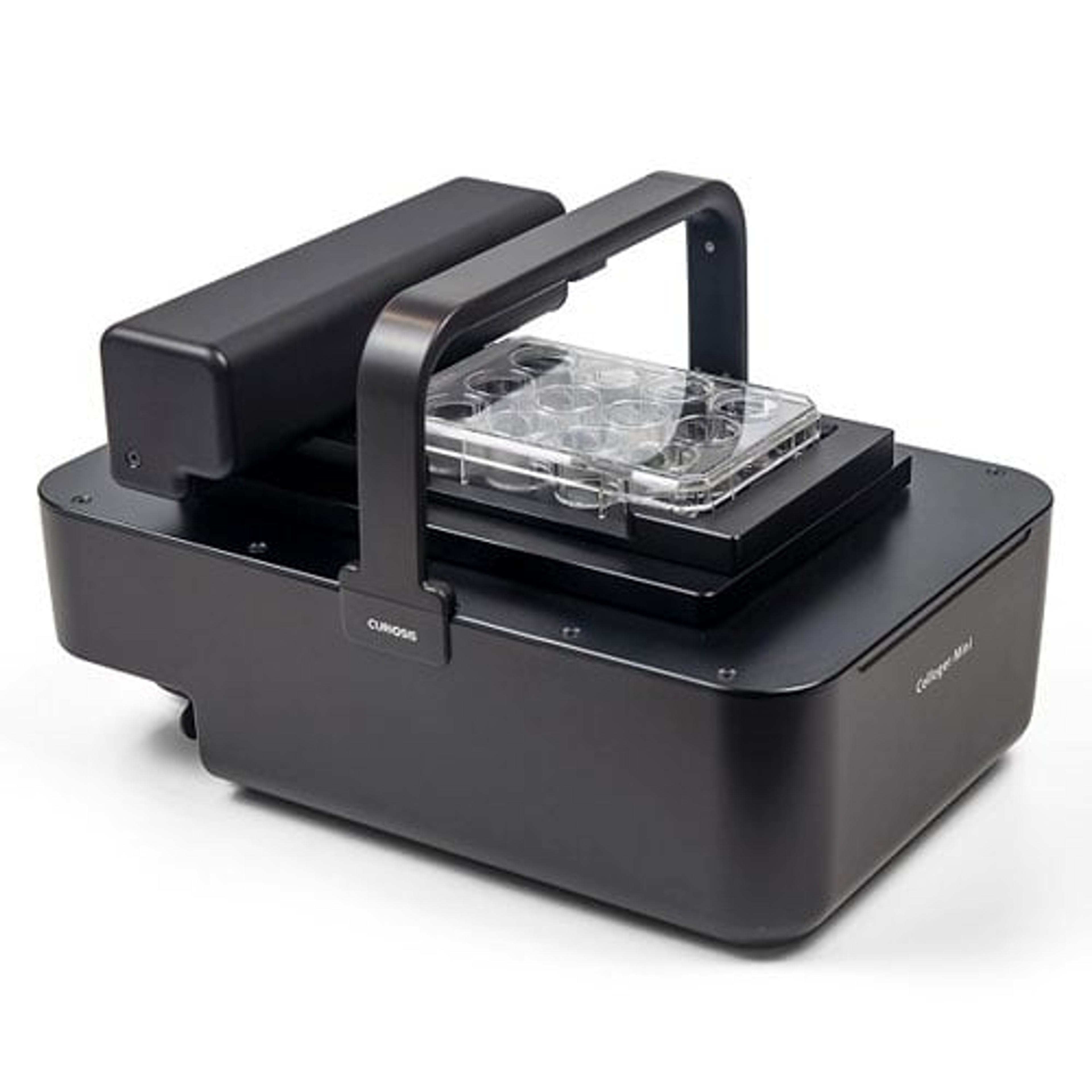
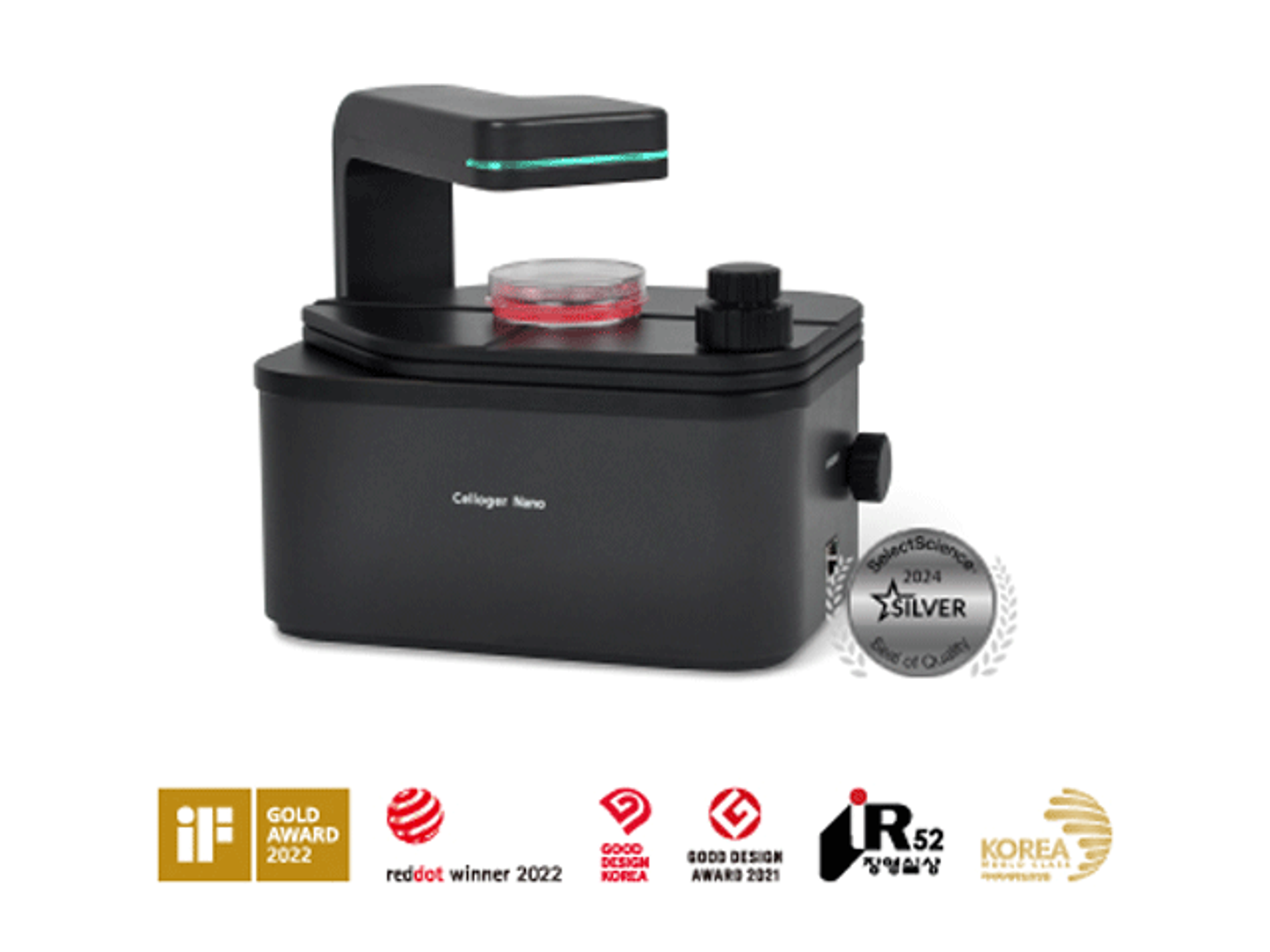
Companies focused on cellular research are developing live cell imaging technology and conducting cutting-edge experiments. Among them is the South Korean based company Curiosis, which is an innovative equipment manufacturing company that specializes in cell-based analysis and diagnostics. As a company, they are committed to making a positive impact by inventing and providing new ways to explore beyond what the eyes can see. Their recently launched line of equipment, the Celloger® Series, provides a variety of user-friendly automated live cell imaging systems including the Celloger® Mini Plus, Celloger® Nano, and the newly released Celloger® Pro. In the conventional method, live cell imaging requires many steps: loading cell samples, culturing cells, acquiring multi-point images according to a specified time interval, and analyzing the images. In addition, many of these steps must be repeated to provide accurate results. The Celloger® Pro greatly diminishes the workload for researchers.
Celloger® Pro: The New Automated Live Cell Imaging System
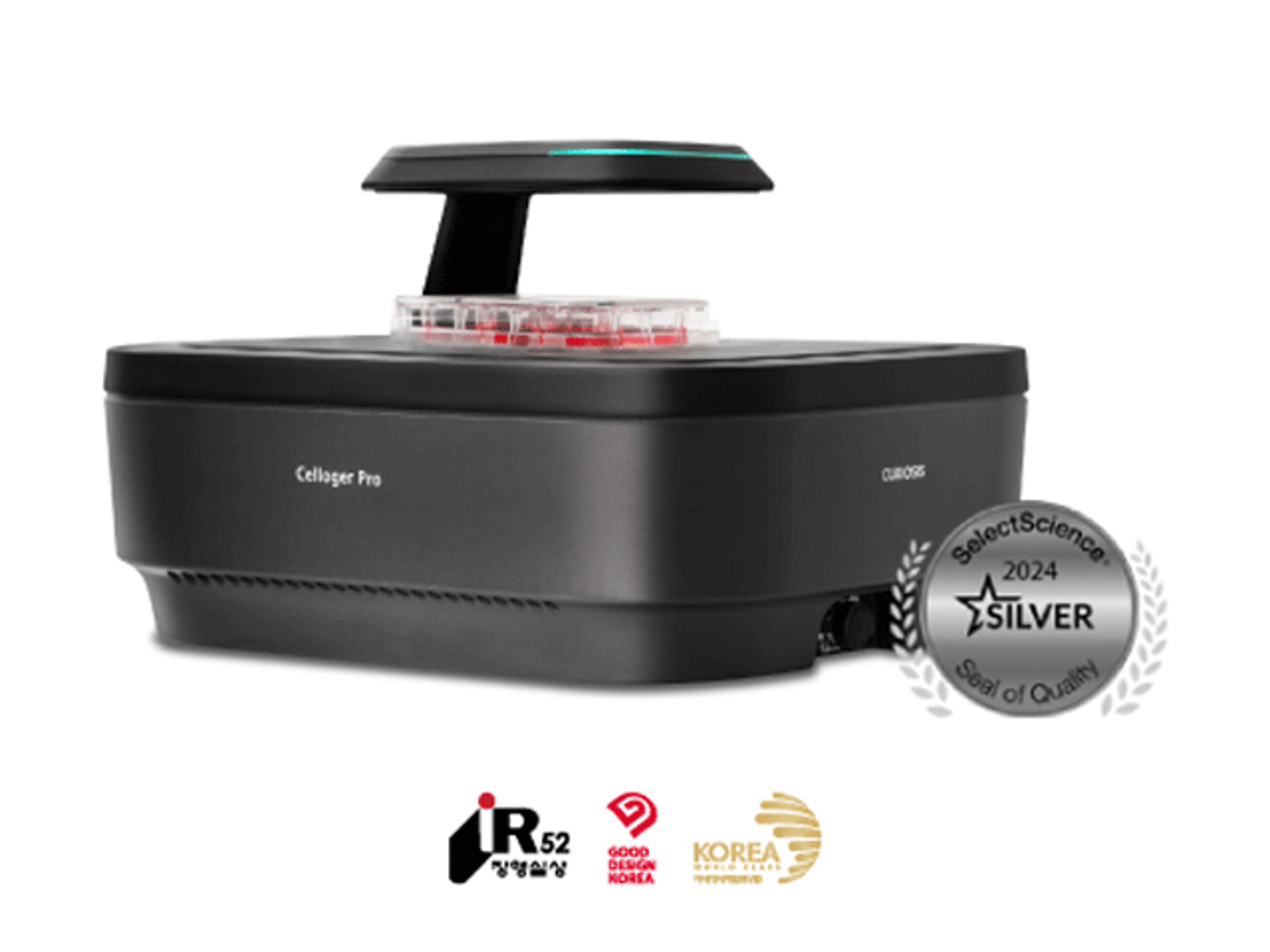
The latest product in their line-up is the all-new Celloger® Pro. This revolutionary automated live cell imaging system redefines research capabilities and provides multi-position imaging in both brightfield and fluorescence microscopy. Its useful features, such as its moving camera and scheduled time-lapse function, enable easy, consistent, and reliable results. Monitoring live cells in real time in an incubator or under regulated environmental conditions allows researchers to analyze them in their optimal state. In addition, it provides advanced visualization and further insight into intracellular events and mechanisms. By using images acquired by the Celloger® Pro, the dynamic processes and structures of cells can be analyzed in a whole new way.
Real-time cell monitoring inside an incubator
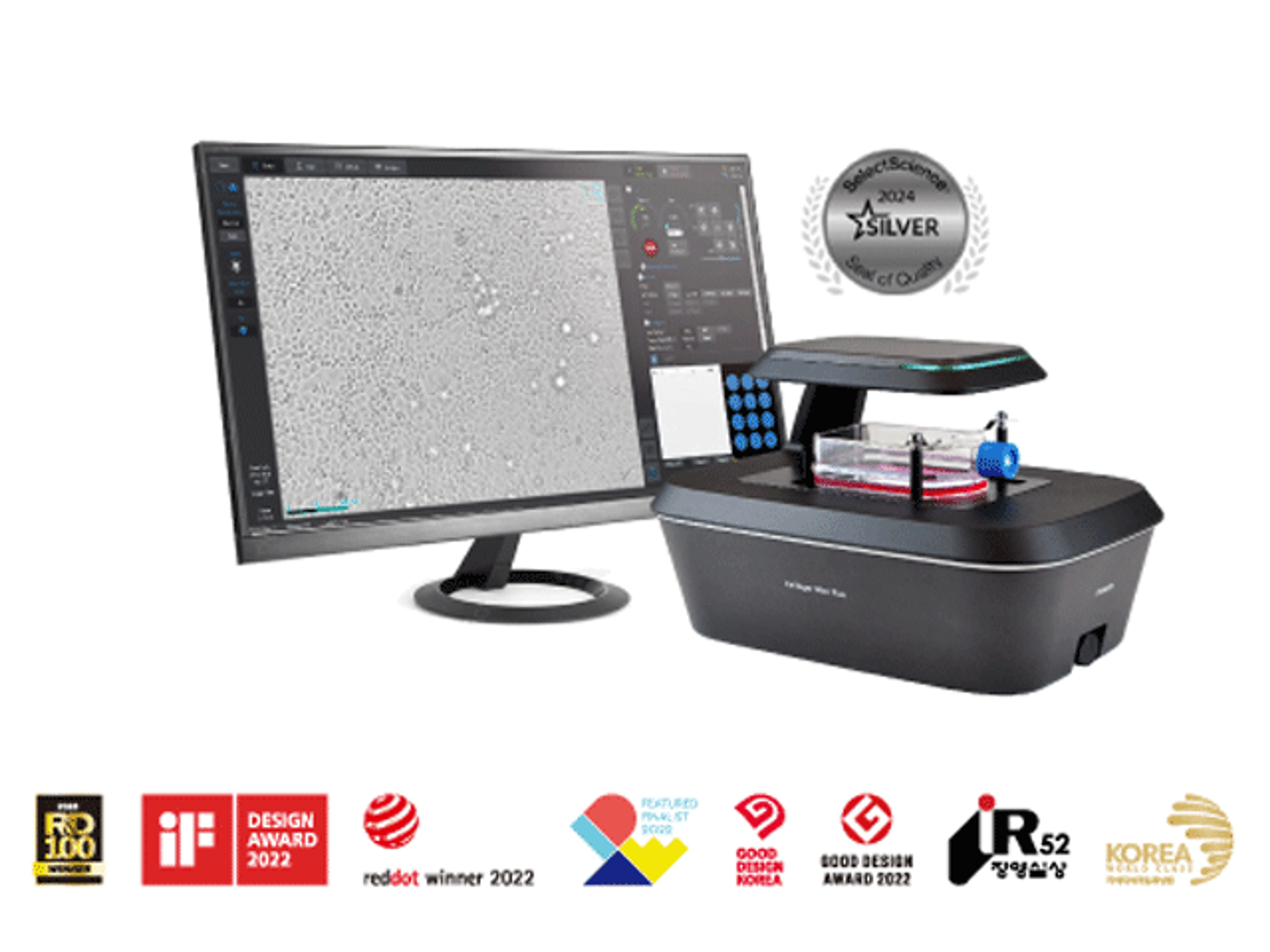
The Celloger® Pro is incubator-compatible, allowing cells to be observed in real time. In conventional imaging methods, researchers observe cells by individually sampling them at various time intervals. However, this method comes with sampling variability and requires a significant amount of effort. Monitoring the subtle changes in individual cells is also difficult. The Celloger® Pro eliminates this tedious task by directly monitoring live cells within the incubator. This maintains the composition of the cells and provides them with a hospitable environment to grow. In other words, it provides more accurate data regarding the functions and development of live cells. The Celloger® Pro connects to an external PC from inside an incubator to display cellular images live on a monitor.
Dual fluorescence microscopy for enhanced imaging
The Celloger® Pro has multicolor fluorescence and brightfield imaging allowing researchers to produce high quality and high-resolution images that can be used to analyze cellular processes with clarity and efficiency. This imaging set enables simultaneous visualization of multiple markers and can be utilized based on the user’s needs. Brightfield microscopy, which is the standard in the laboratory, is used to observe morphological changes in cells. However, it lacks in its ability to provide information about the intricacies of the cell; this is when fluorescence is a great tool to have. Dual fluorescence imaging fills the hole that bright field imaging leaves allowing researchers to monitor biological processes and dynamics of cells. By tagging cellular organelles with fluorescent substances, users can easily see their internal components. In addition, its spectral information can be merged to create a multicolor image, which can be used to obtain more insightful information on cellular structure. By accommodating for both brightfield and dual fluorescence, the Celloger® Pro can fulfill all your cell imaging research needs.
Multi-point time-lapse imaging capability
The Celloger® Pro utilizes both a moving camera and a fixed vessel to maintain a stable environment for cell growth. This ensures that the cells are not disrupted while capturing images from multiple positions. This allows researchers to study cells in a more elaborate way by providing images of cellular events across different locations and allowing for feature analysis. Thanks to multi-point imaging, stitching, which is a method of capturing multiple images and combining them to generate a larger, whole image, can be used as an insightful function in the Celloger® Pro software. The Celloger® Pro’s compatibility with different vessel types, including flasks, well plates, and dishes, adds to its flexibility and customizability. Researchers can use this product for a wide range of cell-based research without worrying about vessel-related restrictions.
User-interchangeable objective lens
The Celloger® Pro’s convenient user-interchangeable objective lens allows researchers to further customize the product based on their needs. This notable feature sets the Celloger® Pro apart from the other products in the Celloger® Series line up. It accommodates for 2X, 4X, and 10X magnification objective lenses, which can easily be changed by the user. This flexible feature broadens the scope of research applications with a quick, tool-free lens change.
Intuitive interface and user-friendly tools
The software provided with the Celloger® Pro has a wide variety of tools, including autofocusing, stitching, time-lapse, and other functions to accommodate for any experiment. Without the Celloger® Pro and its features, researchers must sift through dozens to hundreds of images. The sheer number of images to analyze can make this process tedious and time-consuming. With the Analysis app, researchers can collectively adjust and process multiple images. It is designed to identify significant results and accurately analyze quantitative data, including confluency, fluorescent coverage, and intensity overtime. This app takes it a step further and turns this data into graphs and charts to assist in research. Z-stacking is also a noteworthy function of this app – it captures samples from different focal planes, allowing researchers to observe the focal planes of large cells such as spheroids. Other features enable researchers to look at their samples comparatively. These features include thumbnail images, which allow the user to look at all the wells at once, and dual view, which allows two samples to be observed simultaneously. This user-friendly analysis app can be installed and used on personal PCs for convenience.
Conclusion
The Celloger® Pro is a newly introduced product in the live cell imaging market. It provides a unique set of features that can cater to every researcher’s needs and acts as an all-around system. It significantly simplifies the cellular research process by streamlining heavy workloads and providing a user-friendly software. This automated imaging system is available for pre-order now.
Want the latest science news straight to your inbox? Become a SelectScience member for free today>>