ZEISS Highlights New and Enhanced Microscopes and Imaging Solutions at SEMICON West 2017
Visit Booth # 5214 to learn about equipment ideal for semiconductor manufacturing
8 Jun 2017
Product news
ZEISS announces they will be highlighting a range of microscopes and imaging solutions at SEMICON West 2017, July 12-14, 2017, at the Moscone Center, San Francisco, CA. Visit Booth #5214 to see product stations featuring ZEISS Xradia 520 Versa 3D X-ray microscope, ZEISS ORION NanoFab helium ion microscope, ZEISS SteREO Discovery.V20 modular stereo microscope, ZEISS Stemi 508 stereo microscope, ZEISS Smartzoom 5 automated digital microscope, and ZEISS Axio Imager VARIO upright microscope for large samples.
ZEISS will be showcasing the new ZEISS Xradia Versa with FPX for extended ‘scout and zoom’ imaging, which further broadens the capability of the versatile ZEISS Xradia Versa 3D X-ray microscopy instrument. The new ZEISS FPX flat panel extension delivers large-sample, high throughput scanning with best-in-class image quality. Combined with the high resolution of ZEISS Xradia Versa X-ray microscopes (XRM), the new ZEISS FPX enhances imaging flexibility and creates workflow efficiencies with an all-in-one system for industrial research.
Also on display is ZEISS ORION NanoFab helium ion microscope, a 3-in-1 multibeam ion microscope which lets users fabricate sub-10 nanometer nanostructures with speed and precision. Seamlessly switch between gallium, neon and helium beams with ZEISS ORION NanoFab. The ZEISS SteREO Discovery.V20 modular stereo microscope showcased at the booth features a motorized 20x zoom, and is designed for optimal depth perception and maximum zoom range. Use the 20:1 zoom range to go from largest overview into the smallest details.
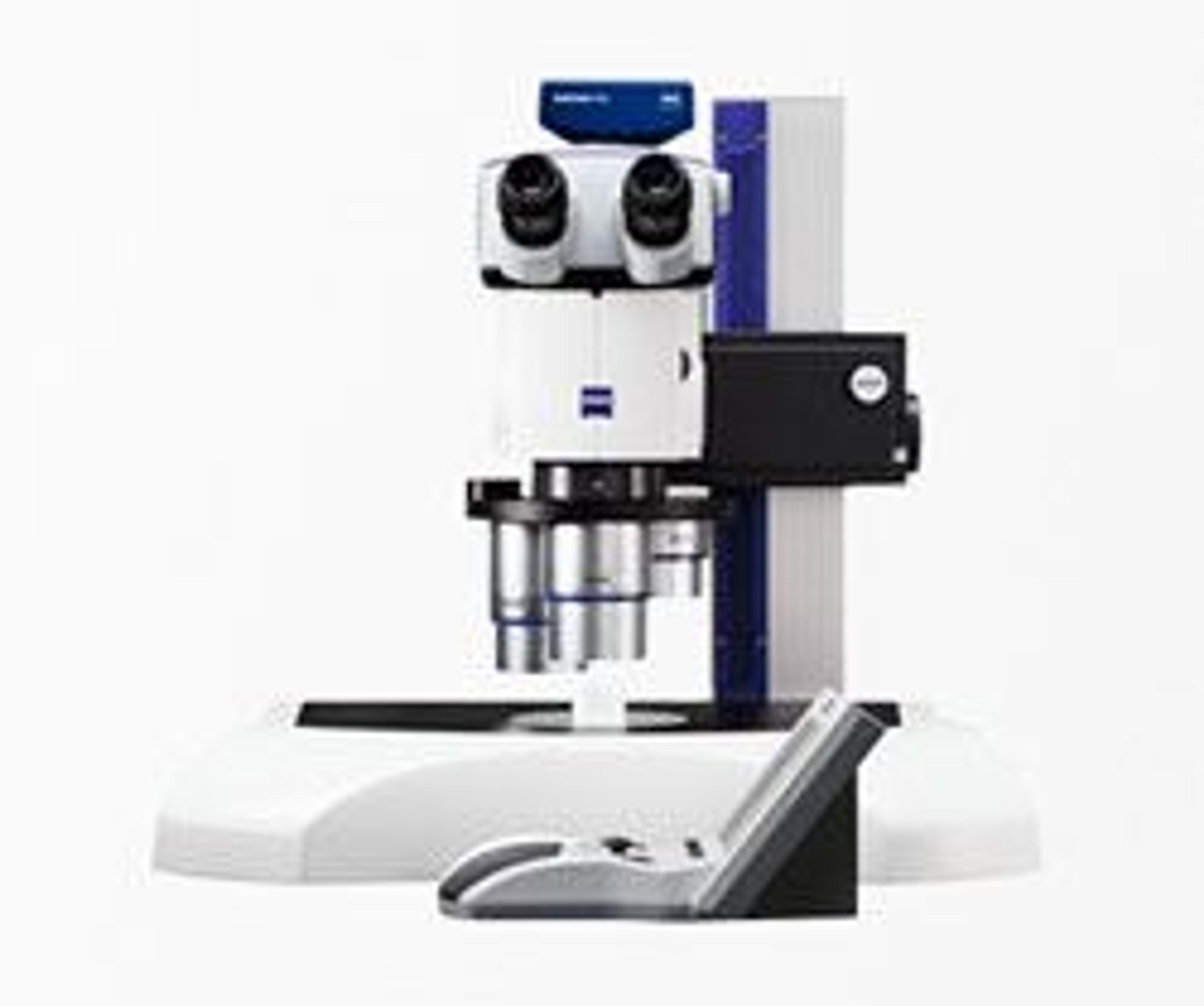
The ZEISS Stemi 508 Greenough-type stereo microscope on display is equipped with apochromatic optics and designed for heavy workloads. The compact, reliable Stemi 508 is great for acquiring images of outstanding image contrast and color accuracy. With the large object field (up to 36 millimeters (mm)), users always keep the overview of their sample.
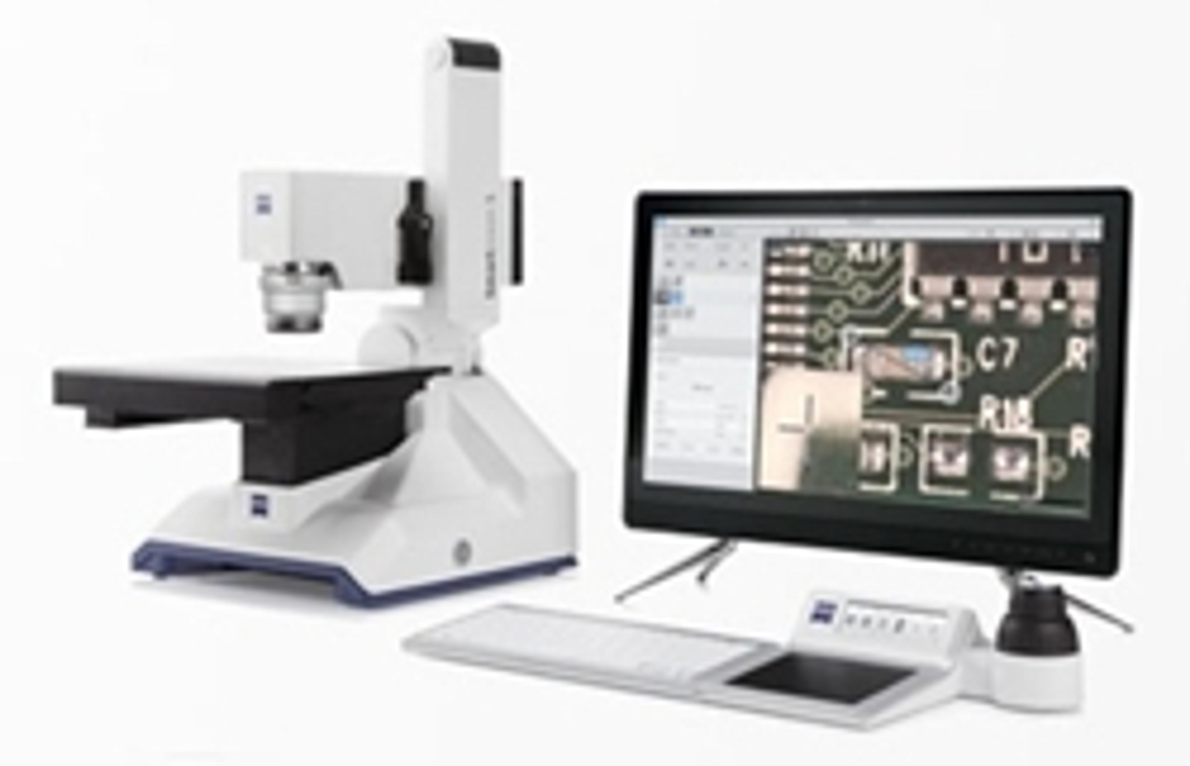
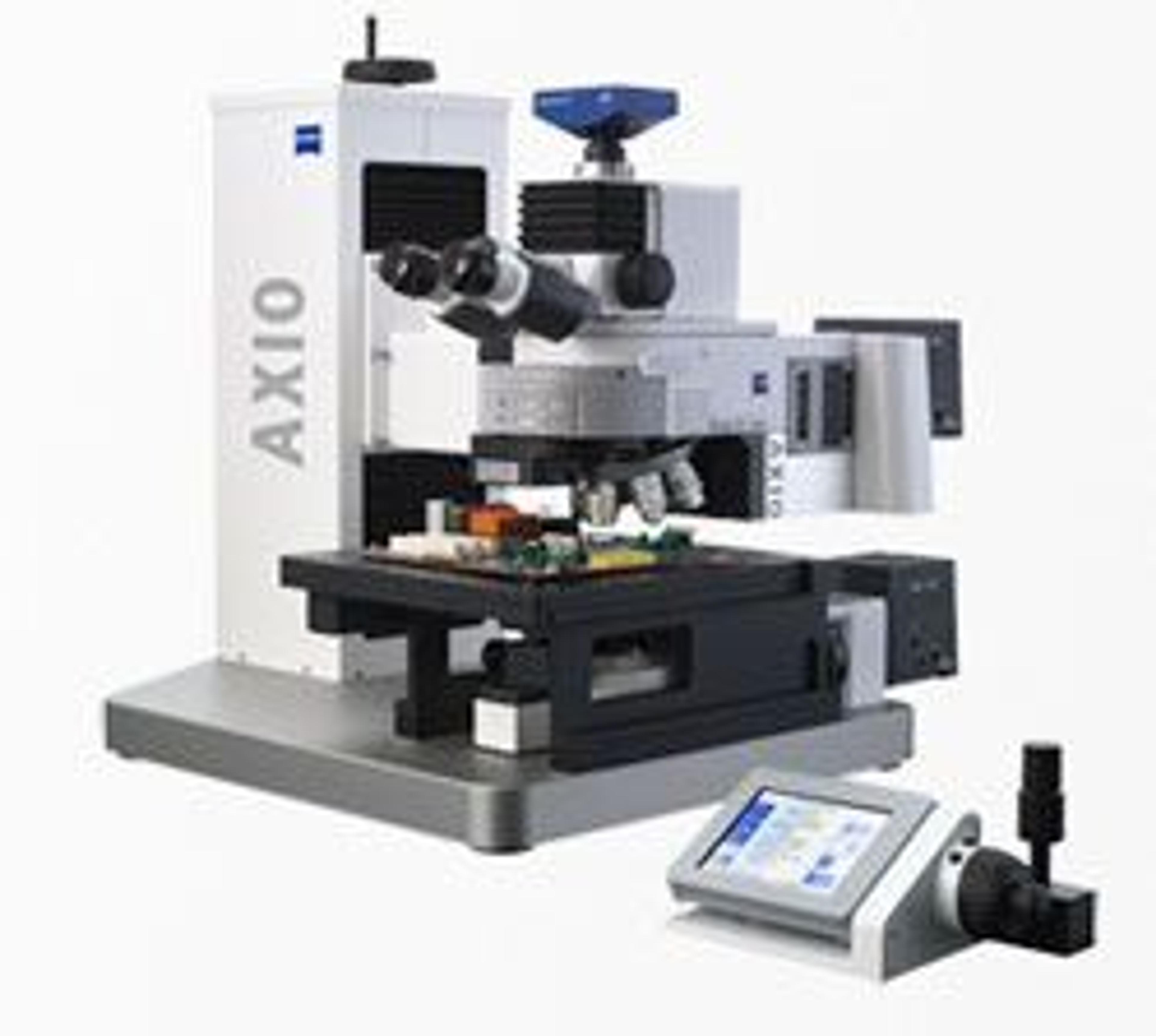
Visitors to the booth can also view ZEISS Smartzoom 5, an automated digital microscope ideal for optical inspection in an industrial environment. Used for production-related quality testing, Smartzoom 5 is the perfect choice for inspecting parts, printed circuit boards, and metal elements and examining them for defects. ZEISS experts will also be on hand to discuss ZEISS Axio Imager VARIO upright microscope, which can analyze samples from the smallest MEMS sensors up to XXL wafers, or even entire flat panel displays, without destroying them. Automated and compatible with clean rooms, Axio Imager VARIO features a motorized z-drive that works with the hardware auto focus to ensure the optimal focus position.