Spatial glycomics meets proteomics in a new multiplex tissue assay
First-of-its-kind assay by Vector Laboratories and Spatomics enables simultaneous mapping of proteins and glycans on a single slide
31 Mar 2026
Editorial article
The holy grail of multiomics is the ability to detect multiple molecular modalities simultaneously, with high sensitivity and spatial context, on a platform that is simple to use and affordable. For many researchers, that goal has long seemed out of reach, but the field is moving steadily closer.
Recent advances in spatial omics technologies have given scientists unprecedented insight into the organization, heterogeneity, and cellular interactions within complex tissues. By revealing not only what is happening in a tissue but where, spatial biology is reshaping our understanding of disease, helping drive the rise of precision and personalized medicine, and paving the way for a new generation of more targeted, effective therapies.
Plus, progress shows no sign of slowing. The sensitivity and resolution of spatial protein and RNA mapping continue to improve, while entirely new classes of molecules are becoming accessible to spatial analysis for the first time. Among these are glycans, the chains of carbohydrates that decorate lipids and proteins on the surface of cells.

(L-R) Rui Zheng, Ph.D. CEO and Co-founder; Jia Guo, Ph.D., Co-founder; Jing Zhou, Ph.D., CSO; and Xiaoshan Wang, MBA, SVP of Business Development, of Spatomics
Spatial glycomics has come of age
Glycosylation, one of the most common post-translational modifications of proteins, plays critical roles in cell communication, immune recognition, and disease progression. The structural diversity of glycosylated molecules, including glycoproteins, glycolipids, and proteoglycans, and the biological functions they perform are therefore becoming an increasingly important focus of scientific research.
Disrupted or dysregulated glycosylation has been linked to a wide range of diseases, including cancer, Alzheimer’s disease, immune disorders, and infectious diseases, sparking growing interest in glycans as potential diagnostic biomarkers and therapeutic targets.
Glycomics technologies have already shed light on the heterogeneity of the glycome but, until recently, the ability to analyze glycosylation in its true spatial context and integrate this information with other multiomics datasets has been extremely limited.
That is now beginning to change, thanks to the combination of two complementary spatial biology technologies: one developed by the multiomic imaging company Spatomics and the other by Vector Laboratories, a long-established provider of bioanalytical reagents and services with decades of expertise in glycobiology.
Mapping proteins and glycosylation on the same slide
Spatomics and Vector Laboratories’ combined platform offers a means to simultaneously detect ten or more protein and glycan targets in the same biological sample, all while maintaining spatial resolution.
The approach marries Vector Laboratories’ Glysite™ Explorer in situ PLA Glycan Detection Kit – the first commercially available kit designed for the spatial detection of protein glycosylation – with Spatomics’ newly developed Cleavable Fluorescent Probe (CFP) for multiplex protein profiling.
Spatomics’ proprietary CFP technology was specifically designed to overcome one of the most persistent challenges in spatial biology: detecting molecular signals in highly autofluorescent human tissue samples.
Many patient biopsy samples are preserved as formalin-fixed and paraffin-embedded (FFPE), a preparation method that often produces strong background fluorescence. Traditional fluorescent probes, which attach only a few fluorophores to an antibody or probe, can struggle to generate signals strong enough to overcome this background.
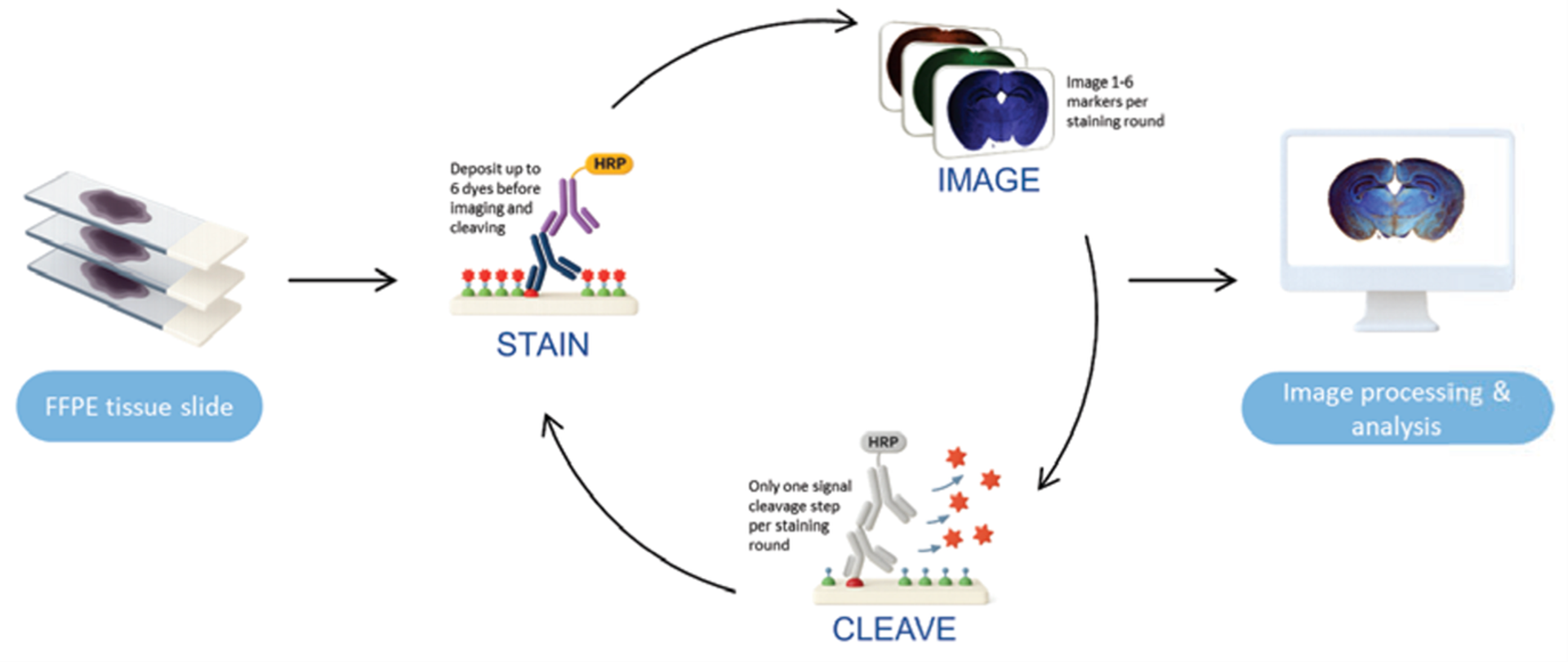
CFP technology addresses this limitation through enzymatic signal amplification. In this approach, antibodies are paired with the enzyme horseradish peroxidase (HRP), which catalyzes the deposition of hundreds of fluorescent probes on or near the target protein. This dramatically increases detection sensitivity.
After staining and imaging, the CFP fluorophores are chemically cleaved, enabling the sample to undergo further rounds of staining, antibody stripping, imaging, and fluorescence signal removal via a patented cleave process. This allows researchers to profile many different proteins, along with RNAs and DNA molecules, within the same tissue sample.
“Having the sensitivity and accuracy to detect low copy number protein molecules in high autofluorescence background patient biopsy tissues is our major advantage,” explains Dr. Jia Guo, co-founder of Spatomics and inventor of the CFP technology. “This is especially important for diagnostic and prognostic applications, where you cannot afford high false-positive or false-negative results.”
But proteomic data is only part of the story. Where this method really stands out is in its compatibility with Vector Laboratories’ Glysite Explorer Glycan Detection Kit. Here, HRP-labelled lectins – proteins that recognize and bind to specific glycan structures – are used to detect glycosylated proteins.
“This enables researchers to interrogate proteins, glycans, and proximity detection all on one slide, something previously not possible on the same section,” explains Xiaoshan Wang, VP of business development at Spatomics.
“This has allowed us to incorporate glycan-associated biology into our spatial biology workflows,” adds Dr. Guo. “So far, it remains the only commercially available solution to visualize glycosylated proteins in FFPE tissues. Vector Laboratories’ antibodies and glycan variations are also well-validated, which is key. This gives us very reproducible and very robust results.”
Revealing glycosylation patterns in disease
This additional layer of omics data enables researchers to identify changes in glycosylation across specific cell populations, for example, comparing pathological and normal tissue states.
One area where this approach has already shown promise is neuroscience. Dr. Guo is also a professor at Arizona State University, where his laboratory is using the platform to investigate the potential role of glycosylation in Alzheimer’s disease.
“First, we use our technology to identify different cell types or cell subtypes within brain tissue, such as neurons, astrocytes, microglia, and oligodendrocytes, through different protein or RNA markers,” he explains. “Then we can use the Glysite Explorer technology to visualize within that individual cell type or subtype how the glycobiology has been altered in the disease through different stages, between different sexes, and so on.”
By combining spatial protein profiling with glycan detection, the researchers can begin to explore how changes in glycosylation interact with broader cellular pathways and disease mechanisms, insights that were previously difficult, if not impossible, to obtain within intact tissue samples.
The future of spatial multiomics
Over the past two decades, there has been a shift in interest from traditional bulk sequencing approaches toward increasingly sophisticated spatial multiomics techniques. “It’s a shift that has been reflected in Nature Methods’ ‘Method of the Year’ selections,” explains Dr. Rui Zheng, co-founder and CEO of Spatomics. “In 2009, it was next-generation sequencing; in 2016, single-cell sequencing; in 2020, spatial transcriptomics; and in 2024, spatial proteomics.”
Now spatial approaches are moving beyond academic research and into the pharmaceutical industry. “Major drug companies are already using high-plex spatial assays throughout the drug development pipeline,” adds Dr. Zheng. These tools are helping scientists to identify biomarkers, discover new drug targets, and even develop companion diagnostics.
At the same time, artificial intelligence is beginning to play a crucial role. As spatial experiments generate ever larger and more complex datasets, AI algorithms are helping researchers interpret the intricate molecular patterns hidden within tissues.
As these two technologies converge, spatial biology (already an invaluable research tool) seems poised to become a cornerstone of translational science and precision medicine in the not-so-distant future.
As Vector Laboratories marks its 50th anniversary, its ongoing commitment to innovation in glycobiology and spatial biology will continue to empower scientists exploring this rapidly evolving field.
Learn moreRelated products
Request Quote for All Products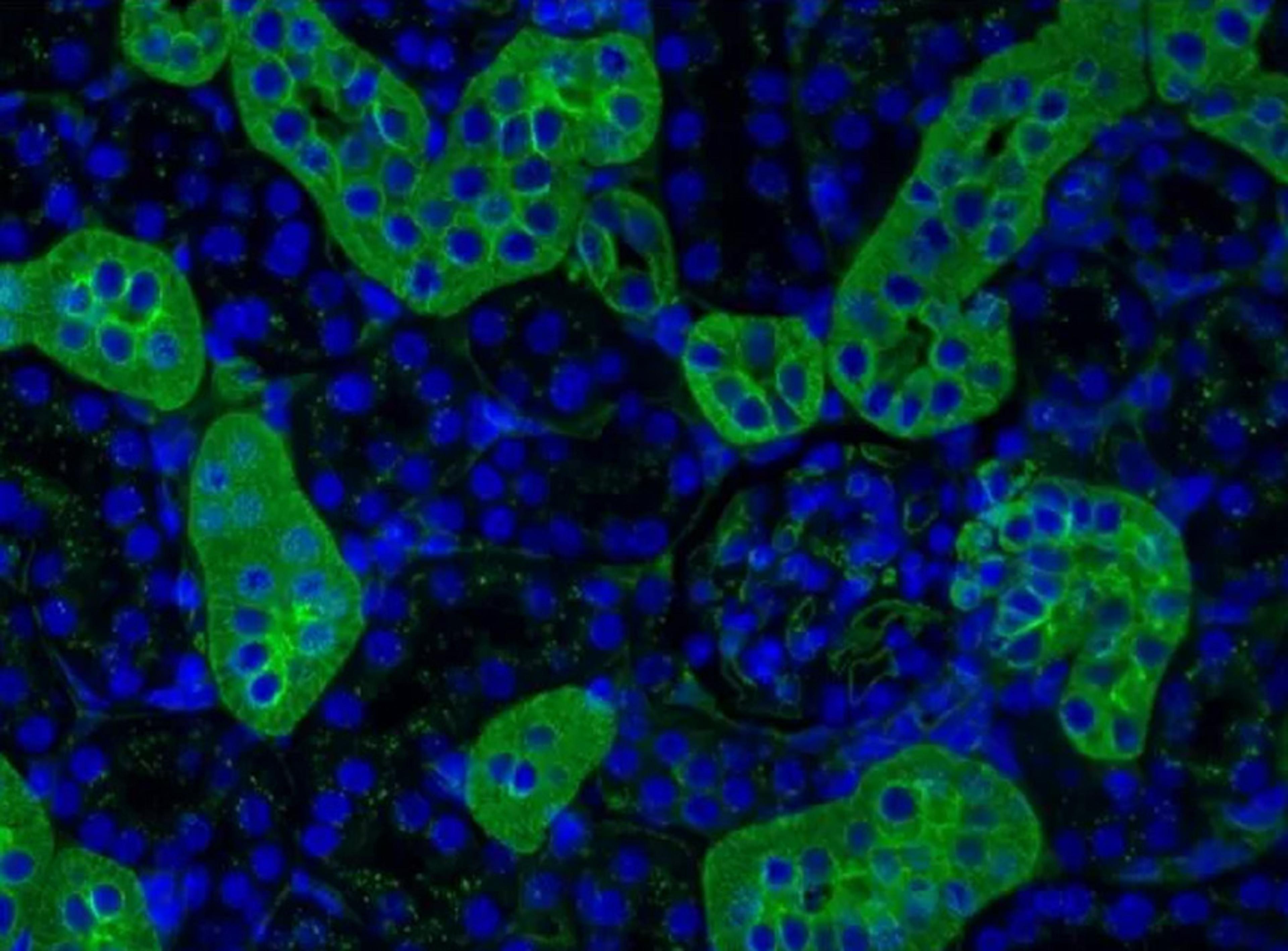
Glycobiology Reagents
Vector Laboratories Inc.Founded on a growing portfolio of purified lectins, lectin conjugates, and lectin screening kits, Vector Laboratories supports the ability to profile, characterize, and capture complex glycans in biological systems.