Highly Automated, Self-Optimizing Micro-CT with Push-Button Ease of Use Requires Essentially No Training to Deliver Top-Notch 3D Image Quality and Productivity for Research and Industrial Markets

27 Oct 2015

Lois Manton-O'Byrne, PhD
Executive Editor

Product news

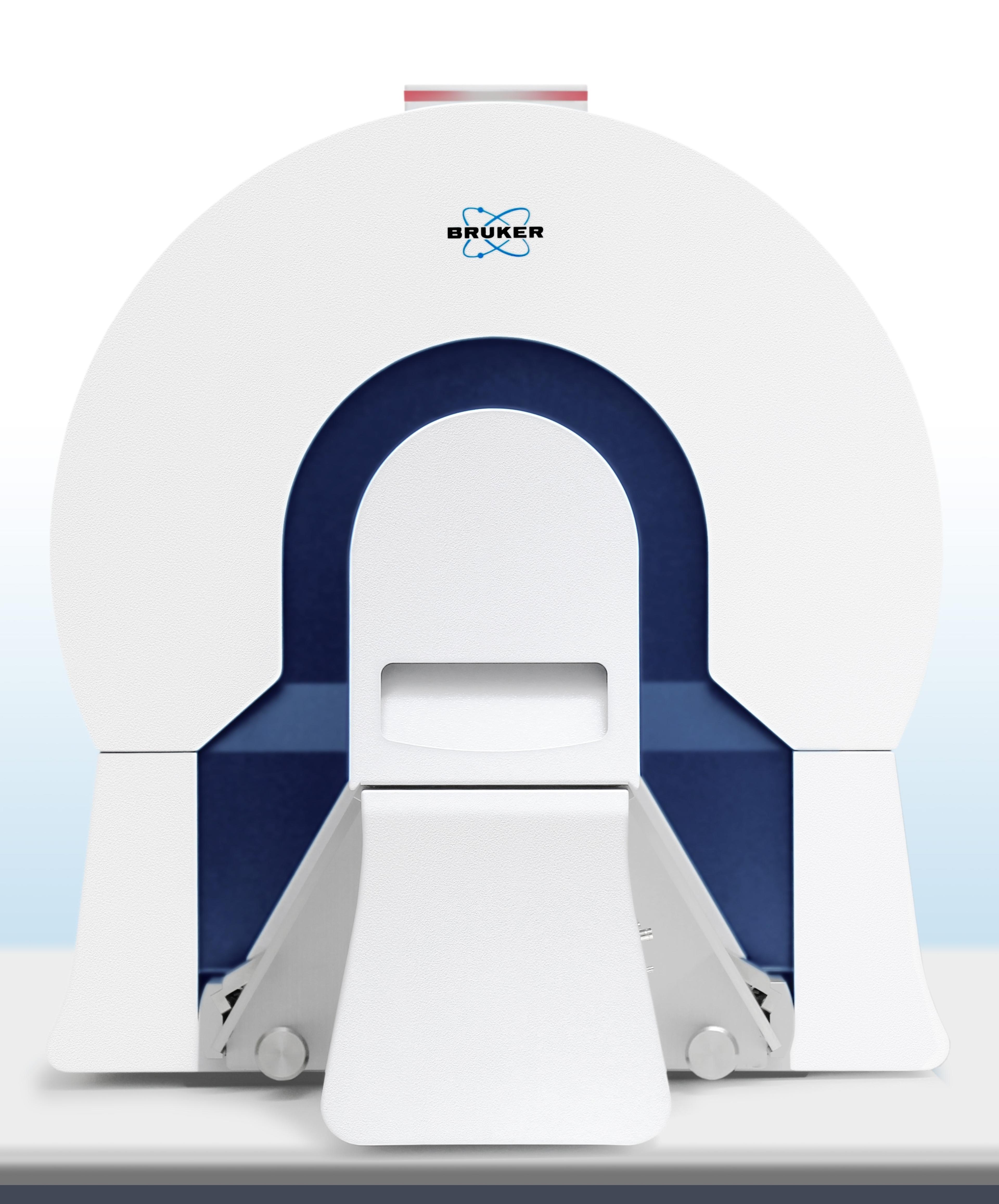
With the introduction of the new SKYSCAN™ 1275 highly automated, self-optimizing desktop X-ray microtomograph, Bruker puts class-leading imaging into the hands of materials and life scientists in both research and industrial applications, such as high-throughput 3D imaging in quality control and production process monitoring. The system takes advantage of new X-ray source technology and efficient flat-panel detectors to reduce scan time to just a few minutes, without compromising image quality.

The SKYSCAN 1275 software features 2D/3D image analysis, as well as a realistic 3D visualization that employs recent developments in reconstruction algorithms, accelerated by new powerful graphics cards to give an additional gain in performance and speed. The outcome is the rapid and realistic visualization of results by volume rendering. Fabulous images of the internal microstructure of an object reveal details by virtual cut or virtual flight around and inside the objects.

Importantly, all these performance advances are delivered in a system that provides an unprecedented level of automation. Push-button operation with a preselected sequence of actions, including scanning, reconstruction and volume rendering, means that even routine users can successfully obtain high-quality results with the SKYSCAN 1275.

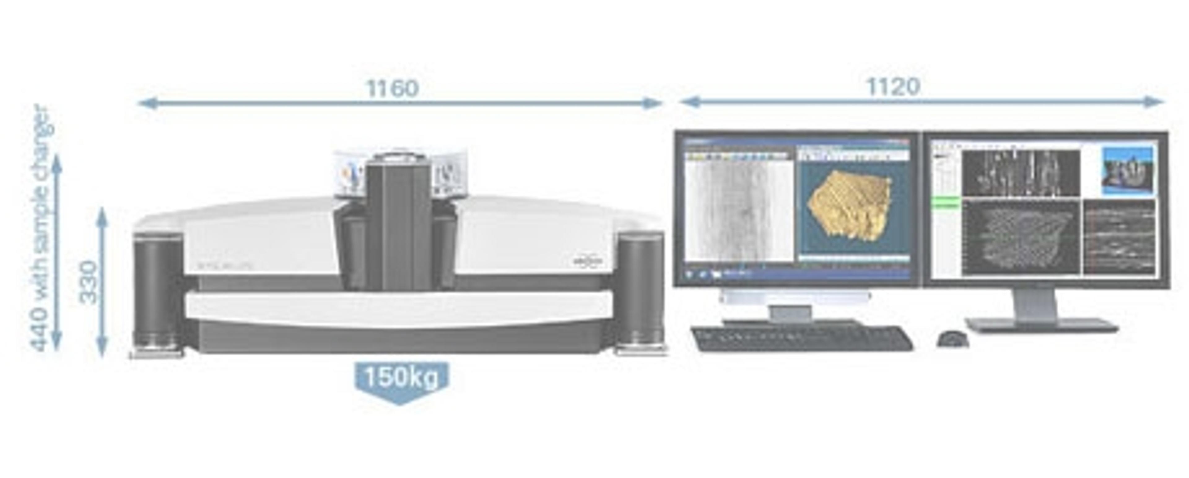
The SKYSCAN 1275 has an object size range up to 96mm in diameter and 120mm in length, which can also accommodate mid-sized samples. Optional stages for micro-positioning and material testing allow scanning of a sample under a range of conditions, e.g. compression, tension, heating or cooling.

Dr. Alexander Sasov, Managing Director at Bruker microCT, explained: “The SKYSCAN 1275 provides our customers a fast, easy-to-use system that is ideal for scanning a wide range of samples. It supports a variety of applications, from analyzing building materials and electronic assemblies for quality control, to geological research for oil and gas exploration, to anatomical pathology research. By providing high-quality 3D non-destructive imaging with significantly reduced scan times and unprecedented ease of use, the new SKYSCAN 1275 delivers increased efficiency for scientific research and industrial applications.”

SkyScan 1278

Bruker BioSpin

The new SkyScan 1278 micro-CT system for in vivo imaging is addressing the needs of scientists working in the exciting areas of physiological response to disease and regenerative medicine. Many researchers strive to reduce the levels of radiation used in animal studies. Lower levels reduce the risk of associated complications, but any system that offers this advantage must not compromise on the quality of images produced.
(0)

Links

Tags

X-ray CrystallographyX-ray crystallography is an analytical technique used to determine the arrangement of atoms in a crystal. Monochromatic x-rays are produced from a synchrotron or x-ray generator. An x-ray crystallography system uses a detector to measure the x-ray diffraction from the crystal. The information is used to generate a 3D image of the crystal.X-Ray Diffraction and SpectroscopyX-Ray diffraction & spectroscopy are used in material characterization to discern the structure and elemental composition of a sample. X-Ray diffractometers (XRD) are superior instruments in elucidating the dimensional atomic structure of crystalline materials, including powders, thin films and single crystals. For large unit cells or ordered macromolecules, consider small-angle X-ray scattering (SAXS). X-ray spectroscopic techniques include X-ray fluorescence (XRF) and X-ray photoelectron spectroscopy (XPS), both providing simple and accurate methods for determining the elemental composition of a material. Energy dispersive (EDXRF) and wavelength dispersive (WDXRF) XRF spectrometers are available, as well as handheld/portable devices. High-resolution, 3D microstructure characterization of materials can be achieved with X-ray microscopes combining sub-micron resolution imaging with 3D computed tomography. Find the best XRD and XRF spectrometers in our peer-reviewed product directory: compare products, check customer reviews and receive pricing direct from manufacturers.Quality ControlQuality control is needed in all production processes. Quality control is a monitoring procedure or set of procedures that are put in place to ensure that a manufactured product adheres to a defined set of quality criteria. AutomationAutomation in laboratories and manufacturing processes enhances efficiency, precision, and scalability by reducing the need for manual intervention. It plays a critical role in improving productivity, minimizing human error, and accelerating workflows in fields like diagnostics, drug development, and industrial testing. Automation technologies include robotic systems, automated liquid handlers, and process control systems that streamline complex tasks and ensure consistent, reproducible results. Explore our peer-reviewed product directory to discover the best automation solutions, compare options, read user reviews, and get prices directly from manufacturers.3D Imaging3D imaging technologies allow for the visualization and analysis of three-dimensional structures at high resolution. These systems are used in fields like molecular biology, material science, and medical diagnostics. 3D imaging can be applied to visualize cells, tissues, and organs, providing valuable insights into their structure and function. Browse our peer-reviewed product directory to find the best 3D imaging solutions, compare products, check reviews, and get pricing directly from manufacturers.X-Ray SourceProcess MonitoringTomography