High-throughput live cell imaging transforms organoid research
Discover how advanced imaging technology is enabling continuous drug safety monitoring in liver organoids
28 Oct 2025
Editorial article
Organoids have revolutionized preclinical drug development by modeling tissue responses much more accurately than traditional cell cultures. These 3D models have become essential for assessing drug candidate efficacy and safety, as they enable researchers to use miniaturized organ systems that can demonstrate how potential therapeutics behave in a biological environment.
Although organoids have become a staple in research, methods of analyzing them have remained unchanged. Typically, researchers take organoids out of the incubator, observe them under a microscope, and then manually analyze the images. This time-consuming manual process poses problems because each handling causes temperature fluctuations, atmospheric variations, and mechanical disturbances. These issues affect data quality, limiting throughput, and lead to inconsistencies across experiments.
As organoid-based screening increasingly plays a key role in drug discovery pipelines, it is imperative that the field develops analytical methods that match the complexity of the models.
Real-time organoid response monitoring without disruption

Researchers at the Suzhou Institute of Biomedical Engineering and Technology, Chinese Academy of Sciences, are tackling this challenge head on. The use of advanced live cell imaging technology enables them to monitor liver organoid responses in real-time and, most importantly, they are doing so without ever removing samples from their optimal culture conditions.
Jinxian Wang, Associate Researcher at the institute, has integrated the Countstar Spica M1 imaging system from ALIT LifeTech into his organoid cytotoxicity studies. This system enables automated monitoring of organoid drug response inside an incubator, capturing changes as they happen rather than at isolated snapshots in time outside an incubator. "It enables continuous imaging to reflect the status of organoids at different time points, providing convenience for the experiment," Wang explains.
While traditional methods require researchers to repeatedly handle samples, Wang's approach completely eliminates these disruptions. He can maintain environmental stability while significantly cutting down on hands-on time.
Capture dose and time-dependent effects of drug targets on organoids
Wang's research focuses on understanding how drug candidates affect liver organoid viability and function. His team treats liver organoid models with varying drug concentrations and monitors them continuously for 24 hours using fluorescent probes to track what's happening inside the organoids.
"During the 24-hour incubation period of organoids with drugs at various concentrations, specific cytotoxic reagents were added simultaneously, and appropriate fluorescent probes were used for long-term dynamic monitoring organoids under different time points and drug concentrations," Wang explains. This method provides comprehensive data on both timing and dose-response, information that's difficult to obtain using traditional methods.
Integration, automation, and image stitching with Countstar Spica M1
The Countstar Spica M1 features ultrafast Z-stacking technology that captures sharp organoid images across multiple focal planes, even within 3D Matrigel environments. Its AI-driven analysis algorithms automatically detect and characterize organoids with high accuracy.
Wang highlights the system’s advanced imaging capabilities: “The Spica M1 generates maximum intensity projections from multiple Z-layers, producing brightfield and fluorescence images that are exceptionally clear and precise.” He also emphasizes the strength of its large-area image stitching: “This technology enables expansive field-of-view imaging, capturing more data per well and maximizing experimental insights.”
These integrated capabilities have allowed Wang’s team to conduct comprehensive drug evaluations using liver organoid models. As organoids evolve into more physiologically relevant systems, the technology used to study them must advance accordingly. The Countstar Spica M1 demonstrates how live-cell imaging and automated analysis can enable organoid-based drug screening to become a robust, standardized component of modern development pipelines.
Related products
Request Quote for All Products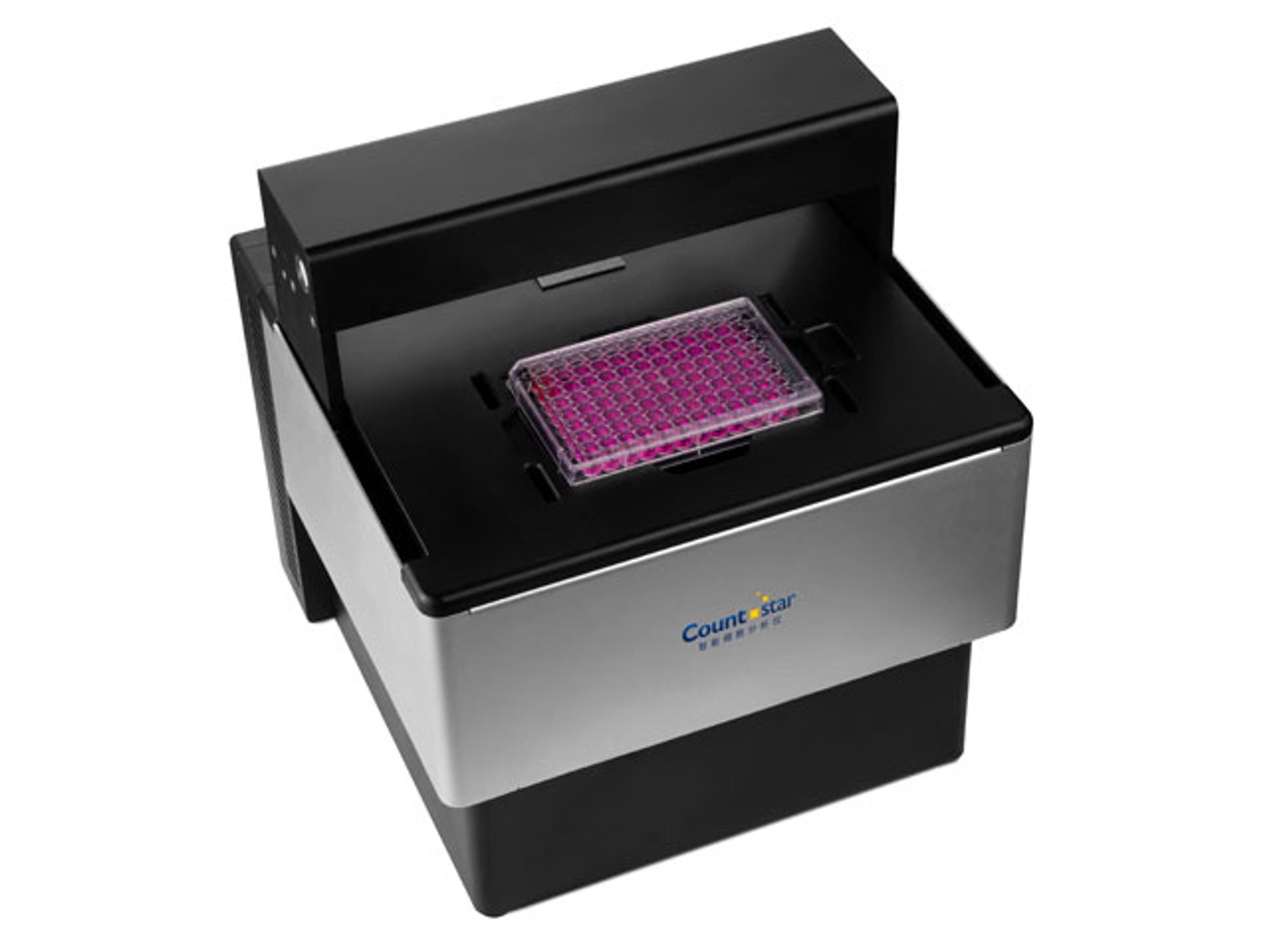
Frequently asked questions
How are liver organoids transforming preclinical drug development and cytotoxicity testing?
Organoids are 3D miniaturized organ systems that model tissue responses more accurately than traditional 2D cell cultures, making them essential tools in preclinical drug development. In liver organoid cytotoxicity studies, they allow researchers to assess drug candidate efficacy and safety in a biologically relevant environment, capturing how potential therapeutics affect organoid viability and function. This improved physiological relevance supports more predictive drug screening and strengthens organoid-based pipelines in modern drug discovery.
What advantages does the Countstar Spica M1 imaging system offer for real-time liver organoid drug response analysis?
The Countstar Spica M1 imaging system from ALIT LifeTech enables automated, real-time monitoring of liver organoid drug responses directly inside an incubator, eliminating the need to remove samples from optimal culture conditions. Its ultrafast Z-stacking generates maximum intensity projections across multiple focal planes, producing clear brightfield and fluorescence images even in 3D Matrigel. AI-driven analysis algorithms automatically detect and characterize organoids, while large-area image stitching provides expansive field-of-view imaging, capturing more data per well and maximizing experimental insights.
How does Jinxian Wang’s live-cell imaging workflow improve organoid-based drug screening data quality and throughput?
Jinxian Wang and his team at the Suzhou Institute of Biomedical Engineering and Technology, Chinese Academy of Sciences, use advanced live-cell imaging with the Countstar Spica M1 to monitor liver organoids continuously for 24 hours under stable incubator conditions. By treating organoids with various drug concentrations and adding specific cytotoxic reagents and fluorescent probes, they achieve long-term dynamic monitoring across different time points and doses. This workflow reduces manual handling, minimizes temperature and atmospheric fluctuations, improves data consistency, and delivers comprehensive timing and dose-response information that is difficult to obtain with traditional, snapshot-based microscopy methods, enabling more robust and standardized organoid-based drug screening.