Award-Winning Cytation™ 3 Now Offers Expanded Imaging Features

3 Dec 2013

Sarah Thomas
Associate Editor

Product news

BioTek’s innovative Cytation™ 3 Cell Imaging Multi-Mode Reader combines automated digital microscopy and conventional multi-mode microplate detection in one modular, compact instrument, and now offers even more new features to drive workflow efficiencies and data collection.

“We’ve seen an overwhelming response to the introduction of Cytation 3, as evidenced by the SelectScience Drug Discovery Product of the Year, and MipTec New Product Innovation awards that it’s received so far,” noted Gary Barush, BioTek’s Director of Marketing and Sales. “And we’ve been able to react quickly to customer feedback with new imaging tools to further streamline imaging research.”

40x and 60x objectives are now available options for any Cytation 3 application requiring increased magnification. These higher magnifications allow easy imaging of bacteria, yeast, and sub-cellular structures such as mitochondria. Additionally, 35 mm, 60 mm and 100 mm Petri dishes are now compatible, for expanded sample vessel convenience.

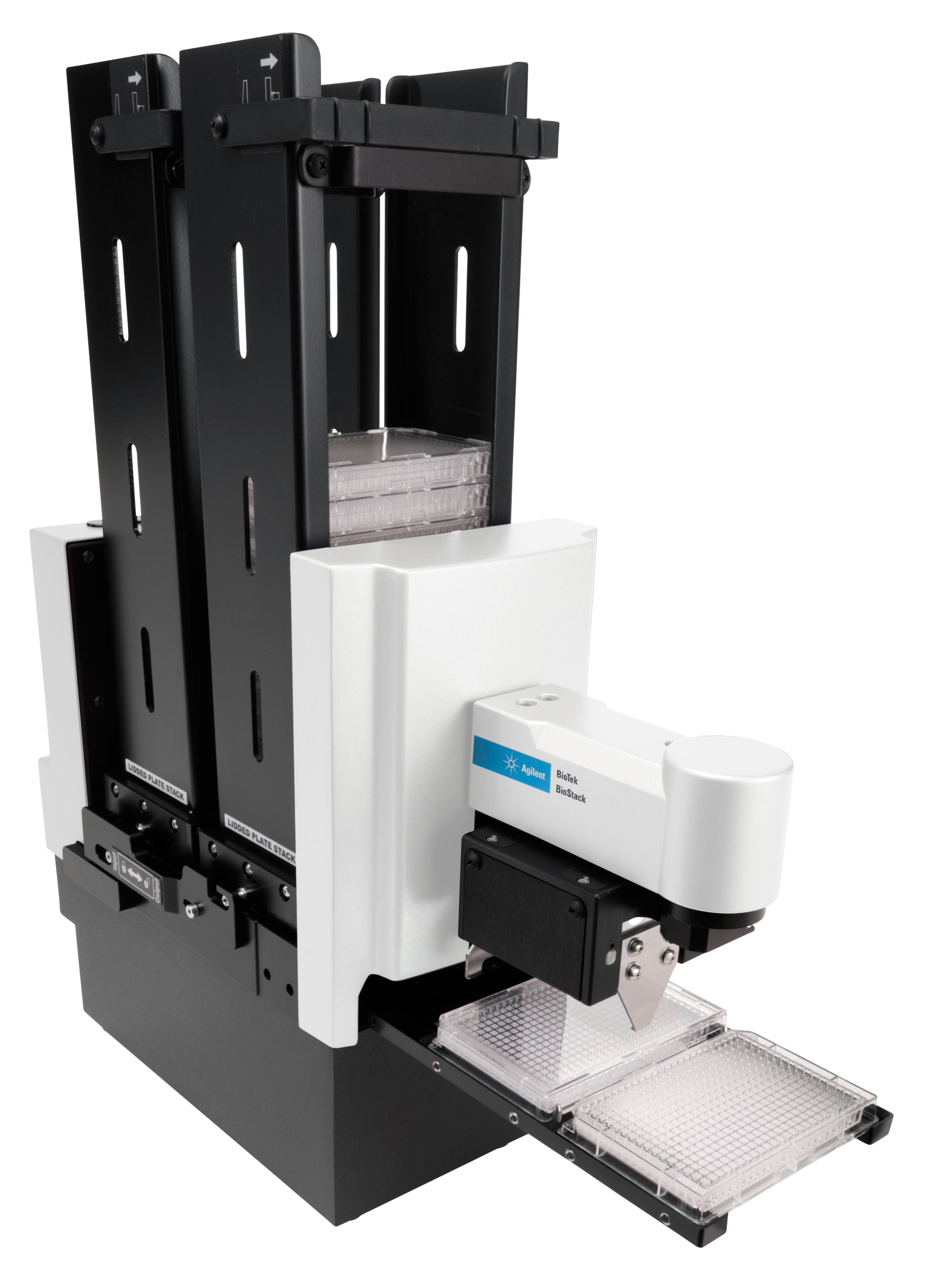
Cytation 3’s Gen5™ Data Analysis Software offers additional tools to take your research to another level. With its broad image analysis capability, Gen5 software editions include options for 21 CFR Part 11 compliance or In Vitro Diagnostics use. Cytation 3 can be integrated with BioTek’s BioStack™ 4 Microplate Stacker to increase throughput of lidded and unlidded microplates.

Agilent BioTek BioStack Microplate Stacker

Agilent Technologies

The Agilent BioTek BioStack microplate stacker is a compact and versatile microplate stacker, compatible with Agilent BioTek washers, dispensers, plate readers, and imaging systems. The rapid plate exchange speeds deliver increased throughput and enhanced efficiency and greater productivity.

(29)

Links

Tags

Microplate Readers / DetectorsMicroplate readers are used to automate the detection and analysis of labeled or label-free components in microplates during assays or live-cell monitoring. Microplate readers are generally distinguished by their mode of detection. Types include absorbance, luminescence, fluorescence intensity, fluorescence polarization, TRF / FRET and multimode microplate readers. Microplate readers deliver a high throughput of samples by reading multiple wells simultaneously, with the 96-well format the most commonly used. As a result, microplate readers are often used in the drug discovery, bioassays, research and pharmaceutical industries for screening applications. Microplate loading can also be automated, with robotic microplate stackers to increase throughput. Find the best microplate readers in our peer-reviewed product directory: compare products, check customer reviews and receive pricing direct from manufacturers.Cell-Based AssaysCell-based assays are used to monitor the presence, quantity and activities of a desired cellular analyte including drug molecules or biomarkers. This can reveal information on cell health (apoptosis, cytotoxicity, viability and proliferation assays), cell metabolism, cell migration and cell signaling mechanisms. Find the best cell-based assay products, kits and equipment with our peer reviewed product directory: compare products, check customer reviews and receiving pricing direct from manufacturers.Light MicroscopyLight microscopes or optical microscopes are used to visualize microscale objects under magnification, including cells, clinical specimens and materials. Lab equipment for light microscopy includes confocal microscopes, fluorescence microscopes, zoom and stereo microscopes. Microscope slides and imaging reagents are available for visualizing samples, as well as various microscope stages and incubators for large or temperature-sensitive samples. Find the best light microscopes in our peer-reviewed product directory: compare products, check customer reviews and receive pricing direct from manufacturers.Cell ImagingCell imaging can be achieved using a number of techniques including confocal microscopy, transmission electron microscopy, atomic force microscopy, and light sheet microscopy.