Top Life Sciences News this Month
28 Nov 2014Editorial article
Read a summary of this month’s top ten news articles from the Life Sciences community.
FEI Announces Teneo VS™ for 3D Volume Imaging of Cells and Tissues
FEI has announced the launch of the Teneo VS™ Scanning Electron Microscope for fully-automated, large-volume reconstructions with dramatically improved z-axis resolution.
My Lab Essentials: Dr Tom Brown (Jr)
In this first article in the new My Lab Essentials series, Dr Tom Brown (Jr), Director and Head of Oxford R&D Laboratory at ATDBio, discusses the lab equipment used by his team for their research and development in the production of highly modified DNA oligonucleotides.
Scientifica Introduces LASU, the 'Laser Applied Stimulation & Uncaging' System, Perfect for Optogenetic and Uncaging Studies
The Laser Applied Stimulation & Uncaging (LASU) system has been introduced by Scientifica to enhance the combination of electrophysiology, optogenetics and uncaging, for advanced studies of neuronal processes.
Leica Microsystems’ Ion Beam Milling System Leica EM TIC 3X Now Available With Vacuum Cryo Transfer Docking Port
Leica Microsystems’ ion beam milling system Leica EM TIC 3X is now available with a docking port for vacuum cryo transfer, to enable transfer at vacuum and/or cryo conditions with the Leica EM VCT100 vacuum cryo transfer system.
How to Achieve Publication-Quality Images
Discover how Datacolor’s ChromaCal Color Calibration System can help you to generate publication-quality images in five simple steps.
EMD Millipore's New SNAP i.d.® 2.0 System for Immunohistochemistry Minimizes Slide Handling
EMD Millipore has introduced the new SNAP i.d.® 2.0 Protein Detection System for Immunohistochemistry (IHC), which streamlines immunohistochemistry workflows and significantly decreases slide handling time.
Five Essential Neuroscience Imaging Applications
Access five useful papers which give an insight into the currently available technology for use in neuroscience imaging.
BioTek Introduces Cytation™ 5 Cell Imager at ASCB 2014
BioTek has announced the launch of its Cytation™ 5 Cell Imaging Multi-Mode Reader, which combines automated digital widefield microscopy with conventional multi-mode microplate detection.
Bessel Beam Plane Illumination Microscopy Enables Fast 3D Volume Imaging
ZEISS and the Howard Hughes Medical Institute’s Janelia Research Campus plan to commercialize Bessel beam plane illumination microscopy (lattice light sheet microscopy). The technology allows high-speed 3D fluorescence imaging of living cellular and multicellular specimens with nearly isotropic spatial resolution.
Upcoming FREE Webinar: Trouble Free HPLC Analysis of Creams and Lotions: Beautiful Separations of Beauty Products
Don’t forget to register for this free SelectScience webinar with Dr Egidijus Machtejevas (Instrumental Analytics Senior Product Manager, Merck Millipore) on December 2nd 2014, to learn about the analysis of matrix rich samples using monolithic silica columns.
Related Products
Request Quote for All Products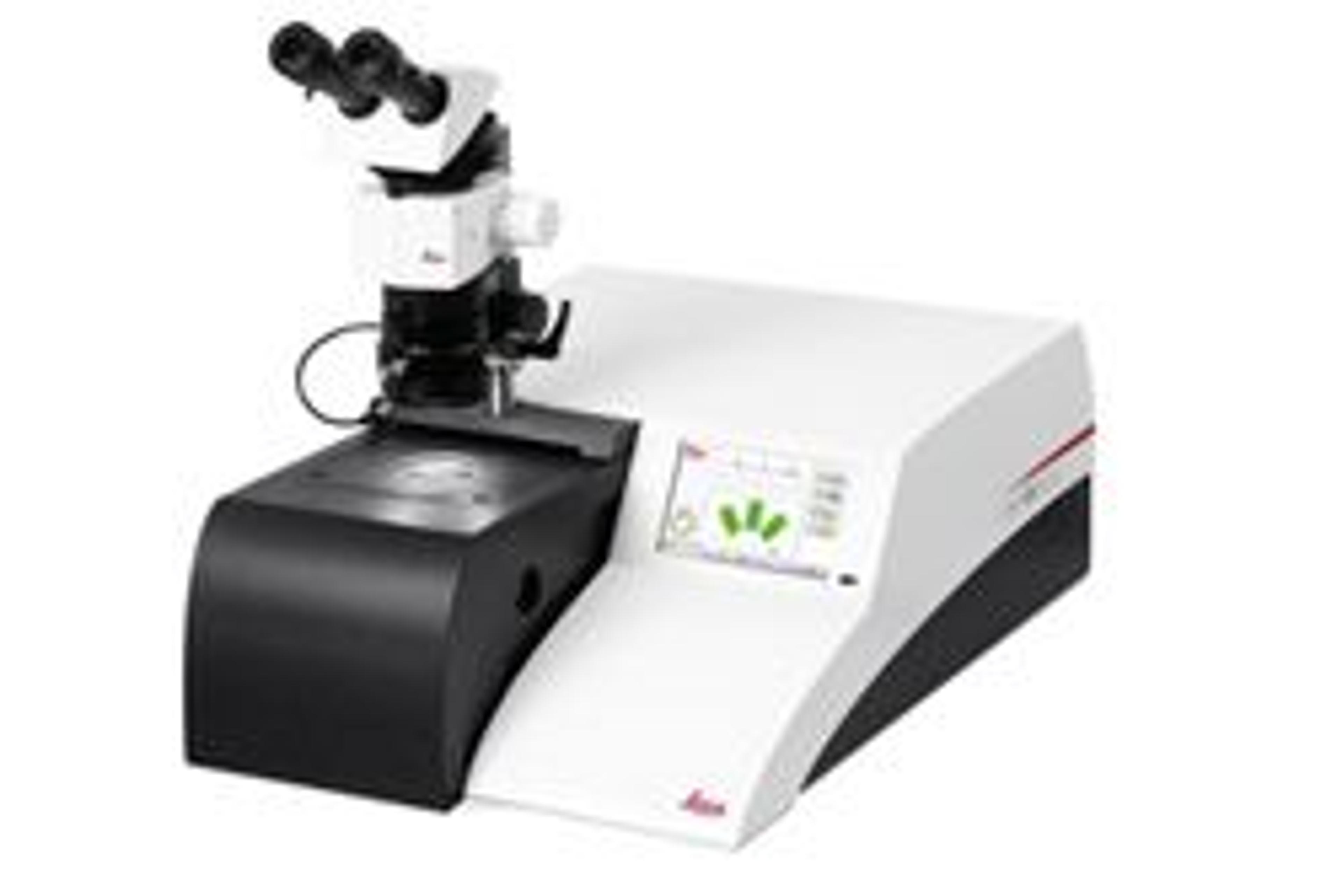
EM TIC 3X Ion Beam Milling System
Leica Microsystems EuropeThe Triple Ion Beam Cutter, EM TIC 3X allows production of cross sections of hard/soft, porous, heat sensitive, brittle and heterogeneous material for Scanning Electron Microscopy (SEM), Microstructure Analysis (EDS, WDS, Auger, EBSD) and, AFM investigations.
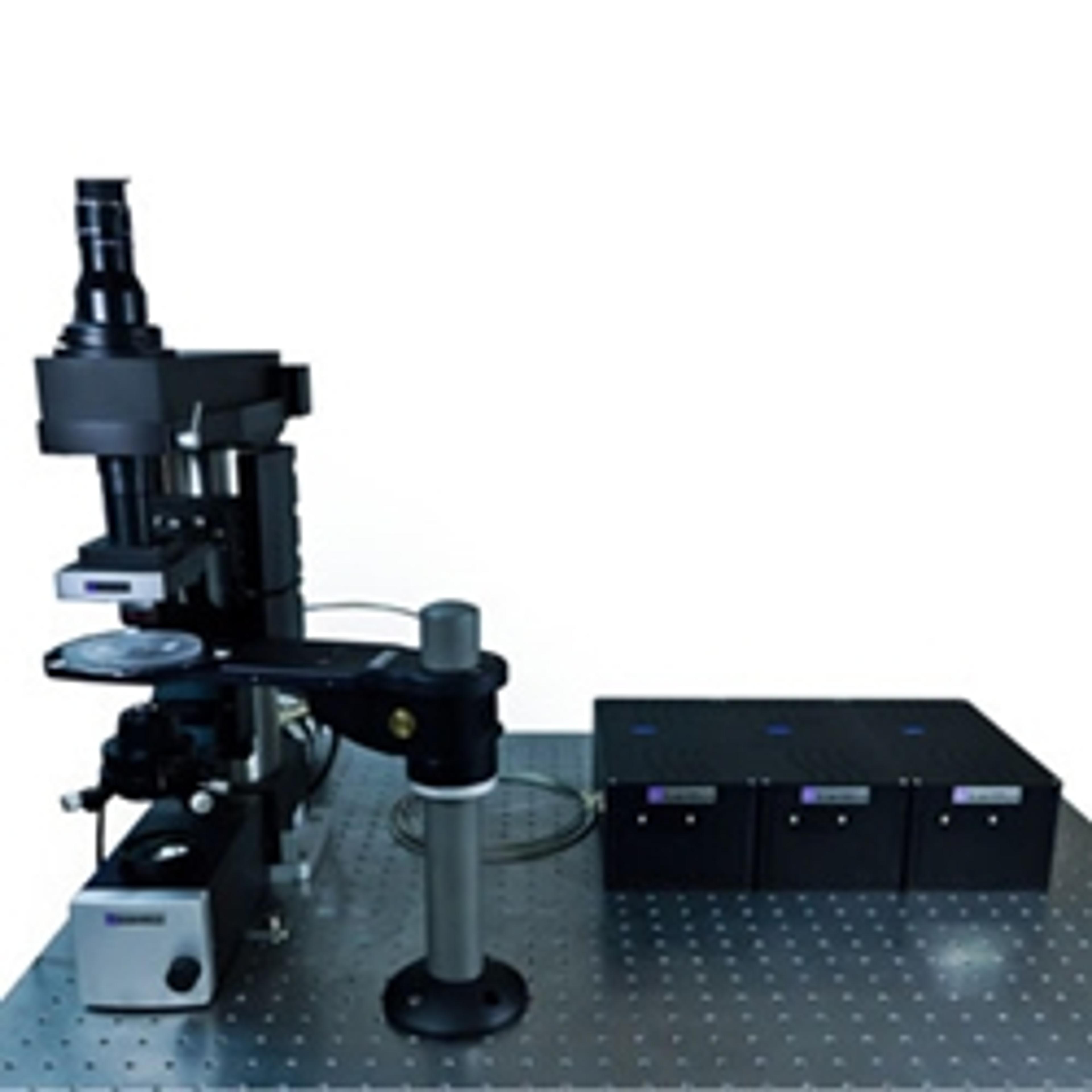
LASU - Laser Applied Stimulation & Uncaging
Scientifica LtdThe LASU system is perfect for users engaging in optogenetics, uncaging or other photo-stimulation experiments. The laser spot positioning is accurate and repeatable enabling fast experimental manipulation of biological systems. Based on the highly successful design of Scientifica's award-winning Multiphoton Imaging System, LASU uses 'Galvo' mirrors for fast, accurate and repeatable laser spot positioning. The wide range of laser modules means you can select one or more wavelengths and add to these as your needs, and the range of 'opsins', develop.The LASU system was developed in collaboration with VU University Amsterdam and provides the ability to carry out uncaging or optogenetics experiments by selecting the specific laser modules. The versatility of LASU means that it is also possible to integrate both techniques into one setup.The reliable hardware and powerful software enables convenient stimulation-spot positioning as well as full control of the laser power and firing frequency. The integrated imaging system also allows for patch clamping and other visibly guided experimentation, making the LASU an extremely versatile system.Benefits: Choice of laser wavelengths For precise control of your experiment, LASU is able to deliver a Laser spot smaller than 2 microns (depending on the objective used). This means you are able to stimulate exactly where you need. This provides much greater experimental control than 'full field' stimulation. Powerful software The LASU software is easy to use enabling control of stimulation site, laser power and pulse duration. With these functions and the live camera feed make this an extremely user friendly system. Simple integration The LASU system fits directly onto the Scientifica upright microscope 'SliceScope' making it an easy upgrade for existing rigs. Proven technology The Galvo mirror driven hardware and optical components are based on the Scientifica award-winning Two-Photon system giving you complete confidence when choosing LASU. Compact The compact nature of the LASU scanhead, SliceScope fram and laser modules mean that it can be mounted on most standard antivibration tables, saving you space and money. Free space launched or fibre guided laser delivery The choice of laser input through either fibre or free-space launch optics means that you can choose which method best suits your experimental setup. Perfect for optogenetics The choice of laser wavelengths available are suitable for 'channelrhodopsin' and 'halorhodopsin'. Allowing compete control over the laser power and stimulation frequency makes the LASU an essential tool for optogenetic experiments. Perfect for glutamate uncaging The 405nm wavelength Laser module is perfectly suited to Glutamate uncaging experiments when used with commercially available caged compounds. Applications: Glutamate uncaging Optogenetics Photo-activation Photo-stimulation Laser uncaging Channelrhodposin activation Halorhodopsin activation In vitro optogenetics Neural network studies
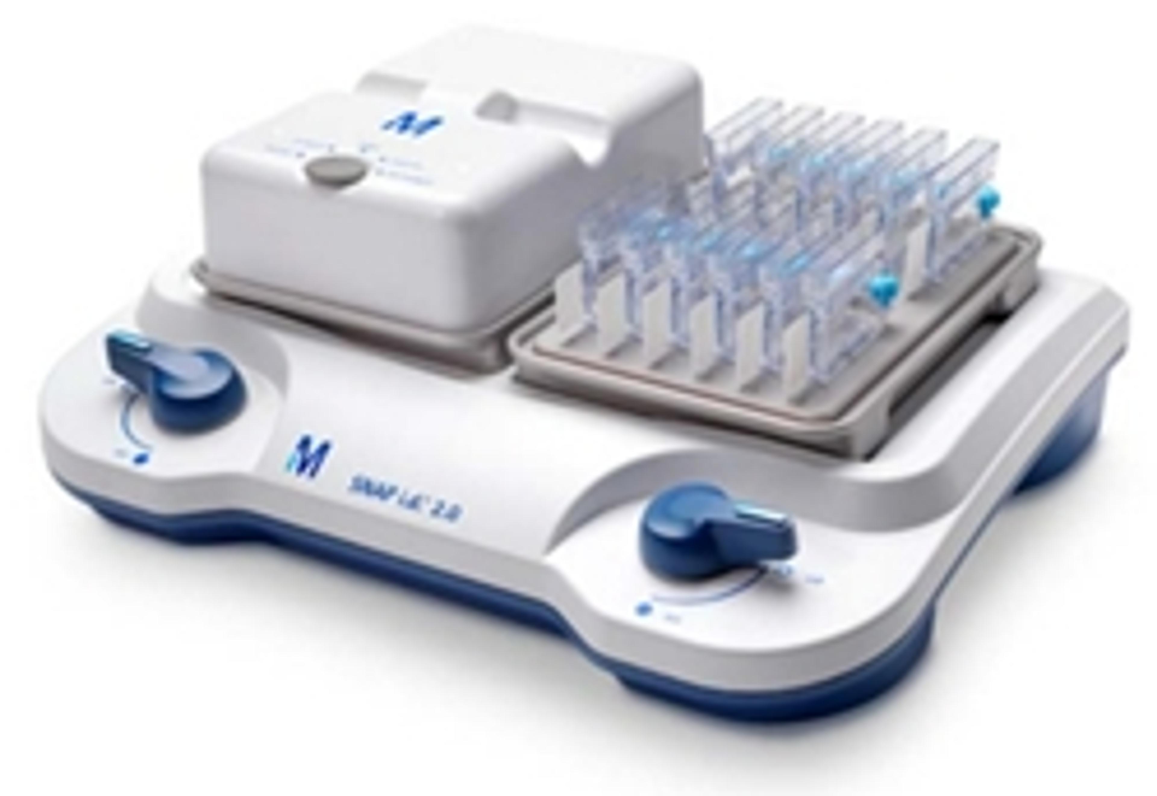
SNAP i.d. 2.0 IHC System
MerckIssues with Your Tissues? Process all Your Slides in a SNAP for Streamlined IHC. The SNAP i.d.® 2.0 Protein Detection System for Immunohistochemistry (IHC) introduces a new capability to the innovative, vacuum-driven SNAP i.d.® 2.0 system. The IHC slide holders allow you to block, probe, and stain up to 12 tissue slides per side (24 slides if both sides are used). Reduced handling time and multiple-slide processing make this system ideal for when you are optimizing antibody conditions and protocols.With two individually controlled sides, the system base allows for independent, vacuum-driven processing of either one or two IHC frames. Each of the IHC frames can process between 1 to 12 glass slides through independent vacuum ports.Each slide holder has an injection/recovery port that enables the manual addition, as well as the removal and recovery, of small volumes of antibodies or reagents; reagents can also be flushed using the vacuum feature if conservation is not a priority.Benefits: Eliminates the need for pap pens Antibodies can be collected and reused Slide handling time is significantly decreased Less time spent on wash steps Parallel processing of multiple slides Features: Flexibility of multiple slide configurations enables the processing of 1 to 24 slides at a time Compatible with standard IHC slides and protocols Compatible with diverse tissue preparations including formalin-fixed or fresh frozen samples Intuitive format Incorporates blocking, washing, and antibody incubation and labelling steps Systematizes handling multiple slides without the cost of automation Test tracker feature on frame cover helps keep track of IHC steps