Perspectives on Raman Spectroscopy of Graphene
30 Jul 2012Read this application note to learn about the ability of Raman spectroscopy to distinguish the number of graphene layers and any defects present. Also learn about the exciting advantages to using graphene as a substrate in Raman spectroscopy.
Related products
Request Quote for All Products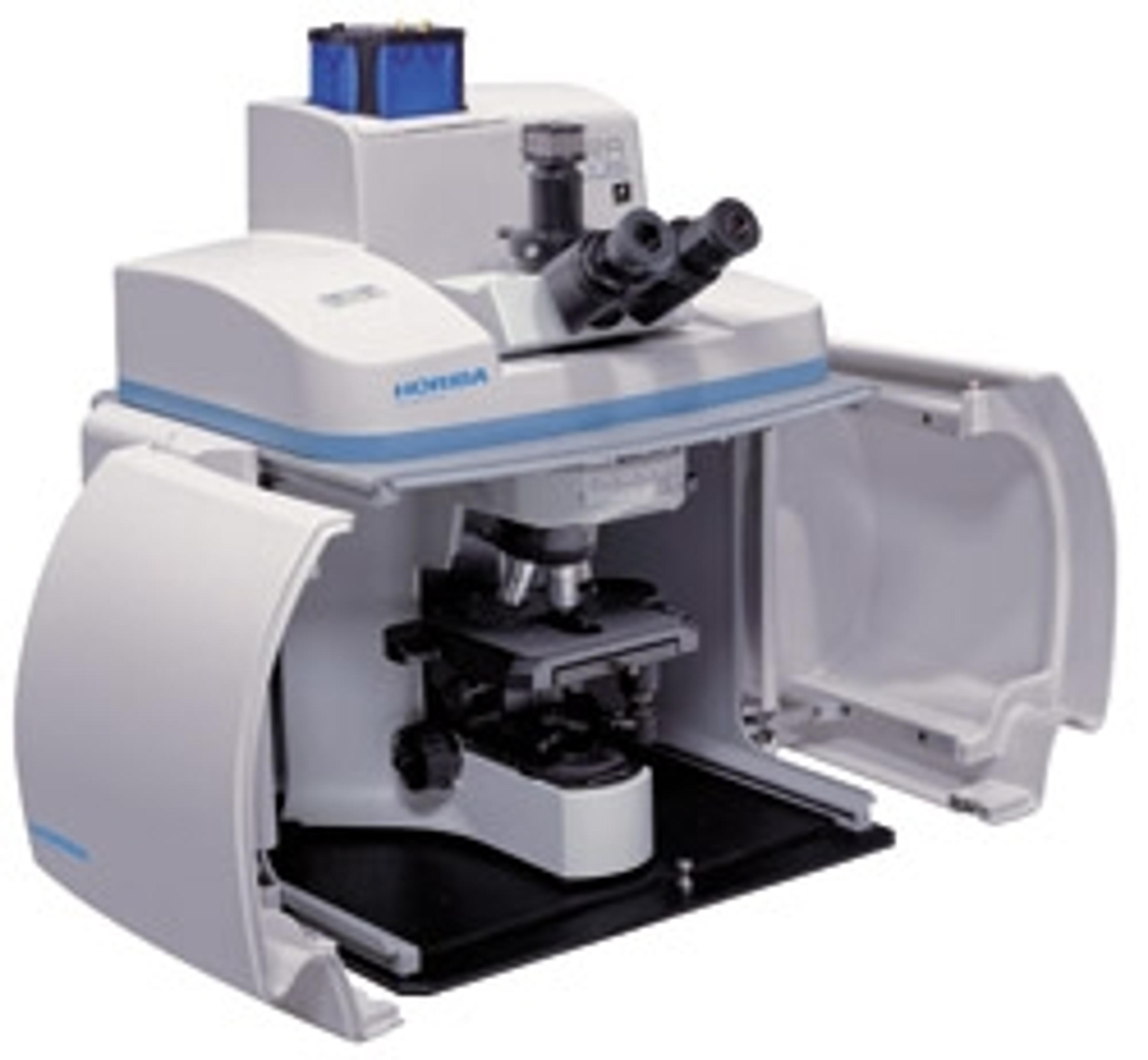
XploRA - Raman microscopy
HORIBA ScientificThe XploRA™ is a new concept in Raman microscopy, bringing Raman chemical identification directly to your microscope. The XploRA can be coupled to both upright and inverted microscopes, allowing analysis of all sample types, ranging from semiconductors and nano-materials, through to biological cells and tissues. The system provides the established performance of HORIBA Scientific Raman microscopes at a surprising price. Combining microscopy and chemical analysis the XploRA™ retains the full functionality of your microscope coupled with high performance Raman spectroscopy. The concept of the XploRA™ is straightforward – a smarter microscopy. A compact, high performance Raman spectrometer is coupled to a research grade optical microscope (upright or inverted geometry), allowing both standard optical microscopy and detailed chemical analysis on a single system. The unique design ensures full capability of the optical microscope, and complete flexibility, full automation and true research performance of the Raman analyzer.
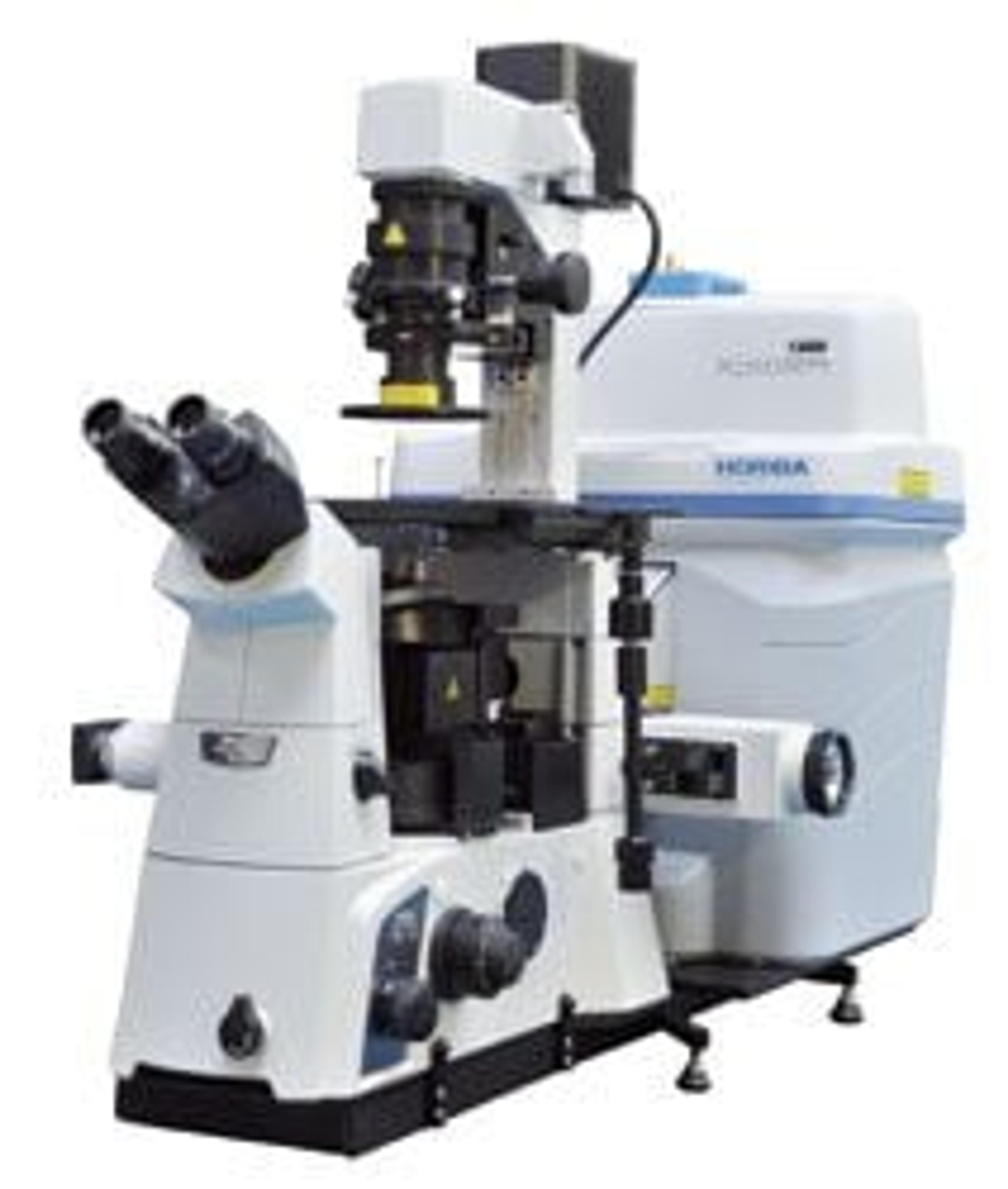
XploRA INV - Inverted Raman Microscope
HORIBA ScientificOptimized Raman Solution for Life Sciences from HORIBA Scientific The XpIoRA INV is an inverted Raman microscope designed specifically for use with biological samples requiring analysis from the bottom and/or open access to the sample from the top. Like all other XpIoRA series of Raman microscopes, the INV features a small footprint, complete automation (autofocus, autoexposure, autovalidation, autocalibration), and ease of use. Applications:• cell research • disease detection • characterisation of drug-cell interactions • pharmaceutical verification of intercellular activities In addition, the inverted geometry allows easy incorporation of AFM units for combined Raman-AFM and TERS (Tip Enhanced Raman Spectroscopy) for highest spatial resolution sample visualisation. The open structure of the XploRA INV also permits the use of virtually all options and add-ons for inverted microscopes, including micro manipulators and “optical tweezers”, specific enclosures for cell applications, etc. The XploRA INV also offers the option to integrate the unique scanning capabilities of HORIBA’s patented DuoScan for rapid spectroscopic multichannel Raman imaging, as well as the new 3D FCI (Fast Confocal Imaging) module, which allows ultra-fast fluorescence imaging for visualisation and verification.