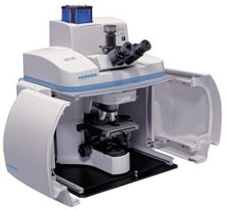
XploRA - Raman microscopy
The XploRA™ is a new concept in Raman microscopy, bringing Raman chemical identification directly to your microscope. The XploRA can be coupled to both upright and inverted microscopes, allowing analysis of all sample types, ranging from semiconductors and nano-materials, through to biological cells and tissues. The system provides the established performance of HORIBA Scientific Raman microscopes at a surprising price. Combin…


The supplier does not provide quotations for this product through SelectScience. You can search for similar products in our Product Directory.
Great results, highly reproducible, very easy to use
Analyzing the different biomarkers in tissue samples
I have used the Xplora Raman microscope for 3 year. It gives very good results that are highly reproducible. The main strength of this instrument is ease of use. Very easy to handle, quick focus and higher resolution. If anyone willing to buy an Raman instrument, then go for it. Fully worth it.
Review Date: 17 Apr 2023 | HORIBA Scientific
Good analytical instrument/hardware. Accessories and software improvable
Semiconductors and Graphene
Conventional system, reliable optics. Compact equipment, which casing is not so robust. LabSpec v6 version quite limited and prone to problems.
Review Date: 8 Jun 2022 | HORIBA Scientific
Amazing results. Trust worthy data analysis
Analyze different nanomaterials
The instrument is easy to operate and handy. It helps us for analyzing several materials in no time.
Review Date: 20 May 2022 | HORIBA Scientific
Clear results with better and multiple options in the software
Analyze different nanomaterials
Effective in analyzing samples and good ease of operation
Review Date: 2 Apr 2022 | HORIBA Scientific
Most reliable results for material characterization.
Material synthesis and characterization
Recently I used this to characterize samples of CVD grown diamonds. I received very reliable data. I recommend this product for Raman Spectroscopy.
Review Date: 17 May 2021 | HORIBA Scientific
Produces great results in an efficient way.
Semiconductor
One can get high resolution spectra on all kinds of 2D materials, very efficient and user-friendly.
Review Date: 4 Dec 2020 | HORIBA Scientific
Great instrument.
2D material characterization
The Horiba Raman spectroscopy helps me achieve high-quality spectra of 2D materials.
Review Date: 26 Oct 2020 | HORIBA Scientific
Great instrument.
2D material
It is easy to obtain high-quality results with the aid of this instrument.
Review Date: 28 Jul 2020 | HORIBA Scientific
Essential equipment for our lab!
Graphene characterisation
Easy to use. Software is very clear. Gives robust data. System is stable in clean room environment. Quick to calibrate. Great for demonstrations. Support via calls/remote sessions very effective. Repairs conducted in a timely manner.
Review Date: 5 Jun 2020 | HORIBA Scientific
The XploRA™ is a new concept in Raman microscopy, bringing Raman chemical identification directly to your microscope. The XploRA can be coupled to both upright and inverted microscopes, allowing analysis of all sample types, ranging from semiconductors and nano-materials, through to biological cells and tissues.
The system provides the established performance of HORIBA Scientific Raman microscopes at a surprising price. Combining microscopy and chemical analysis the XploRA™ retains the full functionality of your microscope coupled with high performance Raman spectroscopy.
The concept of the XploRA™ is straightforward – a smarter microscopy. A compact, high performance Raman spectrometer is coupled to a research grade optical microscope (upright or inverted geometry), allowing both standard optical microscopy and detailed chemical analysis on a single system.
The unique design ensures full capability of the optical microscope, and complete flexibility, full automation and true research performance of the Raman analyzer.

Raman spectroscopy: Rapid QC of healthcare products
The development of Raman spectroscopy has come a long way since its discovery in 1928. Its combination with optical microscopy in the latest, next-generation confocal Raman microscopes has enabled fast, non-destructive, non-invasive hyperspectral imaging of samples without the need for sample preparation in most cases.
In this application compendium, we look at some of the wide-ranging applications of Raman microscopy to address various formulation challenges encountered by the pharmaceutical and cosmetics industries. In particular, we consider HORIBA Scientific’s latest confocal Raman imaging microscope, the LabRAM Soleil™, designed to make Raman imaging faster and easier than ever before.
Download the free eBook to discover how Raman microscopy can yield information on molecular structures, crystal phases, polymorphisms and much more, as we cover:
- Characterization of pharmaceuticals, including compound distribution, chemical homogeneity and more
- Characterization of cosmetics for perfect formulations
- Raman spectroscopy as an essential tool in pharma
Micro-Raman Study of the Sequence of Non-Intersecting Lines for Forged Document Investigation
In connection with investigation of fraud, counterfeiting, blackmail and anonymous letter cases, forensic experts frequently have to find out whether a document was altered after signing. In these cases the sequence of crossing printer and pen ink line often needs to be determined in order to resolve whether any of the printed text was printed after the signature. When there are no intersecting lines between the printed text and the pen ink lines, standard techniques used for sequencing are not applicable. This note demonstrates the application of micro-Raman spectroscopy to investigate the chronological sequence of printed and hand-written features on the documents even if there are no intersecting lines.
Confocal Raman Microspectrometry Imaging Combined with Chemometric Methods for Environmental Applications
The knowledge of the chemical composition of aerosols is of great importance due to their potential adverse effects on the environment and human health. Due to the small size of typical aerosol particles, the challenge is physicochemical characterization of aerosols at the micron scale. This application note explores the use of confocal Raman microspectrometry for the characterization of aerosol particles and other applications.
Perspectives on Raman Spectroscopy of Graphene
Read this application note to learn about the ability of Raman spectroscopy to distinguish the number of graphene layers and any defects present. Also learn about the exciting advantages to using graphene as a substrate in Raman spectroscopy.
The Horiba Xplora: Combining Confocal Microscopy and Raman Chemical Analysis
Watch this video to learn how the XploRA™ from Horiba brings chemical identification directly to your microscope. The XploRA can be coupled to both upright and inverted microscopes, allowing analysis of all sample types, ranging from semiconductors and nano-materials, through to biological cells and tissues.
HORIBA and Digital Surf partner to launch graphYX software range
For the correlative analysis of Raman, AFM, AFM-Raman, cathodoluminescence and fluorescence data and microscopy images

Raman microscopy: Comprehensive characterization of polymers
Learn how to obtain accurate, non-destructive, information on polymers in this on-demand webinar

HORIBA Scientific announces enhanced OEM RAMAN spectral engine for reliable process analytical measurements
Affordable Raman engine aims to provide OEM customers with unmatched detection limit
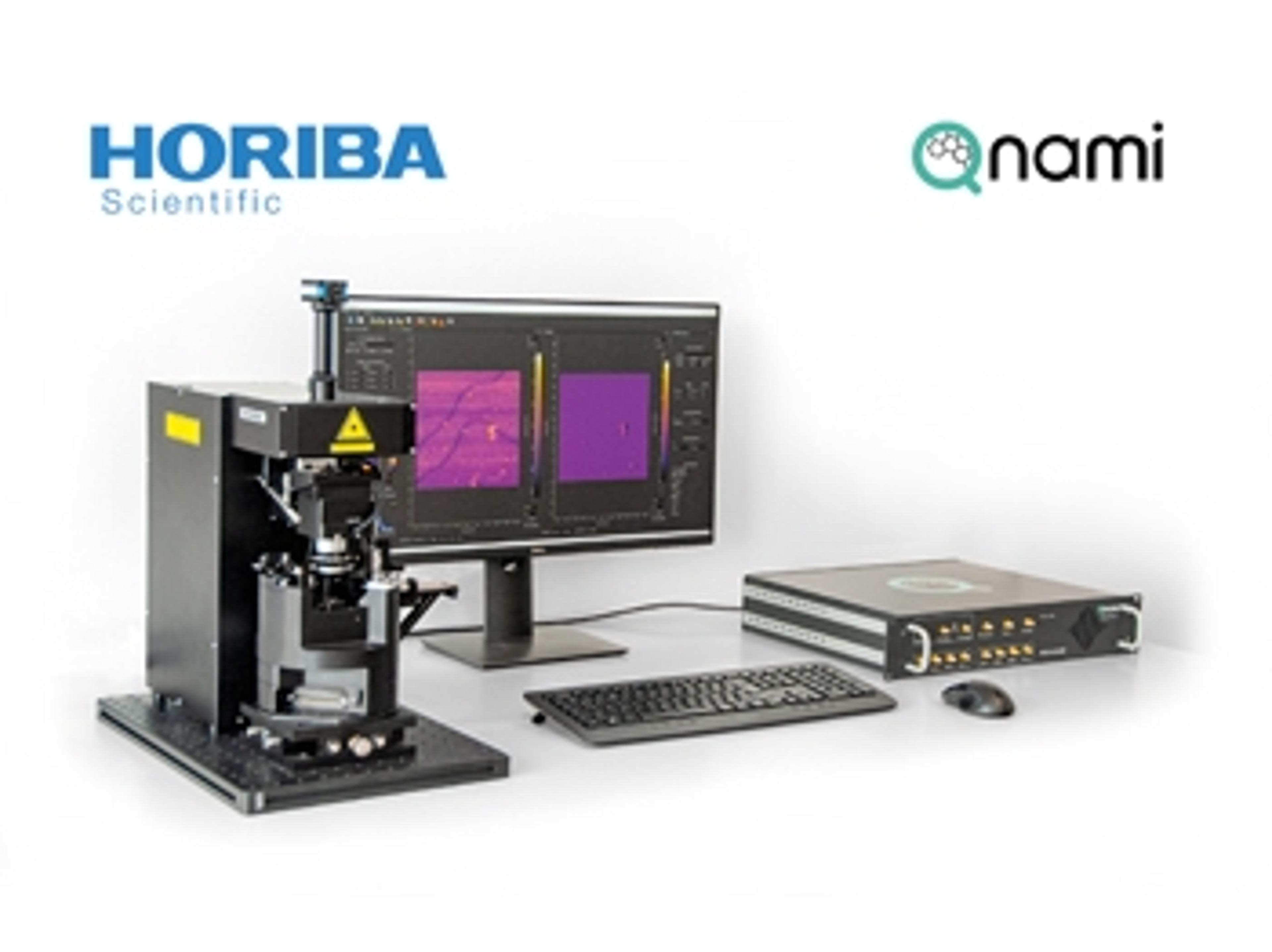
HORIBA collaborates with Qnami to develop scanning NV magnetometry and distribute first quantum microscope
The agreement will allow materials science researchers to benefit from solutions combining HORIBA’s advances in AFM instrumentation with Qnami’s innovation in quantum sensing

Raman microscopy: Comprehensive characterization of polymers
Join us on Monday, September 28, for this expert-led webinar on the intricacies of Raman technology
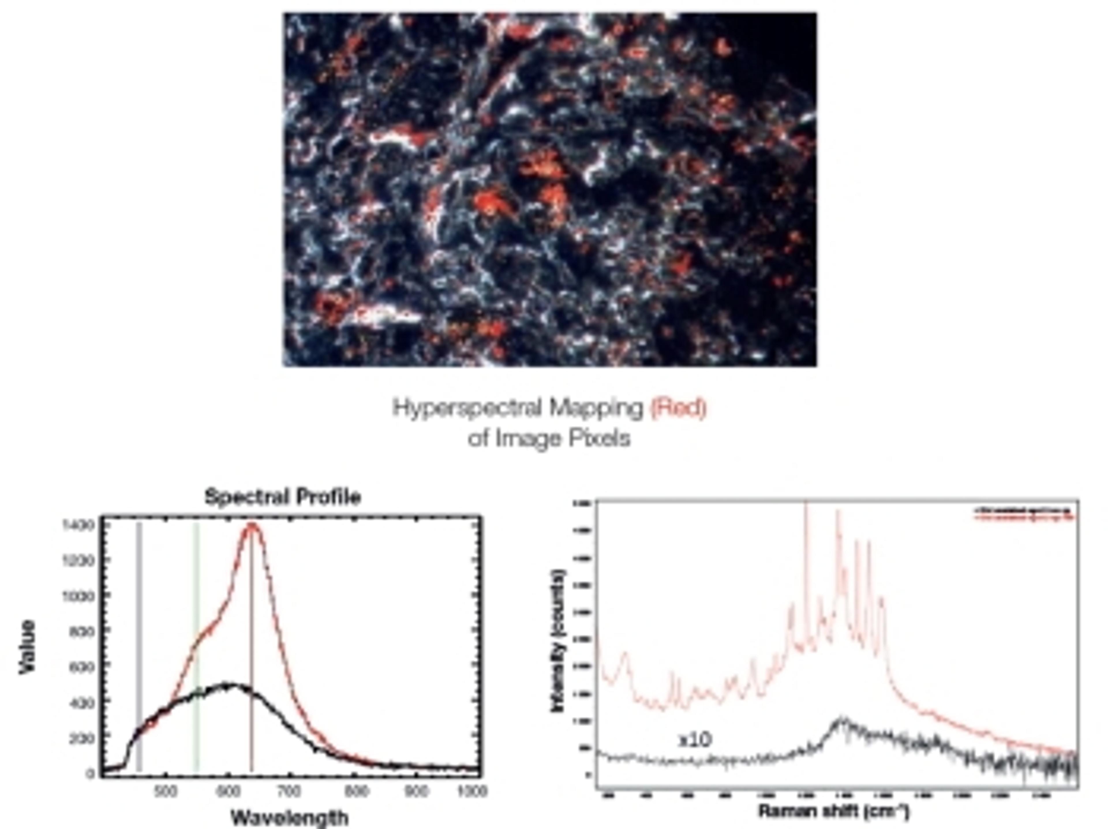
HORIBA combines with CytoViva for more powerful Raman analysis
More powerful nanoparticle analysis with Raman spectroscopy, hyperspectral imaging and enhanced darkfield illumination on the same microscope platform
HORIBA Scientific Introduces New CLUE Series Detectors for Scanning Electron Microscopes
CLUE Series offers scalable platform for imaging and spectroscopic analysis of nano-objects with SEM and dual SEM/FIB (Focused Ion Beam) microscopes.