Fibronectin bovine plasma
Merck KGaA, Darmstadt, Germanysolution, sterile-filtered, BioReagent, suitable for cell culture
solution, sterile-filtered, BioReagent, suitable for cell culture
liquid, 0.1% (Solution), BioReagent, suitable for cell culture
1-2 mg/mL in Tris-buffered saline, 0.2 μm filtered, BioReagent, suitable for cell culture
Phytohemagglutinin PHA-P, lyophilized powder
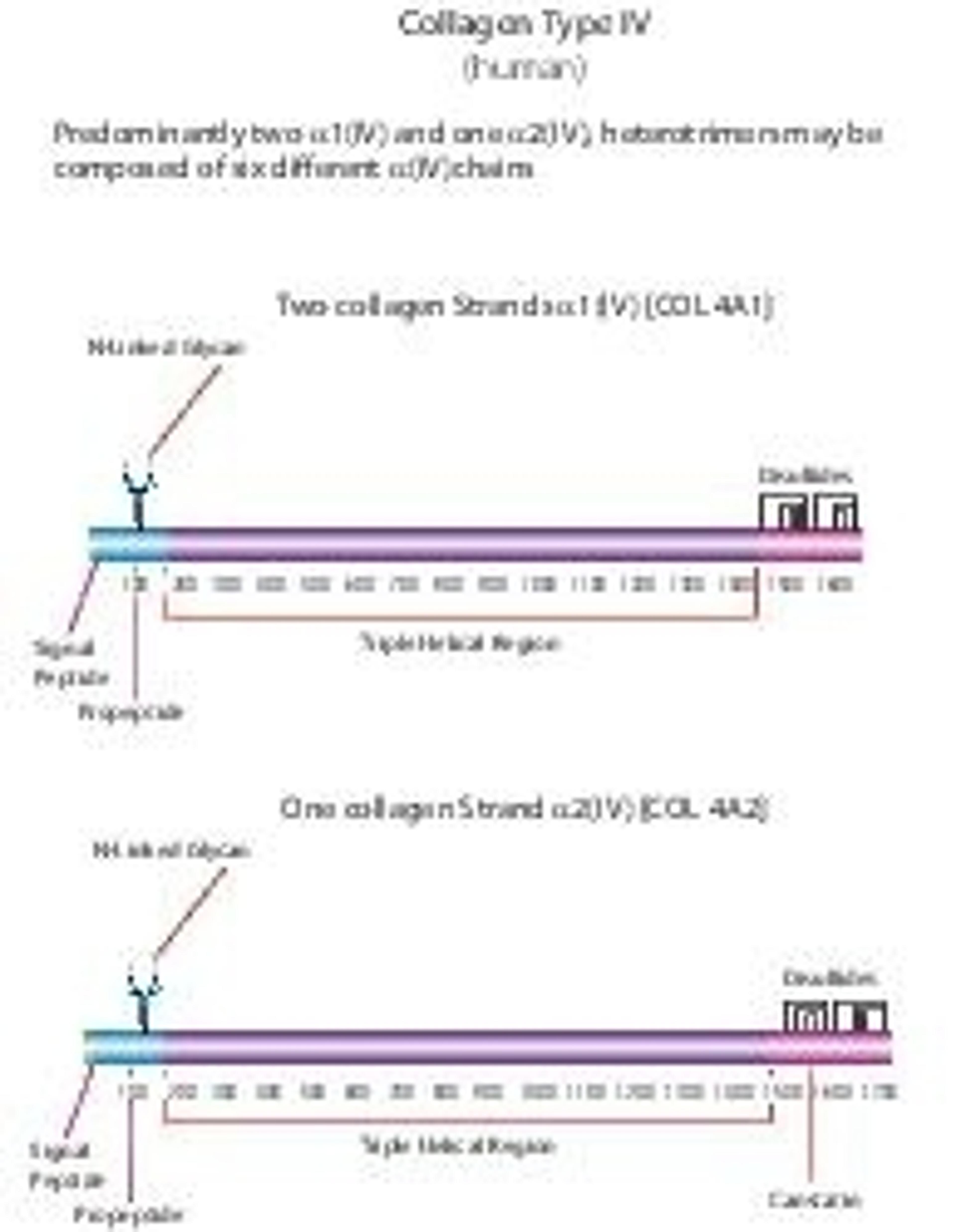
Bornstein and Traub Type IV, powder, BioReagent, suitable for cell culture
50-70% protein (≥80% of protein is clottable)
Optimized for stem cell culture applications
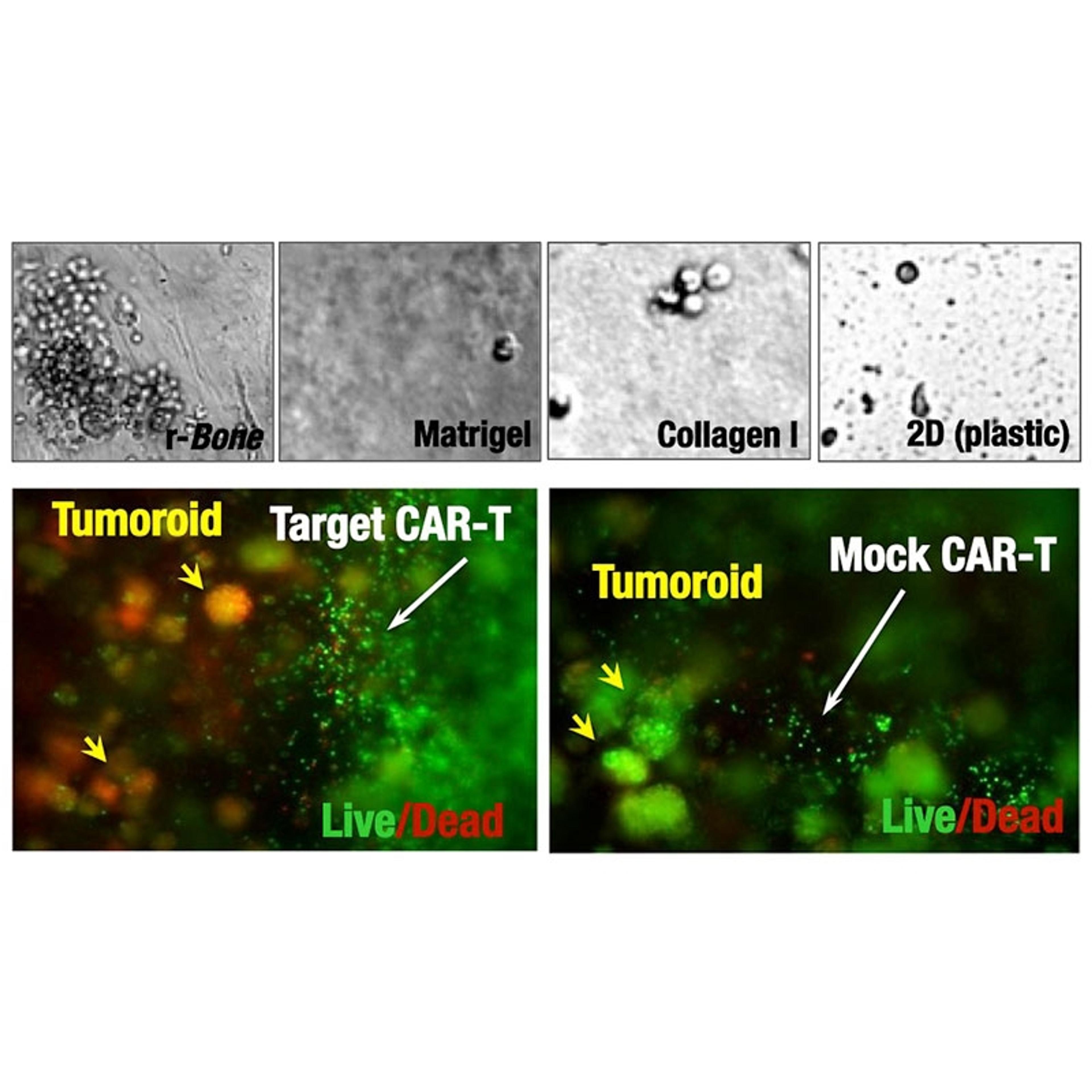
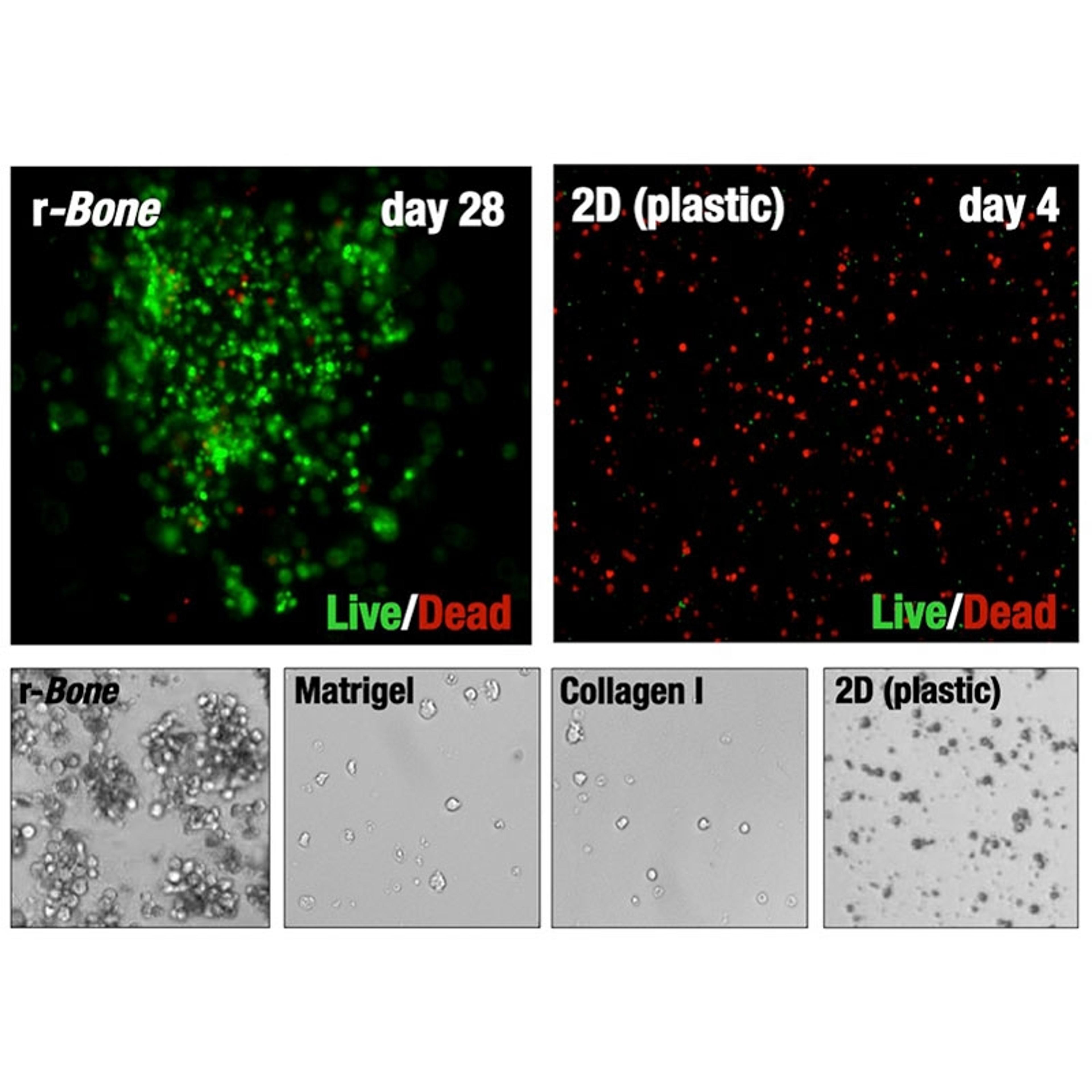
Human, bone marrow 3D culture model enabling accurate testing of therapeutic agents against multiple myeloma.
Human, bone marrow 3D culture model enabling accurate testing of therapeutic agents against acute myelogenous leukemia (AML).
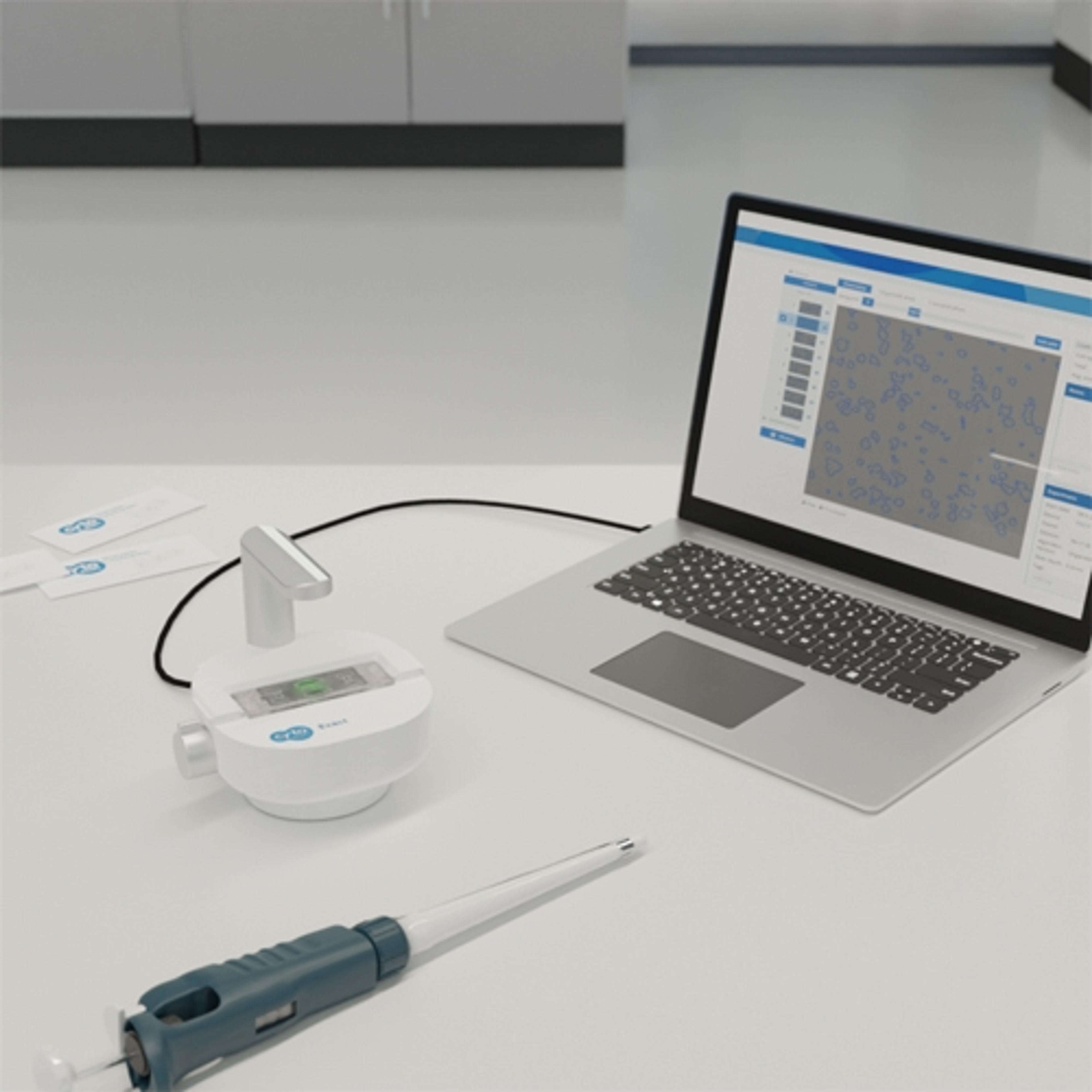
The Organoid Counting module for the CytoSMART Exact platform can detect organoids and spheroids of various shapes and sizes. In addition to counting the total number of organoids, the module can quantify organoid size and distribution. With the latest in image analysis algorithms, quickly and accurately count your organoids and spheroids.
HistologiX is a leading GLP/GCP accredited Contract Research Organisation based in the UK specialising in Histology, Immunohistochemistry (IHC) and Digital Pathology services. HistologiX works with a broad range of clients from big pharma to small and medium biotech companies worldwide. Our team of scientists have in excess of 120 years combined experience and a customer retention rate of 80%.
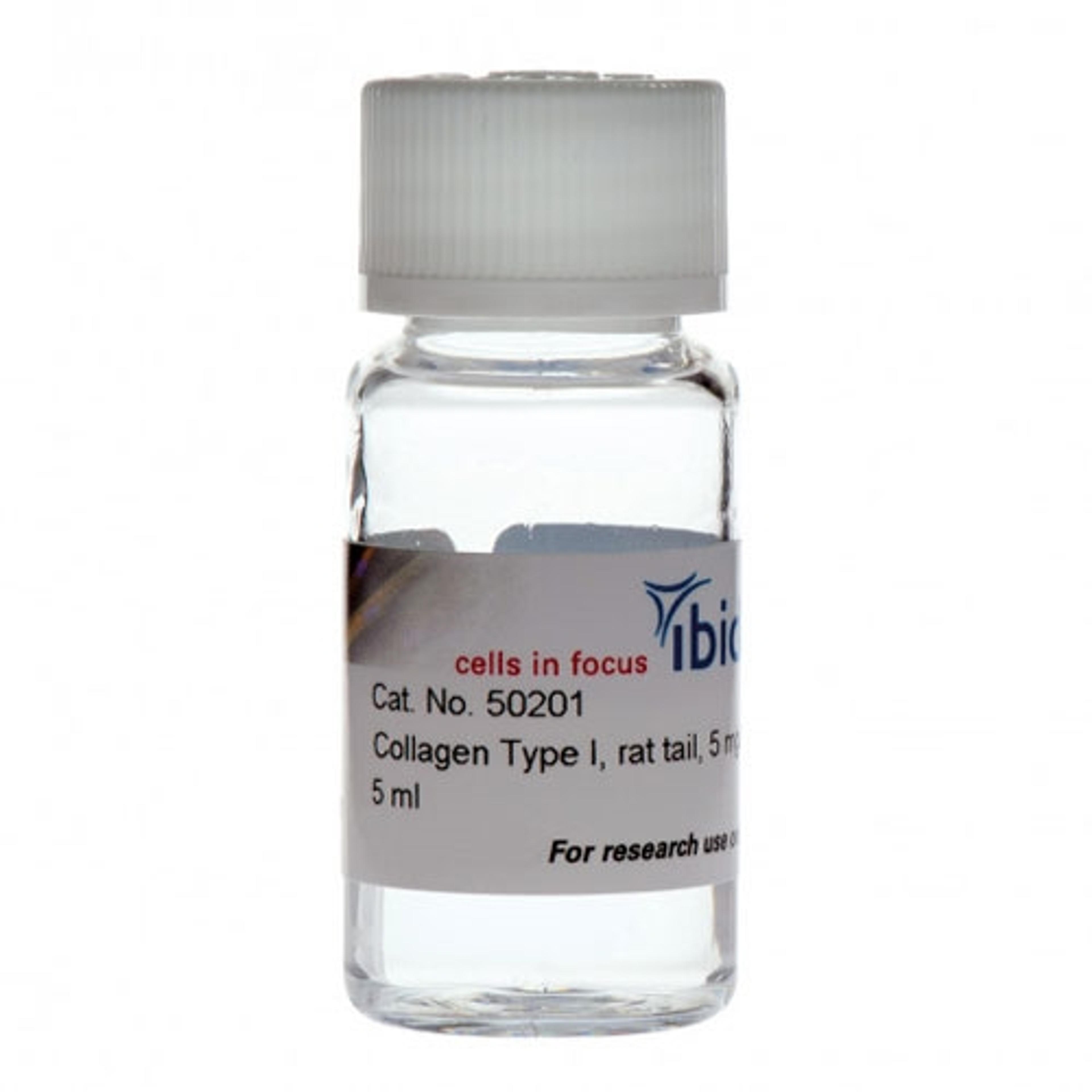
Use this high-quality rat tail collagen type I solution for cell culture applications (e.g., 3D gels, scaffolds, and coating).
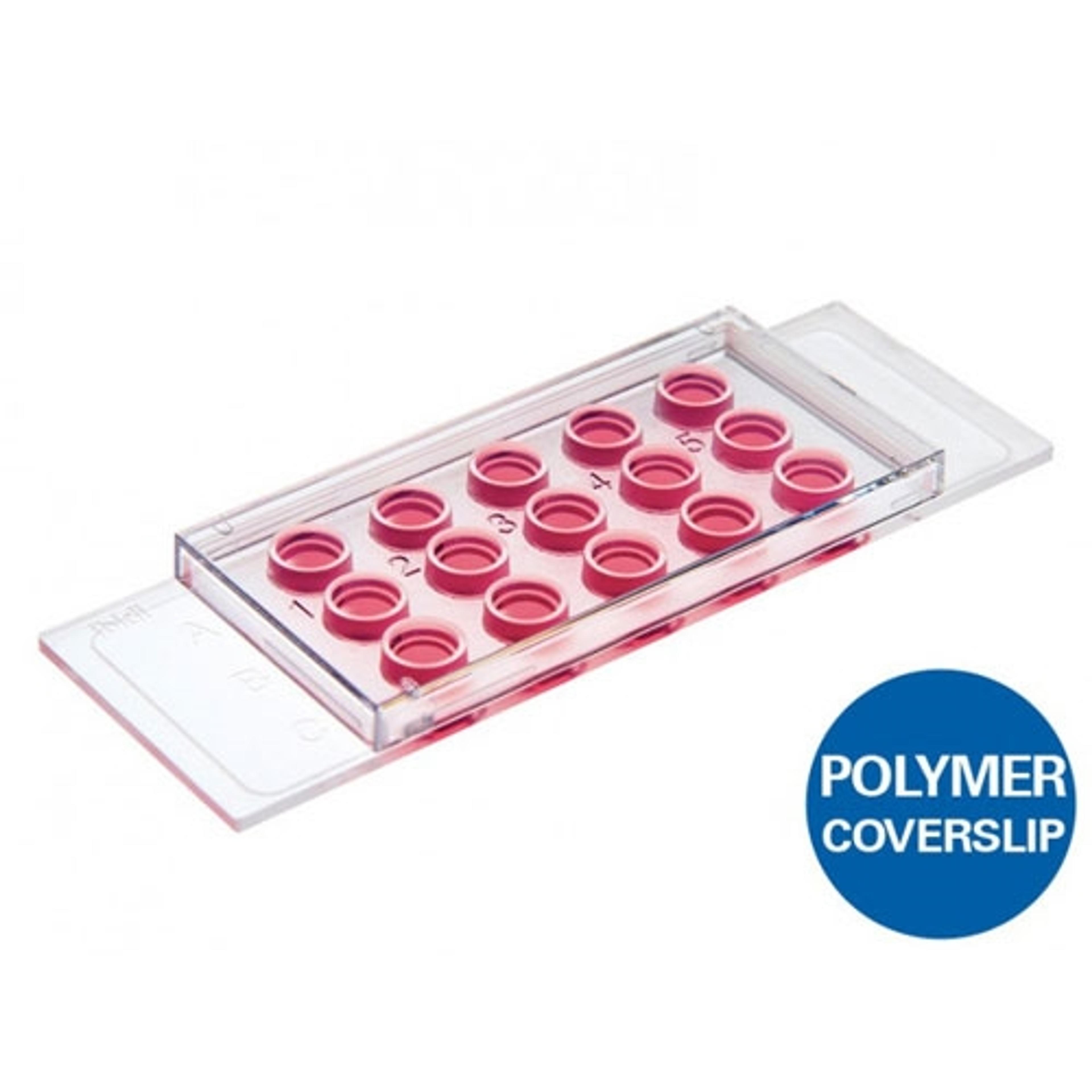
Use this slide to investigate angiogenesis in tube formation assays. Also ideal for 3D cell culture and immunofluorescence staining. Also available in a 96 well format.
Decellularized extracellular matrix (dECM) hydrogels for 2D and 3D cell culture applications.
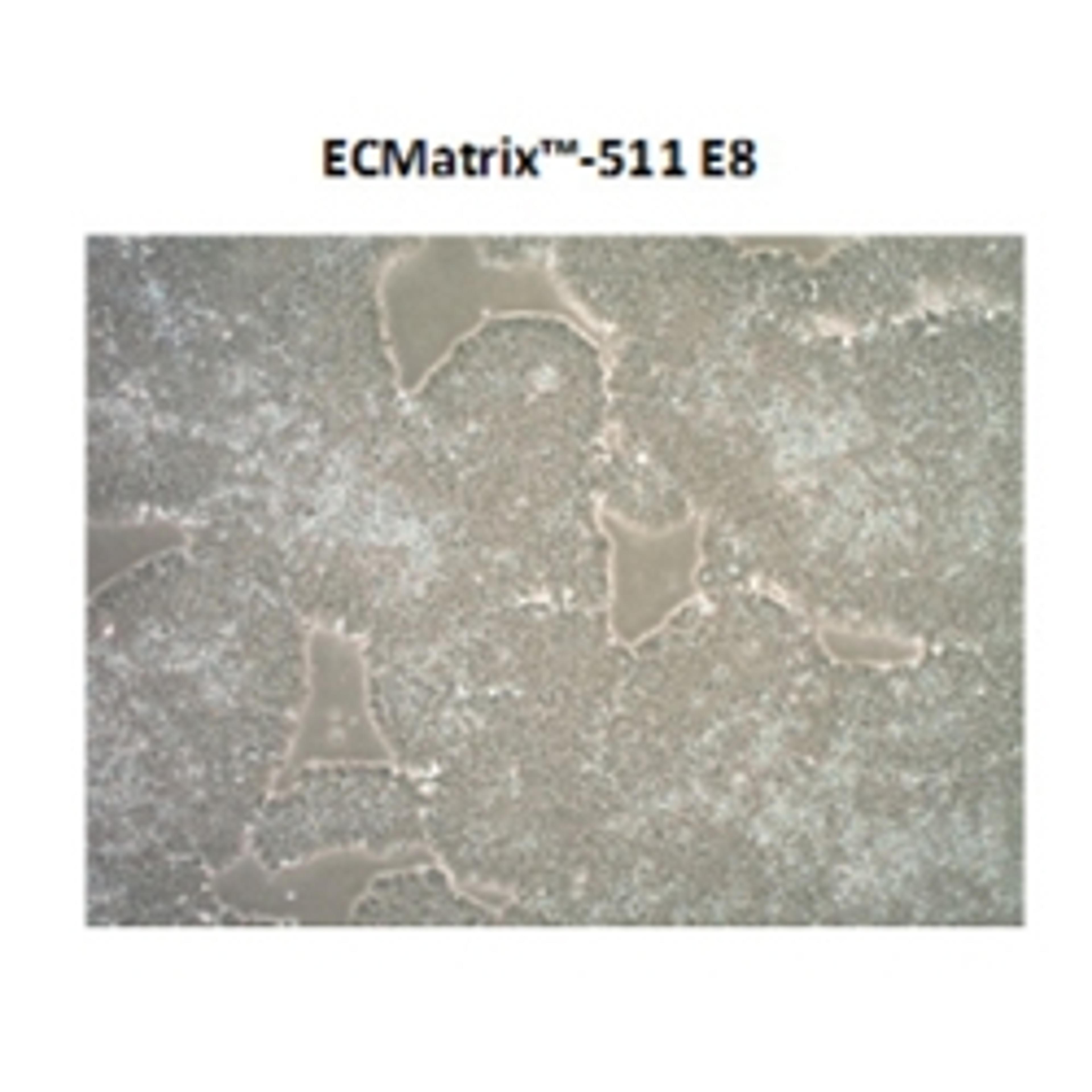
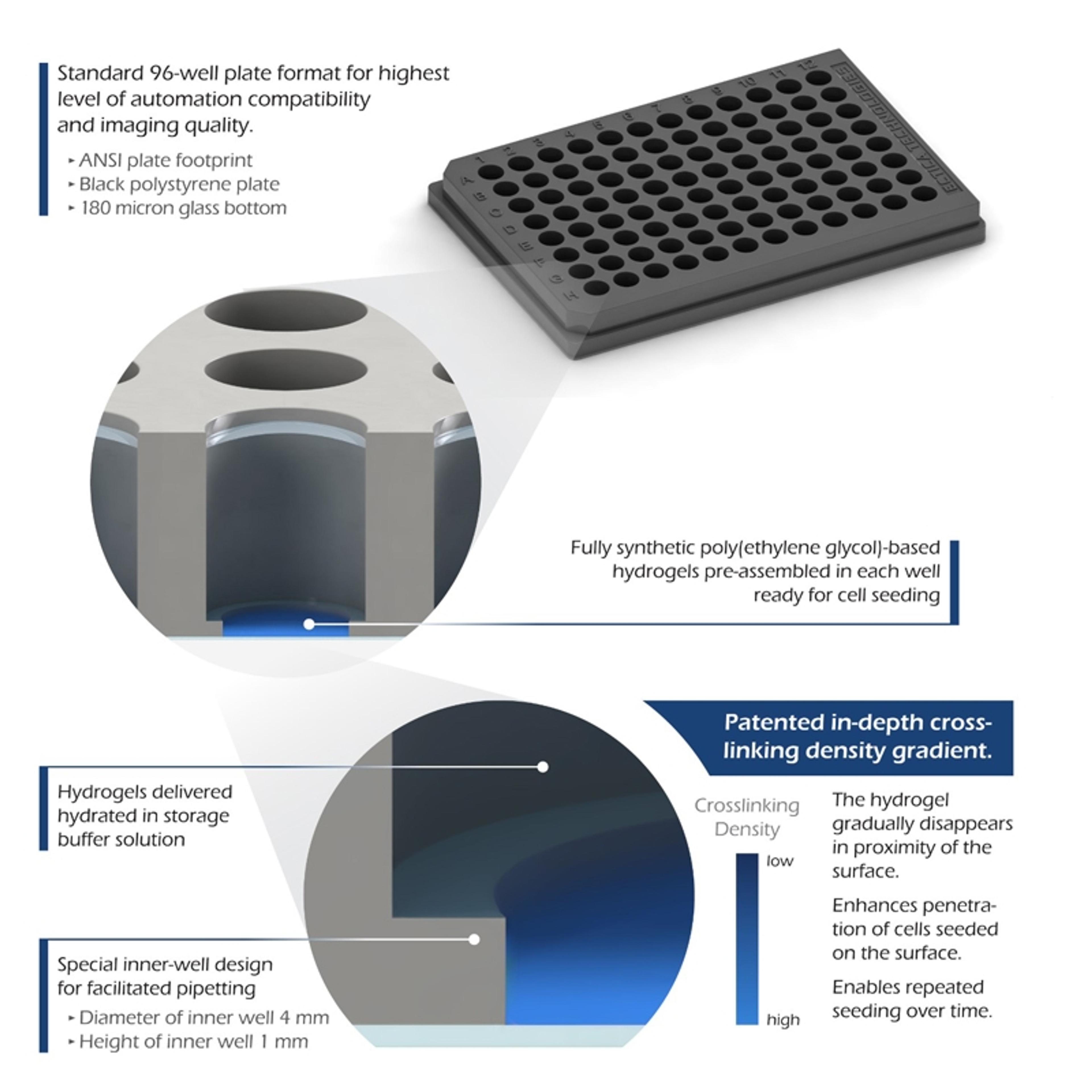
Ready-to-use, 96-well black-walled glass-bottom microplates containing precast synthetic hydrogels designed to easily establish 3D cell cultures by direct seeding of cells.
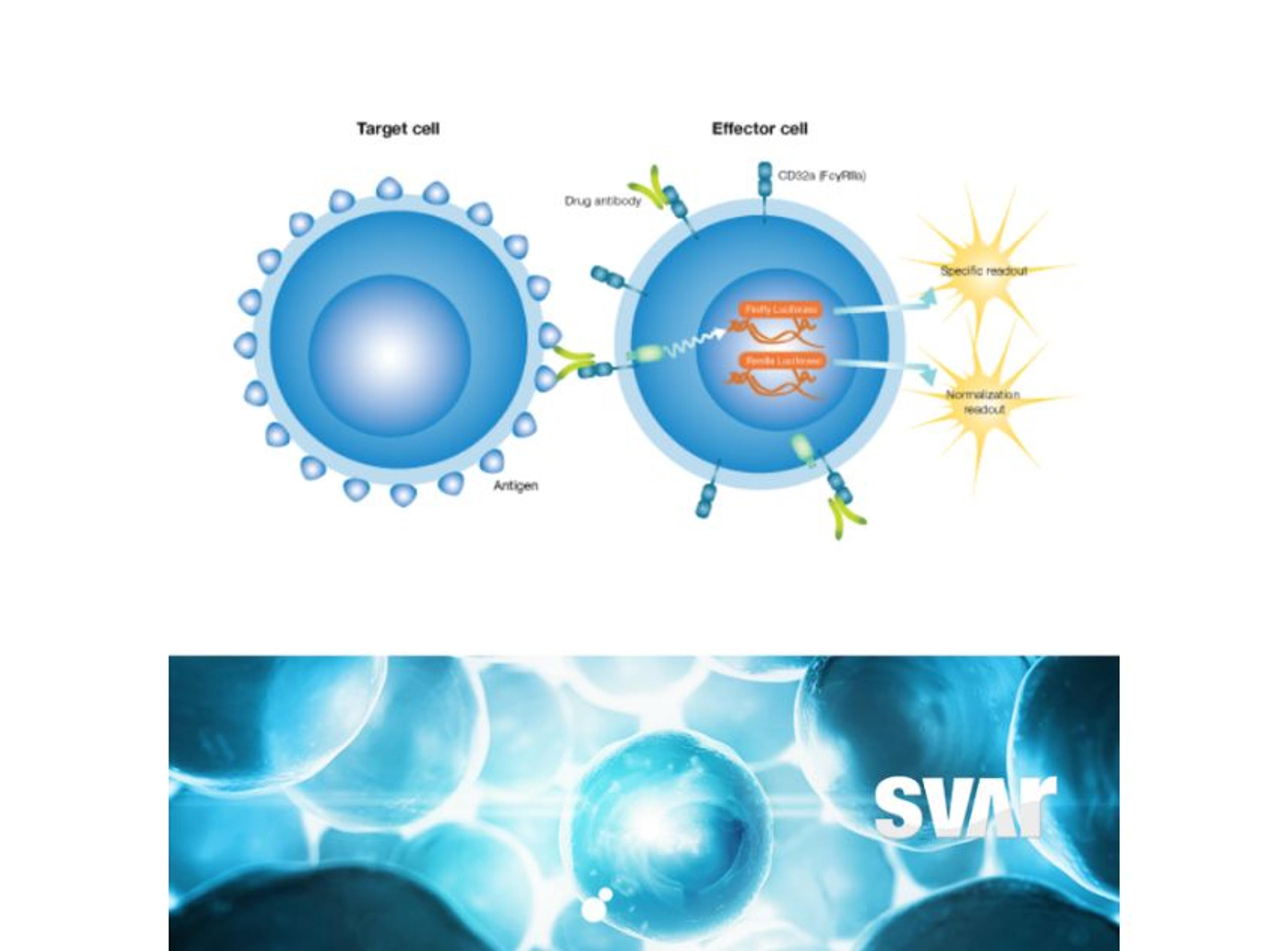
With their proprietary technology, the iLite® technology Svar can develop Report Gene Assays – that area cleverly designed, sensitive and specific cell based reporter-gene system with luciferase readout. Svar can develop assays for virtually any pharmaceutical target that allow an easy, rapid and accurate test format for a wide range of applications, such as Immunogenicity and Potency assays.
Combining the sensitivity of a fluorescence-based assay with a microplate format enables a rapid, quantitative readout suitable for high-throughput analysis. In a microplate well, the fluorescent signal can be generated within whole cells, in cell lysates, or in purified enzyme preparations and may then be analyzed by measuring fluorescence intensity from the well without the need for cellular imaging.
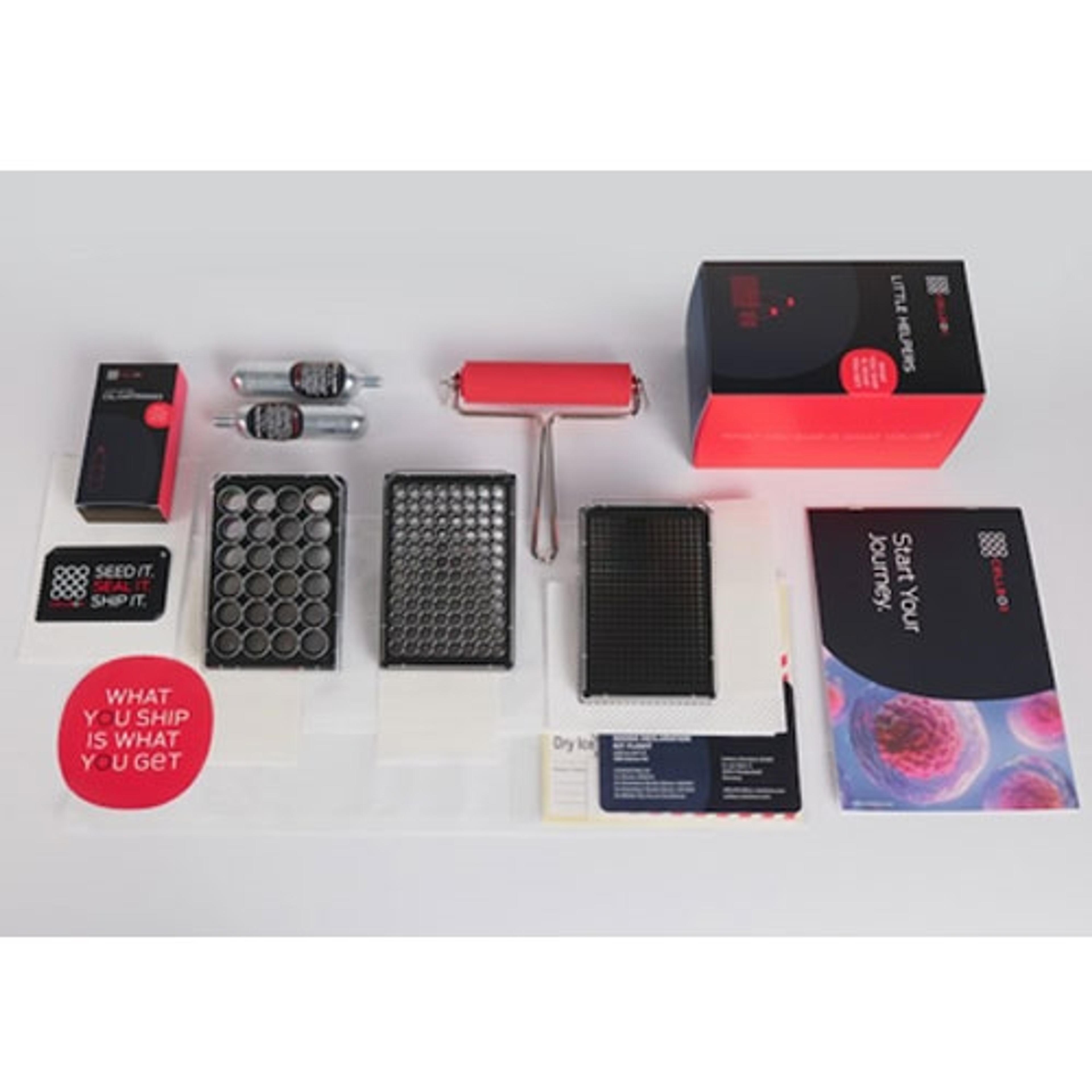
The complete leak proof and gas permeable packaging solution for culturing and shipping in 24/96/384 multiwell plate formats.
Celselect™ Slides capture and isolate individual rare cells, or CTCs based on their size
The iLite® Functional Bioassay portfolio consists of cell lines provided in an “assay-ready” format for a rapid and convenient workflow. The assay-ready format also reduces assay variability, optimizing its use in screening, characterization, stability, and potency studies. iLite cell lines can be tailored to virtually any biopharmaceutical target.
The iLite® Fc Effector Reporter Gene Bioassays Portfolio offers a variety of reporter gene cell lines and target cells that can be used in MOA-reflective assays to measure the Fc effect of antibodies and other biologics that specifically bind and activate Fc receptors. Using a Luciferase reporter gene system offers a convenient and powerful way of measuring antibodies' in vitro efficacy in triggering ADCC and ADCP.
Svar offers assistance in gene therapy projects through robust platforms suited for all phases of drug development and experience from immunoassasy product development as well as their CRO services. Svar currently offer cell-based assays for Neutralizing antibody assessment and custom-made immunoassays for Total antibody assessment.