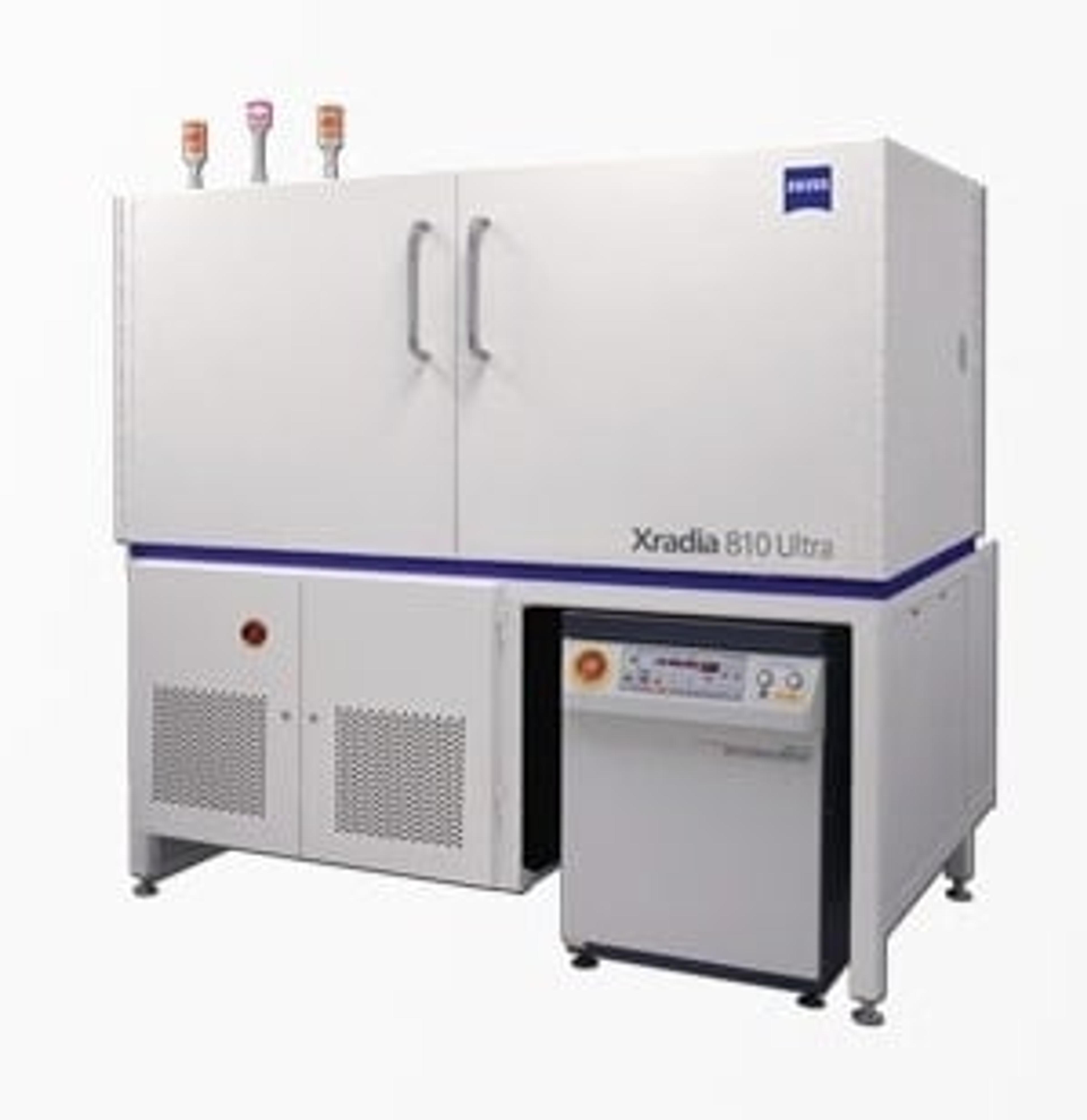
ZEISS Xradia 800 Ultra
Your Ultra-high Resolution, Non-destructive 3D Imaging System.


Receive your quote directly from the manufacturer.
With the ZEISS Xradia 800 Ultra X-ray microscope, achieve spatial resolution down to 50 nm, the highest among lab-based X-ray imaging systems.
With non-destructive 3D imaging playing a vital role in today’s breakthrough research, you will experience unparalleled performance in an ultra-high resolution lab-based system. The innovative Xradia Ultra architecture features absorption and phase contrast imaging modes and X-ray energy of 8 keV, using unique optics adapted from the synchrotron.
With Xradia 800 Ultra, expect to accomplish unrivaled in situ and 4D capabilities for studying material evolution over time and extend the limits of X-ray imaging used in materials science, life sciences, natural resources, and diverse industrial applications.

High-resolution characterization of solid foams
Characterization of the 3D morphology of solid foams is extremely important but has been limited due to the shortcomings of conventional techniques. High-resolution techniques such as physical sectioning coupled with optical or electron microscopy are not only destructive and time-intensive, but can also introduce physical artifacts. ZEISS highlights how the ZEISS Xradia Versa and the ZEISS Xradia Ultra series of 3D X-ray microscopes provide a unique solution for non-destructive submicron resolution and the highest contrast in foam imaging that can be achieved. Foam structures can be imaged to submicron spatial resolutions: down to 500 nm on ZEISS Xradia Versa and 50 nanometers on ZEISS Xradia Ultra.
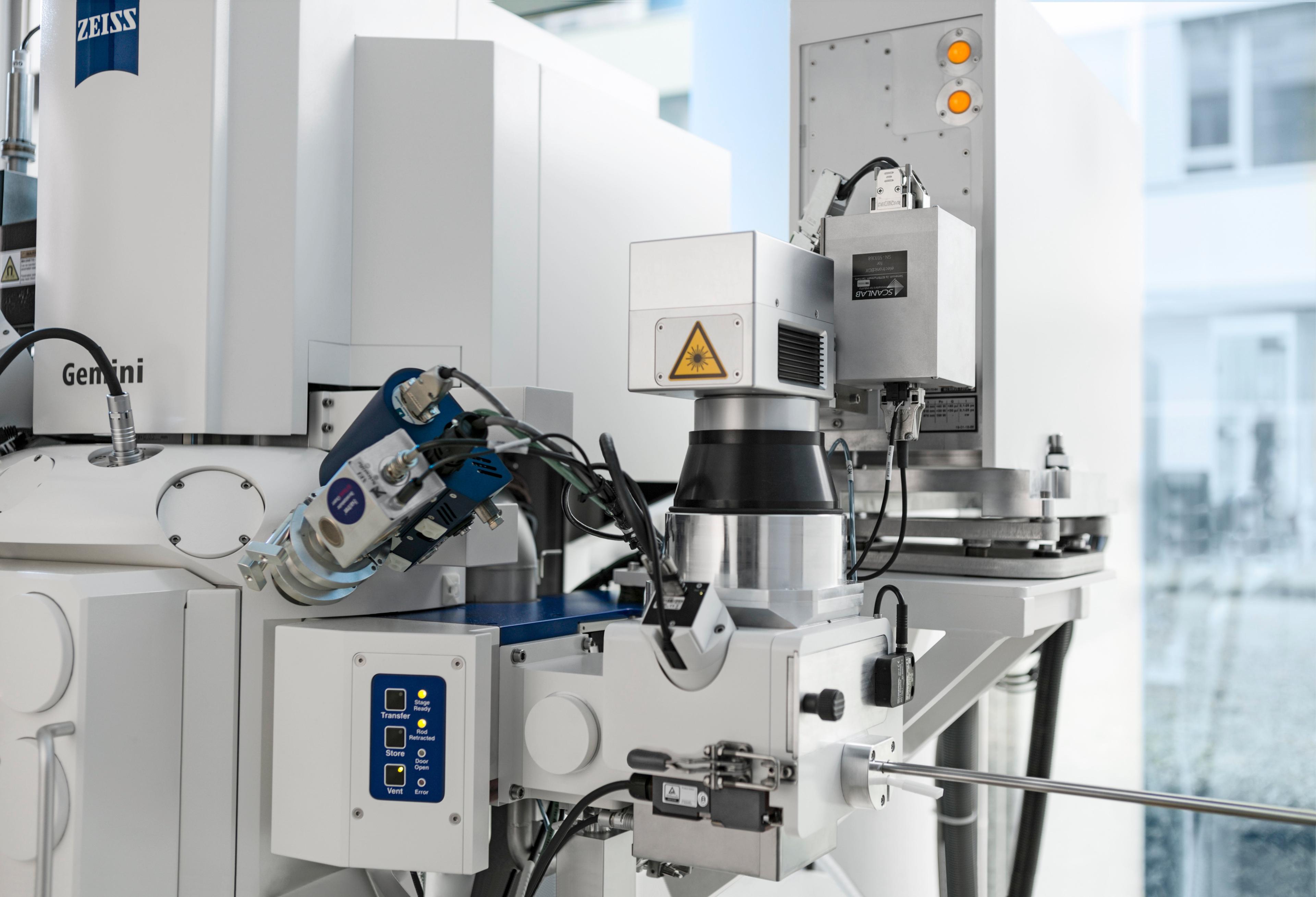
Microstructural Investigation of Austempered Ductile Iron (ADI) with "Shuttle & Find"
Austempered ductile iron (ADI) excels through strength, wear resistance and toughness – characteristics that make ADI the material of choice for use in combustion engines and gear box components. This means that safety aspects are also involved in addition to purely functional aspects. For this reason, changes in the ADI production process need to be monitored with respect to the material's characteristics and must be optimized systematically. For the micro- and nanoscopic analysis of the structure and precipitations, scientists typically use both light and electron microscopes. To date, however, there has been no possibility of relocating regions of interest without doubt when transferring the sample from the light to the electron microscope or vice versa. "Shuttle&Find" – the interface for correlative microscopy in materials analysis — offers an easy-to-use solution, enabling seamless integration of these two complementary technologies for the first time.
Biological Sample XRM Imaging from Selected Publications
The new field of 3D X-ray microscopy (XRM) brings dramatic resolution and contrast improvements to X-ray tomographic imaging of biological specimens for correlative studies and hierarchical structure investigations of hard and soft tissue. This document is a compendium sampling the vast diversity of biological samples and preparation methods published in literature by researchers utilizing ZEISS X-ray microscopes for their research.
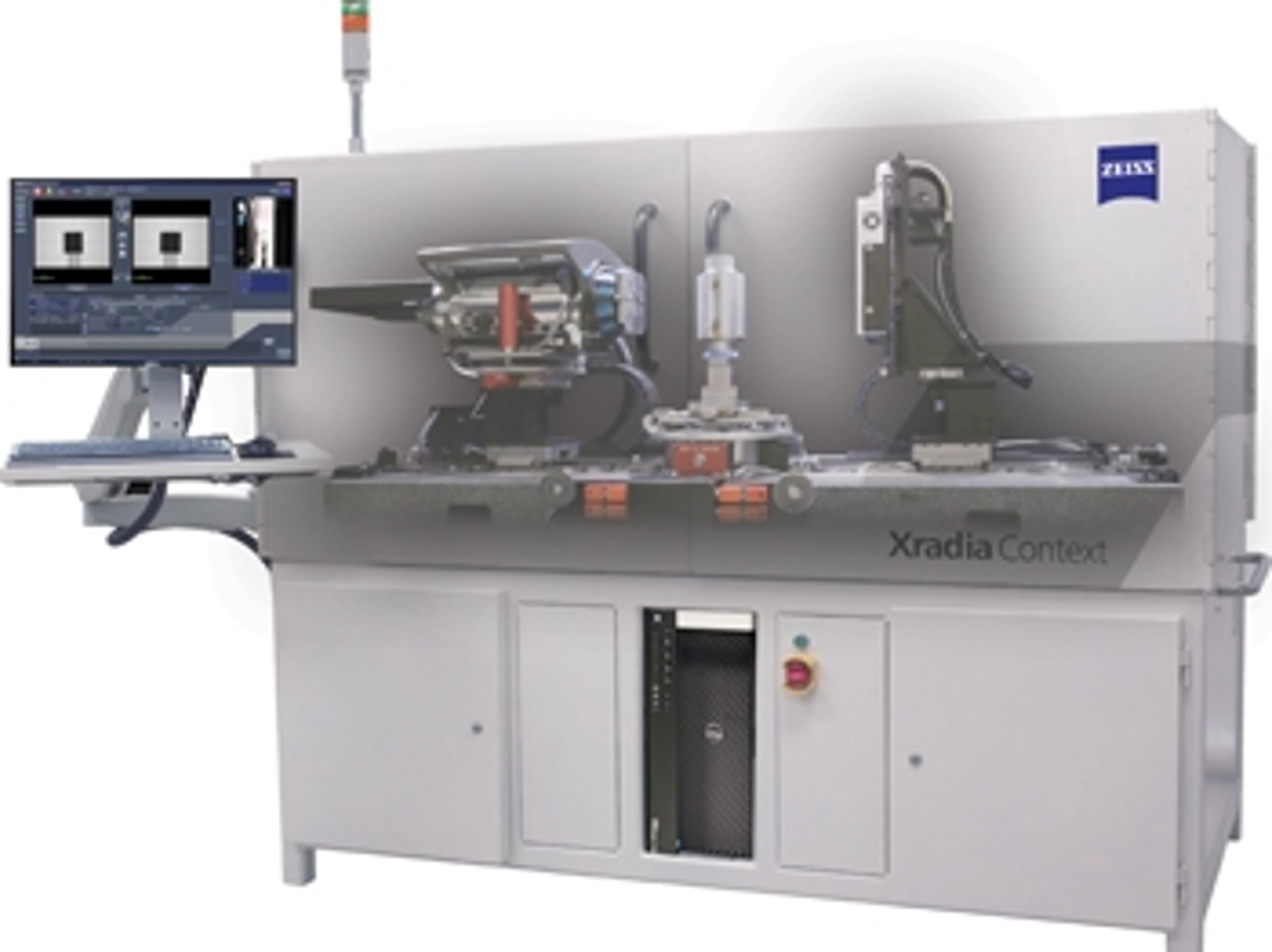
ZEISS Reveals New X-ray Imaging Instrument: ZEISS Xradia Context microCT
X-ray imaging instrument is built on renowned ZEISS Xradia platform and field-convertible to ZEISS Xradia Versa X-ray microscope