MMI CellScan
The most versatile Whole Slide Imaging system
The most versatile Whole Slide Imaging system

The most versatile Whole Slide Imaging system


The supplier does not provide quotations for this product through SelectScience. You can search for similar products in our Product Directory.
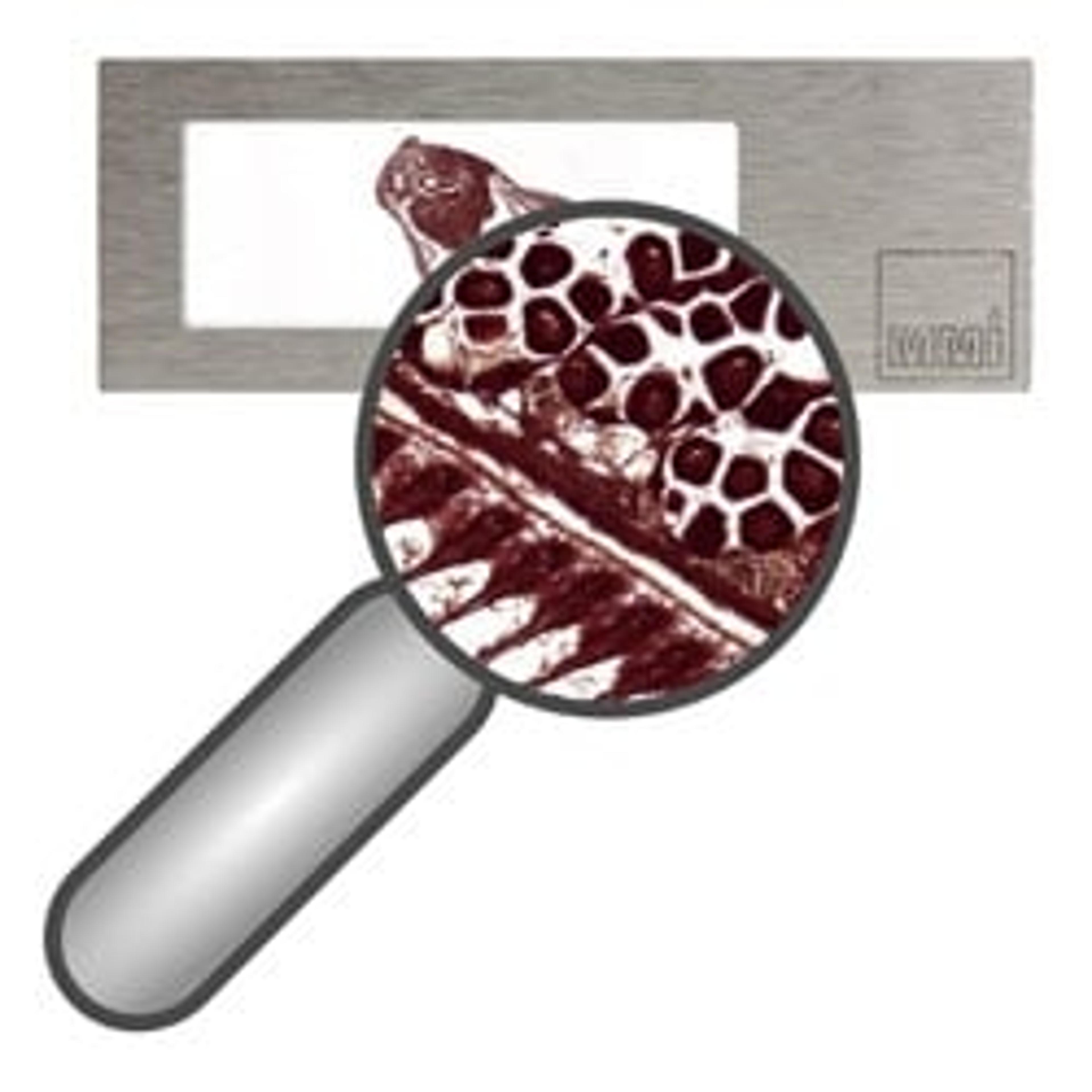
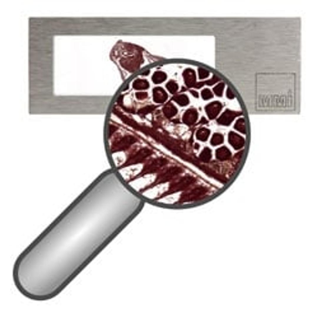
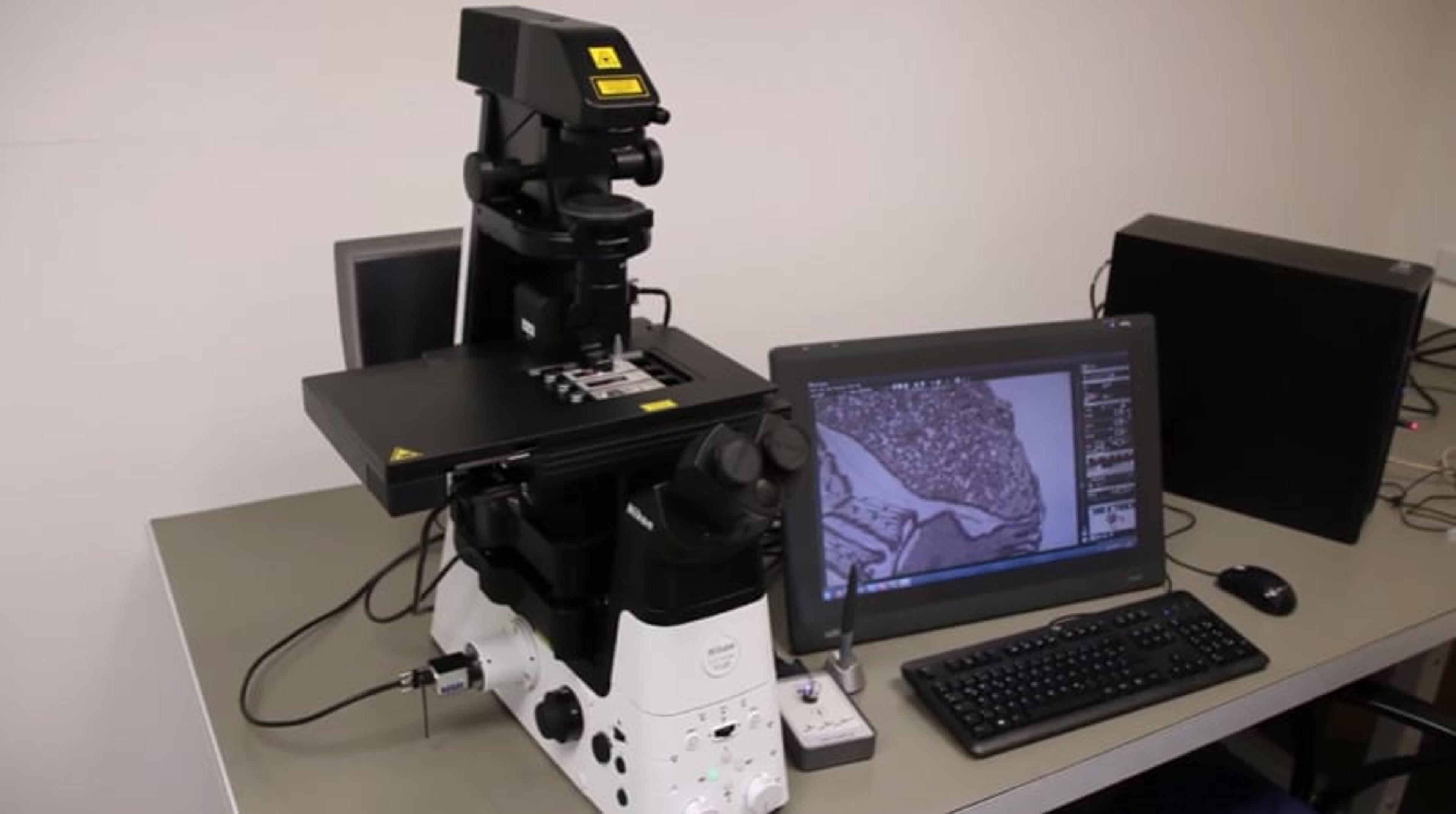
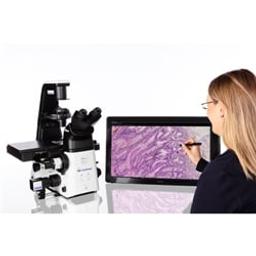
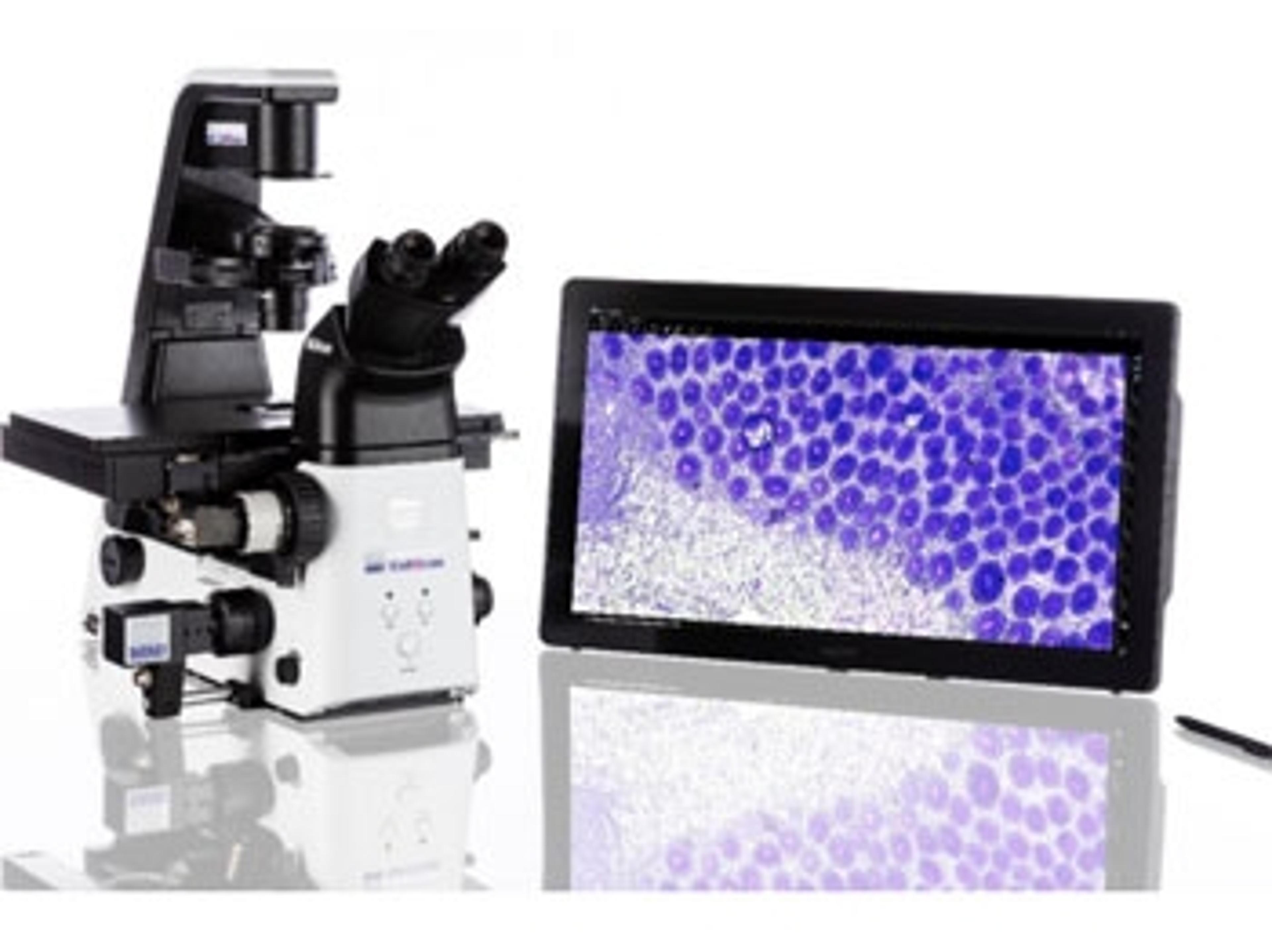
The MMI CellScan allows you to scan full resolution digital slides, to annotate your selection, and to cut cells by laser microdissection - all in one system!
In addition, the MMI CellScan be used as a stand-alone Slide Scanner supporting all functionalities of your microscope. Using the 20x objective of your microscope and our high sensitivity CMOS camera, an area of 15 mm x 15 mm is scanned in less than 75 seconds, which corresponds to 10 minutes for a full slide (75 mm x 25 mm). In addition, the individual focus map ensures that every single image is acquired in the optimal focus point. Thus, the whole slide image is perfectly in focus, even with uneven sample slides.
The image is saved as BigTIFF at full resolution for analysis, documentation and archiving. The digital slides can be accessed with most Slide Viewers. Within the CellScan software, annotations can be applied directly in the scanned slide. Since the system provides absolute positioning and accurate sample localization, the annotations can be precisely transferred to the CellCut to excise the selected cells. The MMI CellScan is compatible with all MMI products, with all microscopy objectives as well as with all standard microscopy imaging modules, including fluorescence.
Features & Benefits
- Combine full resolution whole slide scanning with absolute and uncompromised positioning for subsequent laser microdissection
- Pathologists and researchers reduce their hands-on time but contribute their valuable expertise using MMI CellScan’s easy to use on-screen annotation feature
- Improved handling of sensitive fluorescent samples: annotate your selection without prolonged exposure to photobleaching light
- Flexible and quick slide scanning is compatible with all MMI products, with all objectives, with most microscopes, and most Slide Viewers
- Document all steps of your work with full resolution images
- Ultra-fast data processing and slide viewing at any zoom level,... down to the single pixelThe MMI CellScan module fully integrates into your workflow with applications in Pathology, Oncology, Diagnostics, and many more
Applications
The MMI CellScan can be applied in various different research projects when it’s required to fully document and analyze your work at any time.
Typical applications include:
- Digital Pathology
- Pathology
- Oncology
In addition to the stand-alone functionality, the MMI CellScan module allows you to include Whole Slide Imaging to your CellCut Laser Microdissection workflow:
Scan your slides – full resolution in any magnification
Annotate your selection – directly into whole slide image
Excise your cells – precisely and accurately
MMI CellScan: Next Generation Laser Microdissection
The MMI CellScan is a new module to combine Whole Slide Imaging with Laser Microdissection. Thus, for the first time, researchers are now able to scan full resolution digital slides, to mark their selection and to precisely cut the specified cells by Laser Microdissection, all in one system! This video displays the workflow for Next Generation Laser Microdissection in real time.
"The most versatile whole slide imaging system": MMI launches MMI CellScan
Researchers are now able to scan full resolution digital slides in any magnification

Whole Slide Imaging meets Laser Microdissection
MMI launch their latest solution at the AACR meeting in Chicago: CellScan