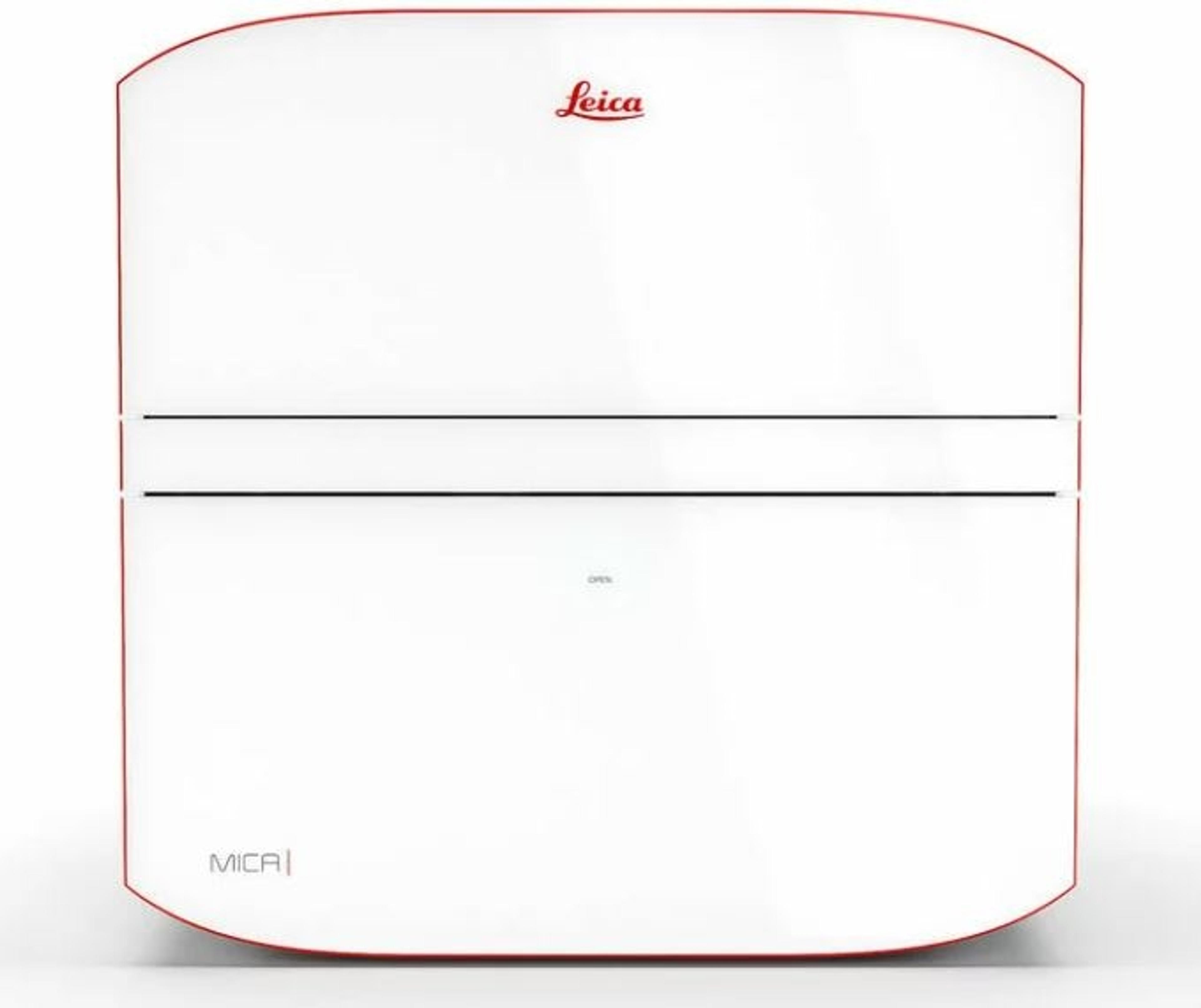
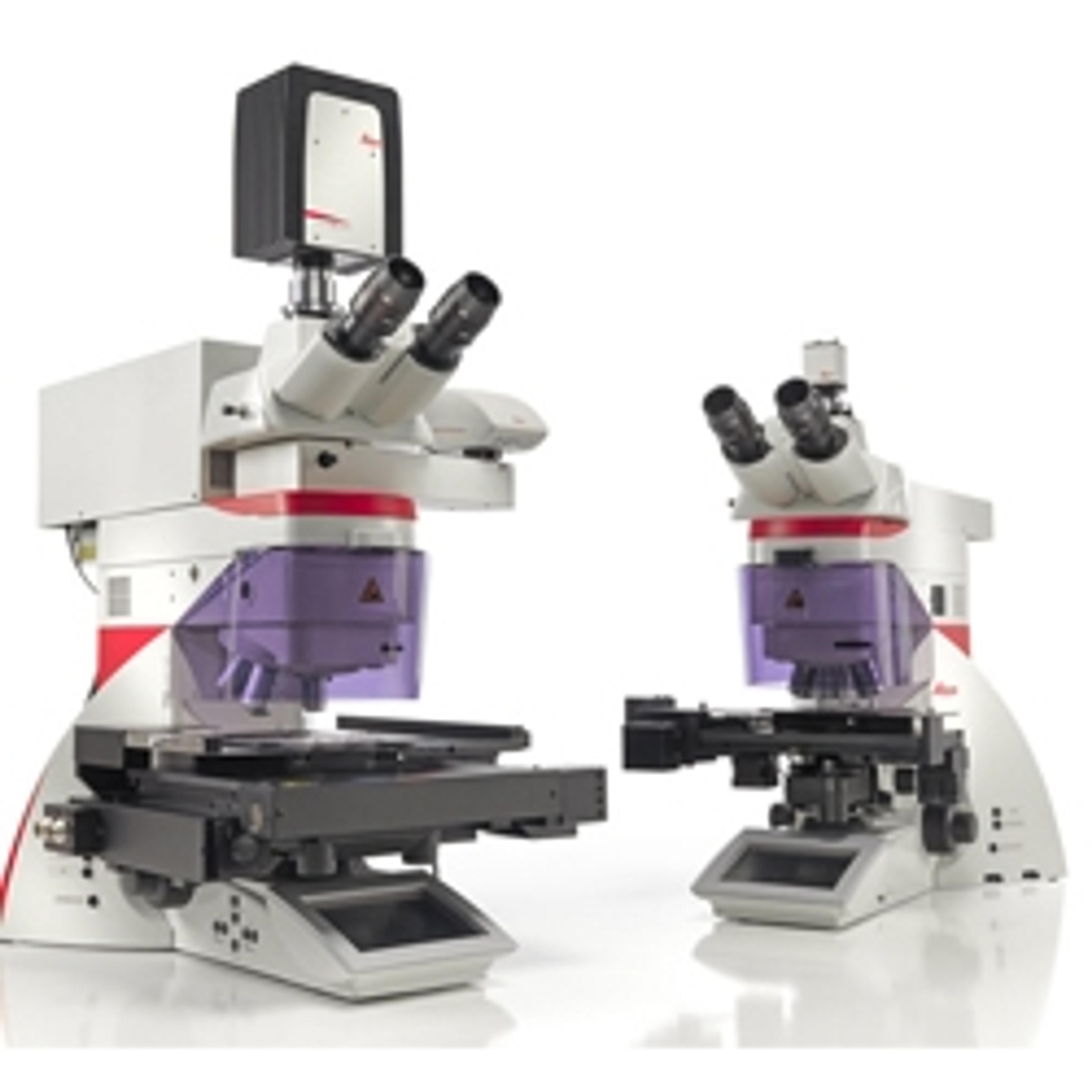
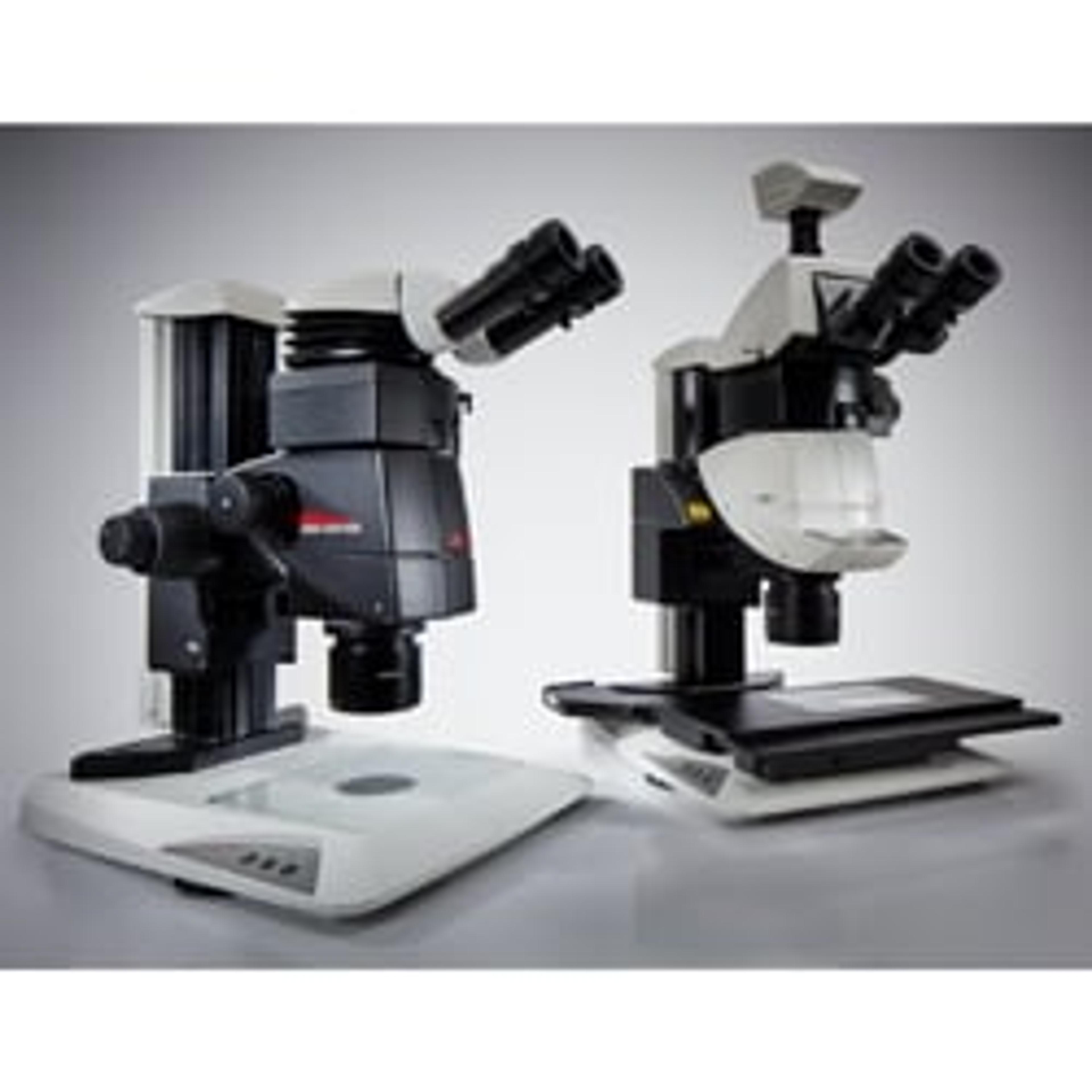
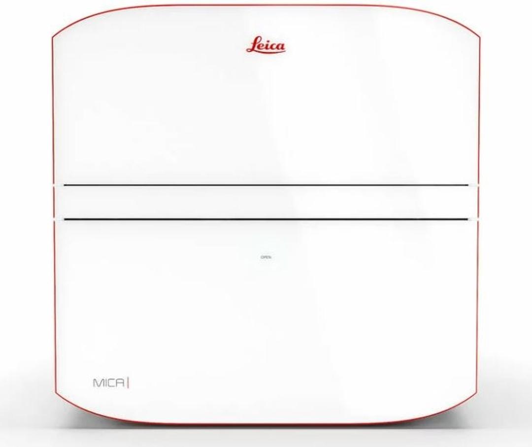
Mica - Imaging Microhub
Mica is the world’s first imaging Microhub. More than a highly automated microscope, it unites widefield and confocal imaging in a sample protecting, incubating environment. With the simple push of a button, you have everything you need - all in one place - to supercharge fluorescence imaging workflows and get meaningful scientific results faster.


Receive your quote directly from the manufacturer.
What if every scientist could access spatial information?
Mica empowers every researcher to move from set up to beautifully visualized results and analysis efficiently, accurately, and confidently. Now you can focus on your science, not figuring out your microscope.
Access for all
Everyone can now leverage microscopy to make more discoveries. Mica provides a clear sample overview and allows you to easily change observation conditions with just a few clicks.
- 85% fewer steps to the first image
- 33% less time to the first image
- 50% of the training time
No constraints
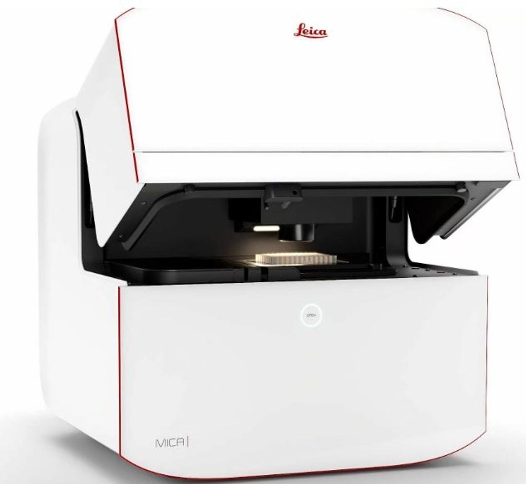
The Microhub has everything you need to enable discoveries, unified in one easy-to-use system.
- Simultaneously capture all 4 labels or different structures in a single acquisition for both widefield and confocal, without ever moving your sample. This overcomes the spatiotemporal mismatch between labels of moving objects during sequential acquisition.
- Select from multiple imaging modalities, including widefield, confocal, THUNDER imaging, LIGHTNING, Z-stacks, time-lapse and more. Seamlessly move from fast overview to high resolution when required by your experiment
- Achieve physiological-like conditions throughout your experiments
Radically simplified workflows
Intelligent automation and AI-supported analysis enables greater efficiency and a faster track to publication.
- Reduce over 60% of process steps through system intelligence
- Reduce time and effort from sample to insight by simplifying your entire workflow
- Enable 100% reproducibility and repeatability throughout your experiment
Key applications include fluorescence multi-well plate assays, 3D tissue imaging and long-term timelapses.

Considerations for multiplex live cell imaging
Live cell imaging experiments are key to understanding dynamic processes. They allow us to visually record cells in their living state, without artifacts that could be introduced by fixation or termination of the various living processes. With live cell imaging, it is possible to observe dynamic processes in cells, tissues, or whole organisms in their natural environment, rather than just obtaining endpoint images and data. For example, you can study co-localization of cells or proteins to see if the different targets are positioned close to one another and observe how they interact. In this application eBook from Leica Microsystems, explore the use of multicolor, or multiplex, microscopy for live cell imaging, some key aspects of setting up a successful multicolor live cell experiment, and how to avoid some of the major pitfalls.

The power of spatial biology: A microscopy guide
Location is key to understanding biological mechanisms, from the inner workings of subcellular components to how cells form and interact across normal and diseased tissues.
Many application areas are seeing a growing trend toward spatial biology, which uses transcriptomics, imaging, and other approaches to put dissociated cellular information into spatial context.
In this free eBook, explore key microscopy techniques, across the spatial biology workflow, including:
- Multiplexing
- Laser microdissection
- Removing the blur to observe fine details
- Super-resolution microscopy
- Ultra-structural context
- AI-enabled spatial analysis
Plus, we look "under the microscope" at how these can be applied to study a wide range of spatial biology questions and gain expert insight into the imaging solutions designed to meet a variety of different research needs and priorities.
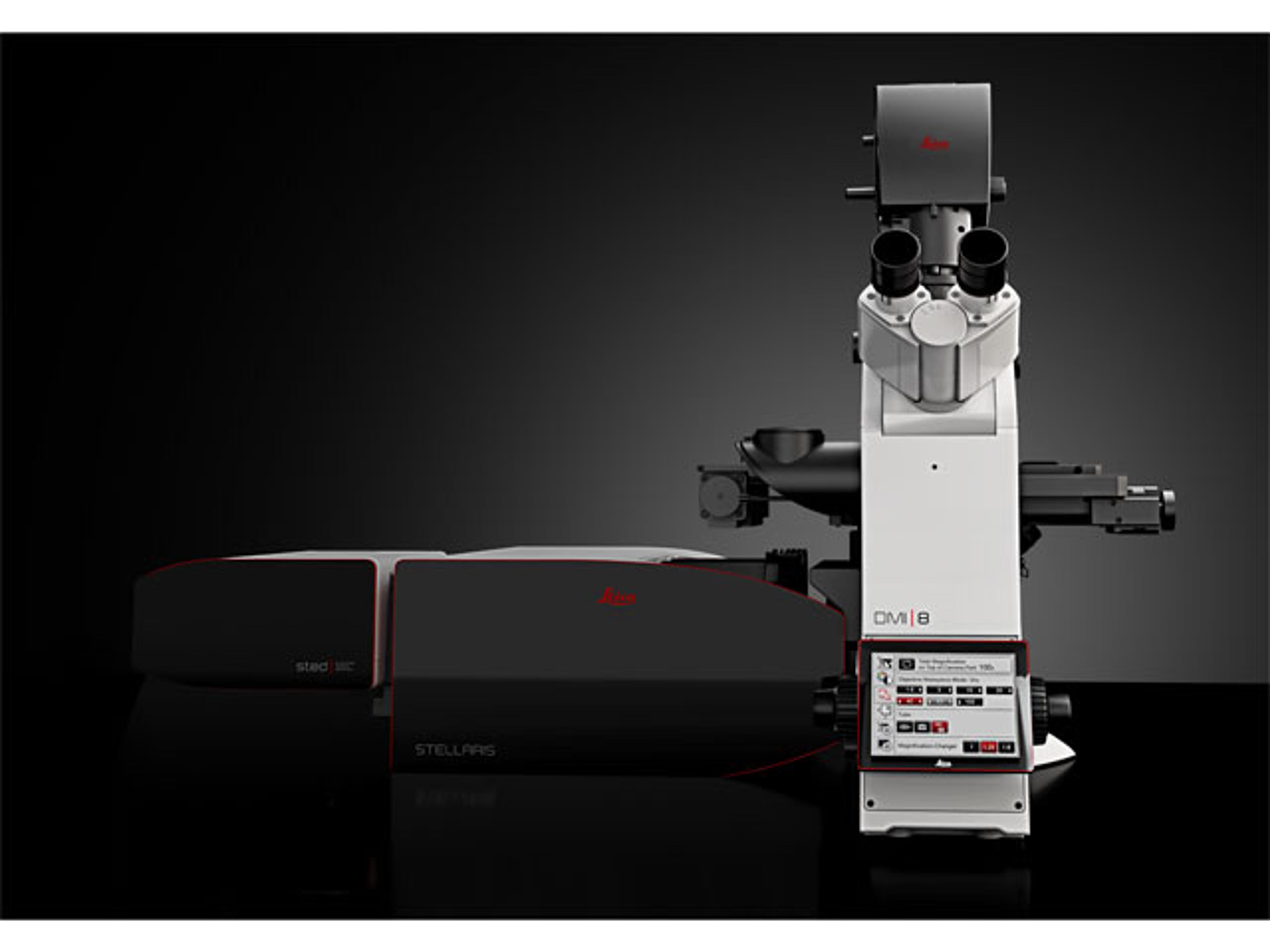
Step into the future of microscopy: Latest innovations from Leica Microsystems
We speak with Leica Microsystems to gain a sneak peek at their latest microscopy products on display at analytica 2022. From the ‘world’s first imaging Microhub’ to cryo-immobilization of aqueous samples, discover the key features and research applications from the product experts themselves.

World's first Microhub makes spatial context accessible for all
New Mica imaging platform from Leica Microsystems brings previously inaccessible experiments to all life science researchers