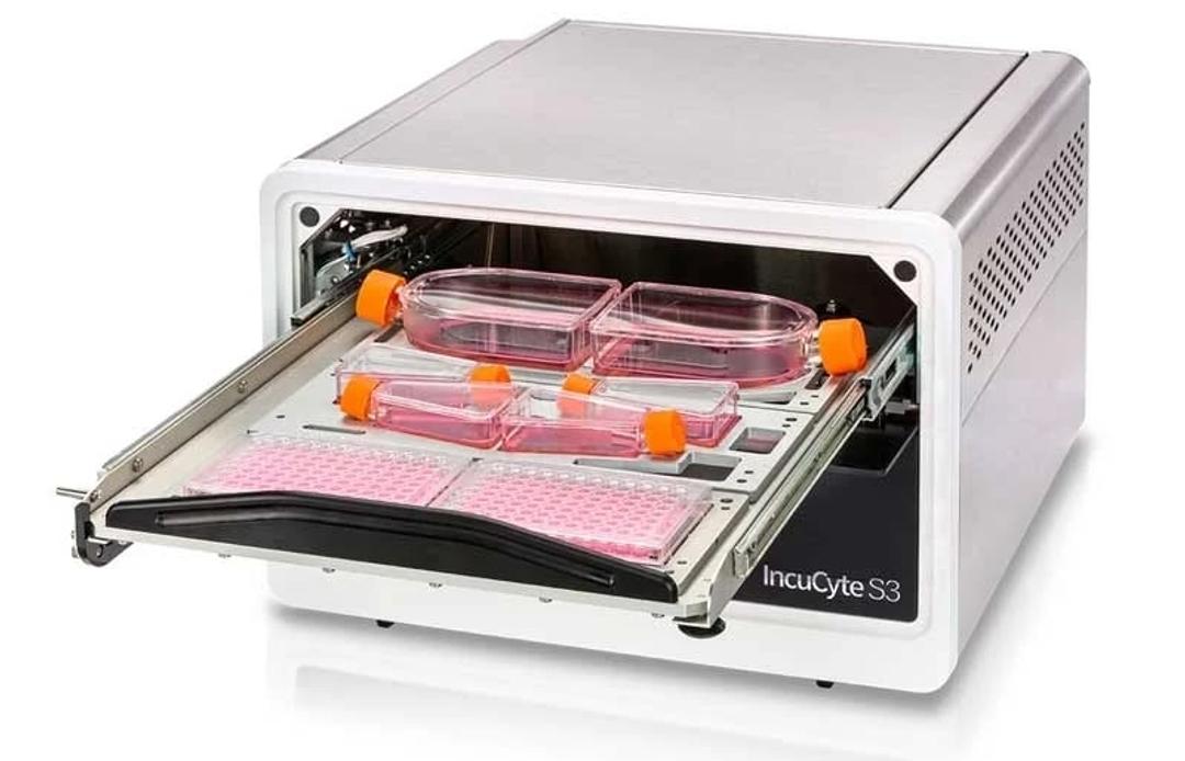
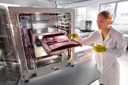
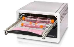
Incucyte® S3 Live-Cell Analysis System
Our flagship, supports the workflow and workload of larger laboratories.


Receive your quote directly from the manufacturer.
Fluorescence cell count
Virology assays
Simply love this microscope. It is the best that I ever used for fluorescence. The fluorescence cell count is the best!
Review Date: 10 Mar 2025 | Sartorius Group
Extremely great results. This is what we expect for and high quality for publication
Analyze Functional assay using plate reader in Incucyte.
We have been using the Incucyte machine for over 3 years now and I have to say that this machine by far is the best and easiest to use for functional assay as well as for screening of libraries. The quality of the output is great and I would surely recommend this to my collaborators and networking conference attendees.
Review Date: 23 Feb 2022 | Sartorius Group
Great potential, but ours was very sub-par for a $100+K machine. Poor reliability.
Analyze cell growth and/or differentiation morphology and markers
Our S3 had service-requiring issues almost immediately. Three total calls within the first 12 months, during a period in my new lab where it was only being occasionally used. Twice for the mechanics of the drawer, and once when they had to replace the motherboard. Then, right after our warranty expired, the machine needed the same exact repair as our second service trip, which would cost $7000. For a $100K machine, this level of service need is completely unacceptable. I cannot recommend this machine or this company to colleagues, which is a shame, because the promise of what the IncuCyte could do is very high.
Review Date: 10 Feb 2021 | Sartorius Group
Great results, we performed drugs testing and viability assay on this platform.
Cell biology
Incucyte S3 imaging system is a user free, reliable, ready to use, excellent instrument for cell biology research.
Review Date: 24 Dec 2020 | Sartorius Group
Can’t live without this equipment!
Neurobiology
Our lab has been using Incucyte for many years. It is particularly useful for studying microglia cells. We mainly use Incucyte to track cell proliferation and cell death, as well as monitor microglial phagocytic activity. The software is easy to use, and the machine can accommodate multiple users at the same time. It is essential to our research.
Review Date: 2 Dec 2019 | Sartorius Group
Workhorse imaging!
Live imaging of drug response in neurons
The S3 is a perfect imager that produces high quality images even in phase contrast mode. We regularly are screening for neuro protective compounds in our iPSC model system where we can gain invalueable real-time data of response with images and graphs of 5 phenotypes of neurite changes. We can set up the images and times ahead of time with analysis in real-time and come back days later with super graphs of data. It is a highly-valued component of our research work.
Review Date: 2 Oct 2019 | Sartorius Group
Powerful and easy to use
Cell cycle, drug response
Intuitive controls and powerful software. Can get reproducible data easily. Companion kits make designing experiments easy.
Review Date: 25 Jul 2019 | Sartorius Group
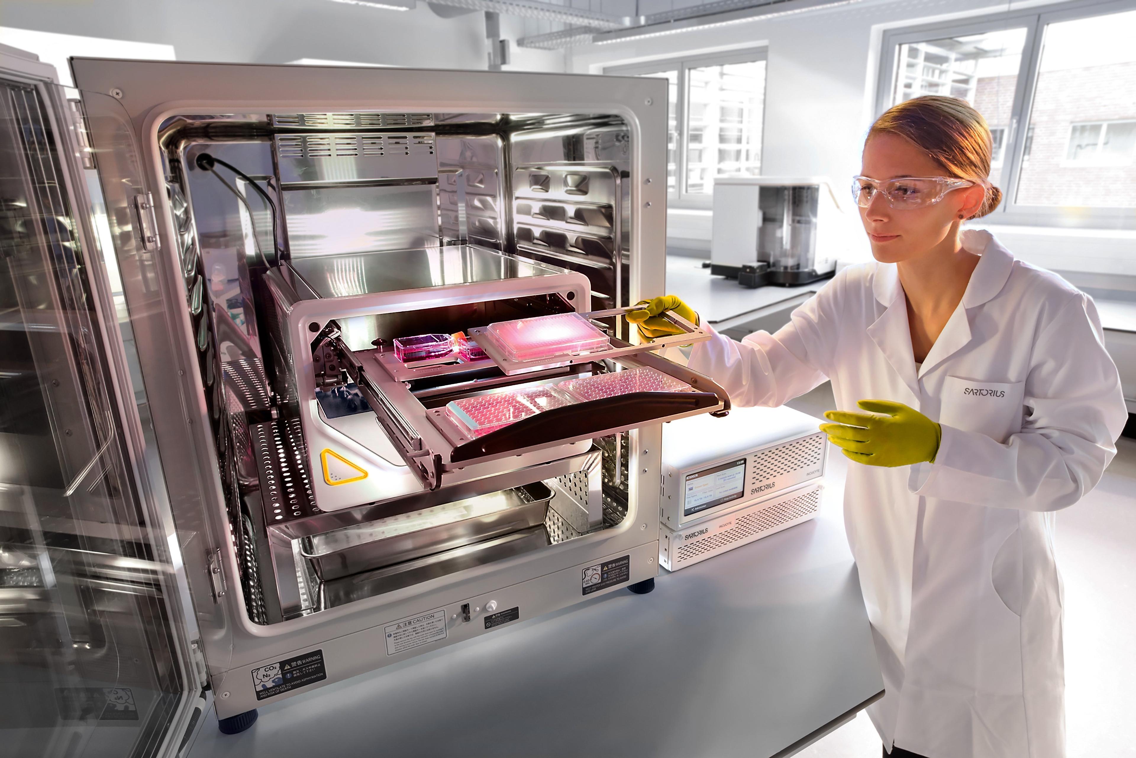
The Incucyte® S3 Live-Cell Analysis System automatically acquires and analyzes images around the clock, enabling you to derive deeper and more physiologically relevant information about your cells, plus real-time kinetic data — without ever removing your cells from the incubator.
Change can happen in an instant. Whether simply assaying cell health or more complex processes like migration, invasion, or immune cell killing, see what your cells are doing and when they do it. The Incucyte® S3 instrument, proprietary assays and reagents provide you with the ability to gain new insights into biological processes via real-time, quantitative analysis of live cells.
Conventional approaches to cell analysis only capture a single time point, enabling only single-point and end-point measurements, and cells are perturbed or destroyed as part of the assay process. The Incucyte® system offers the advantage of performing live-cell analysis without ever having to displace cells or disrupt their surroundings. The system automatically and continually collects and analyzes images throughout the course of an experiment while cells remain unperturbed in a physiologically relevant environment. Furthermore, the Incucyte® S3 accommodates multiple users and applications seamlessly and combines information-rich, image-based analysis with the convenience and throughput of microplate assays.
Incucyte technology is featured in over 3,000 peer-reviewed publications in journals such as Nature, Chemical Society Reviews, Nature Medicine, and Nature Genetics, among many others. The S3 offers a completely reimagined user interface, enabling even first-time users to set-up an experiment and begin acquiring images within minutes. Image viewing, analysis and graphing is similarly streamlined – images from a 96-well experiment can be viewed simultaneously, then converted into movies, metrics, and corresponding publication- and presentation-ready graphs with just a few clicks. In addition to improving productivity for the individual user, the Incucyte® S3 empowers an entire research team. Multiple users can run multiple applications on the Incucyte® in parallel, reducing time spent waiting for an instrument to be available.
In addition, data is accessible remotely to any user via unlimited, free networked licenses. The Incucyte® and its unique features provide a powerful platform that enables researchers to devise new experiments not previously thought possible. In addition to the Incucyte® platform, Incucyte® proprietary reagents and analysis software are also available which enable researchers to perform real-time, quantitative live cell analysis and evaluate a wide variety of cellular processes over time.
Never miss powerful insights again, with the Incucyte® S3 live-cell analysis system, reagents, and consumables.
Key Features of the Incucyte® S3 Live-Cell Analysis System:
Ask new questions
- Devise new experiments not previously possible
- Conduct routine monitoring and get answers to unique scientific questions with kinetic, image-based measurements
Get new answers
- Never miss a data point with real-time continuous analysis
- Profile cell-specific and time-dependent biological activity
- Visualize and validate results with images and movies
Improve productivity
- Enjoy walk-away convenience as images are automatically acquired and analyzed
- Multiplex measurements in 96- and 384-well assay formats
- Accommodate multiple users and applications simultaneously
Protect your cells
- Perform analysis without ever removing cells from the incubator or disturbing cultures
- Maintain cell health and morphology with non-perturbing reagent formulations
Applications:
- Proliferation (confluence and cell counts)
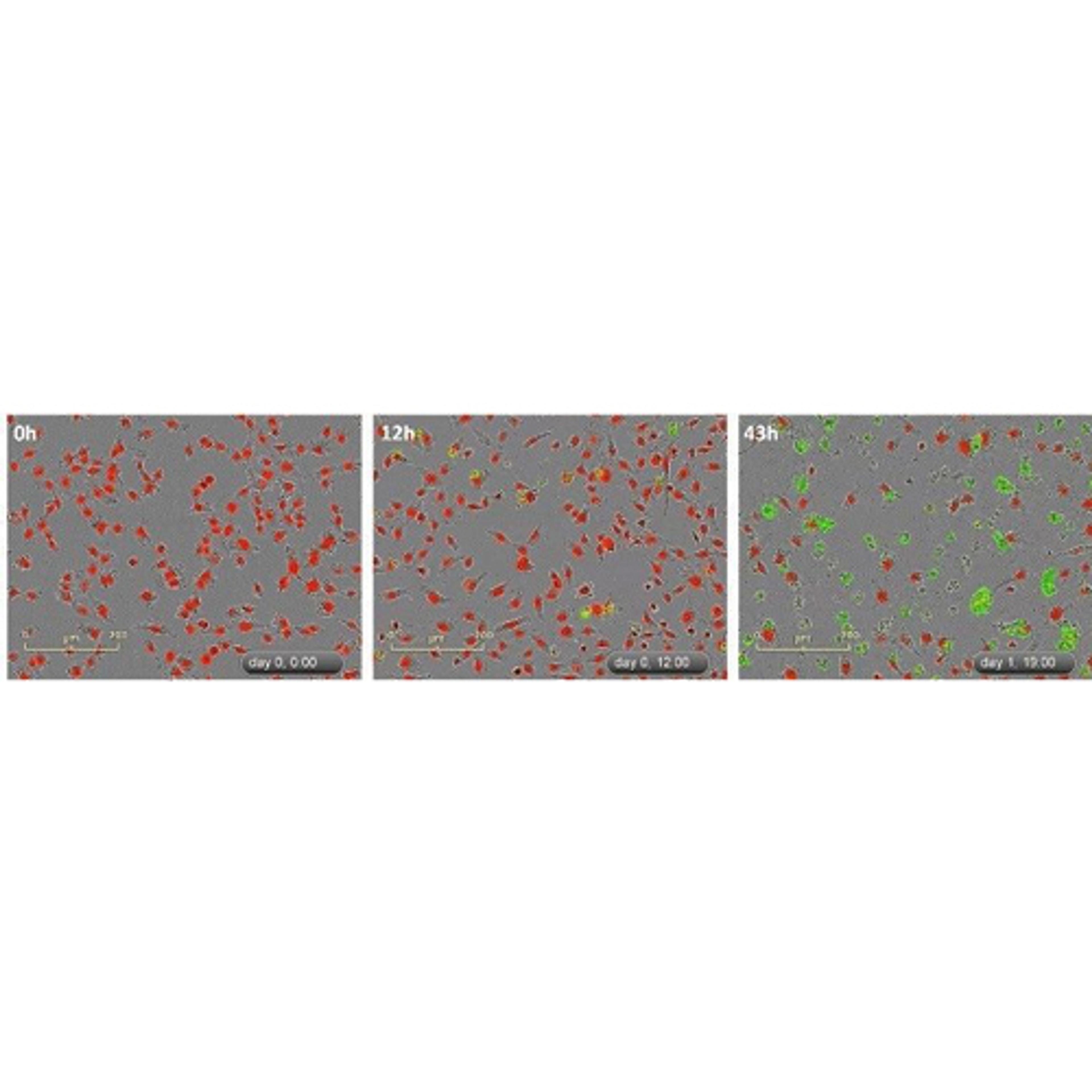
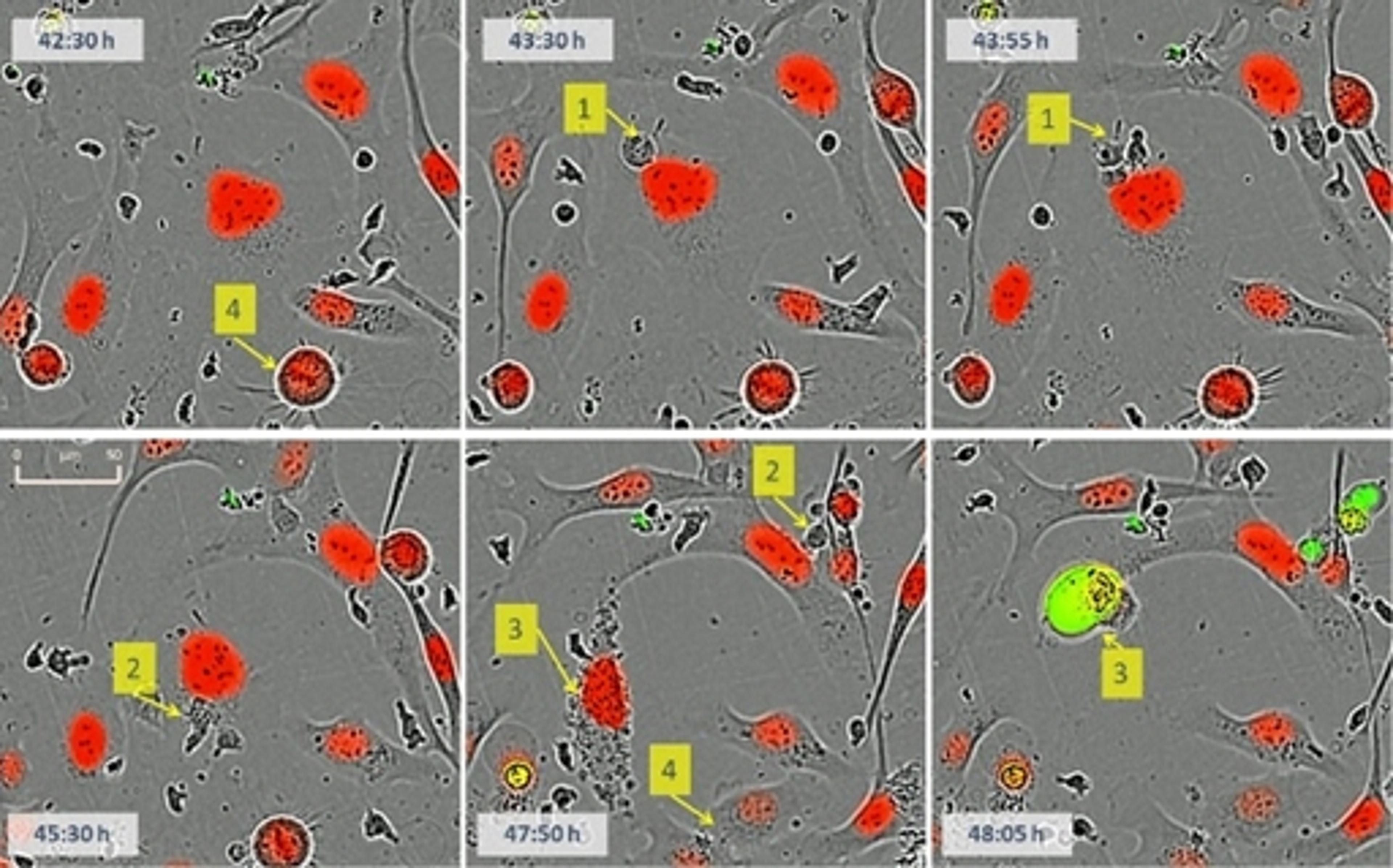
- Apoptosis (caspase 3/7 for live-cell imaging)
- Cytotoxicity
- Dilution cloning (whole-well imaging)
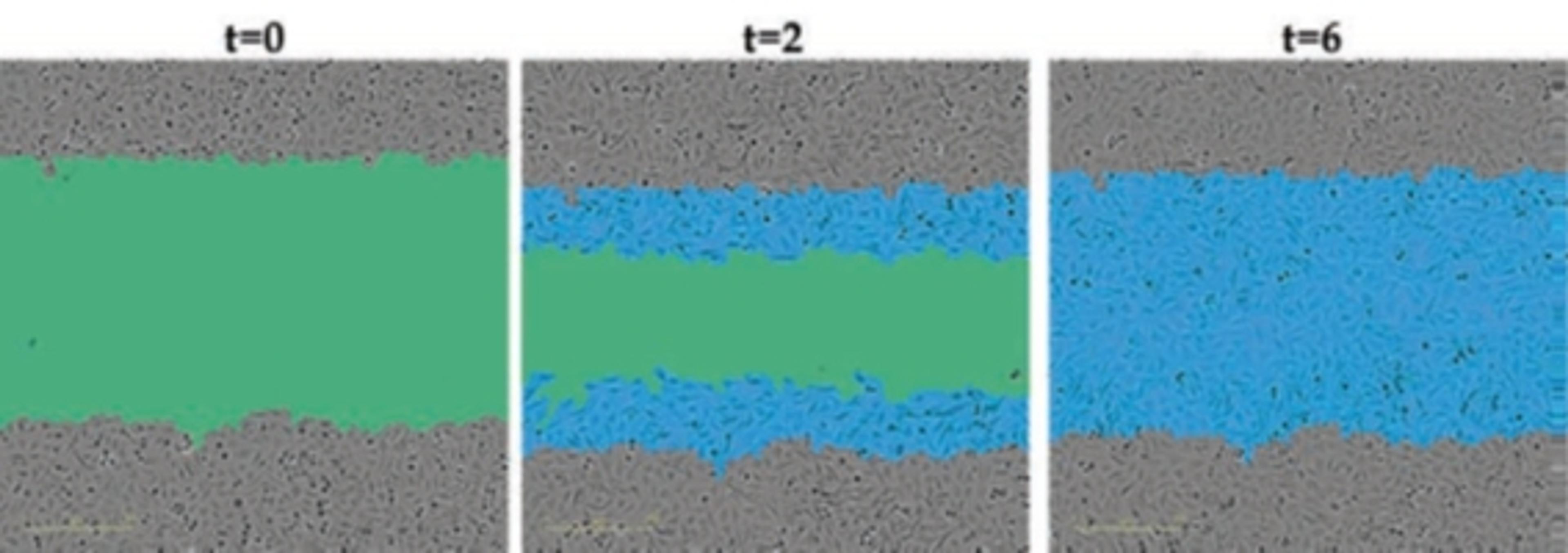
- Migration / Invasion
- Stem cell monitoring and reprogramming
- 3D-Spheroids Angiogenesis
- Neurite outgrowth and dynamics
- Neuronal Activity
- Reporter gene expression
- Viral studies
- Immune response – Immune cell killing
- Antibody Internailization
- NETosis
- Phagocytosis
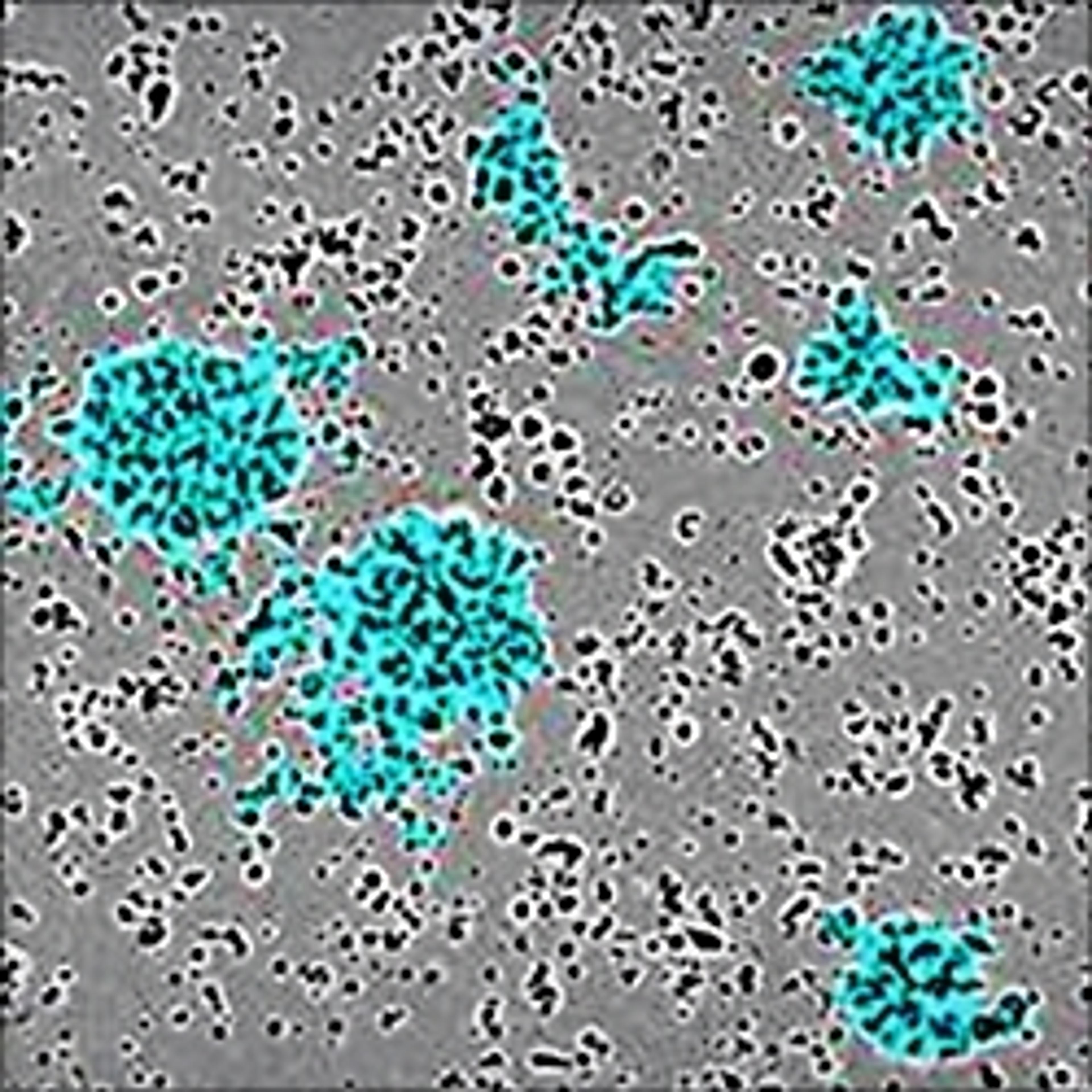
- Immune cell clustering
- Immunocytochmistry
- Cell-by-Cell Analysis
- Cell culture (& QC)

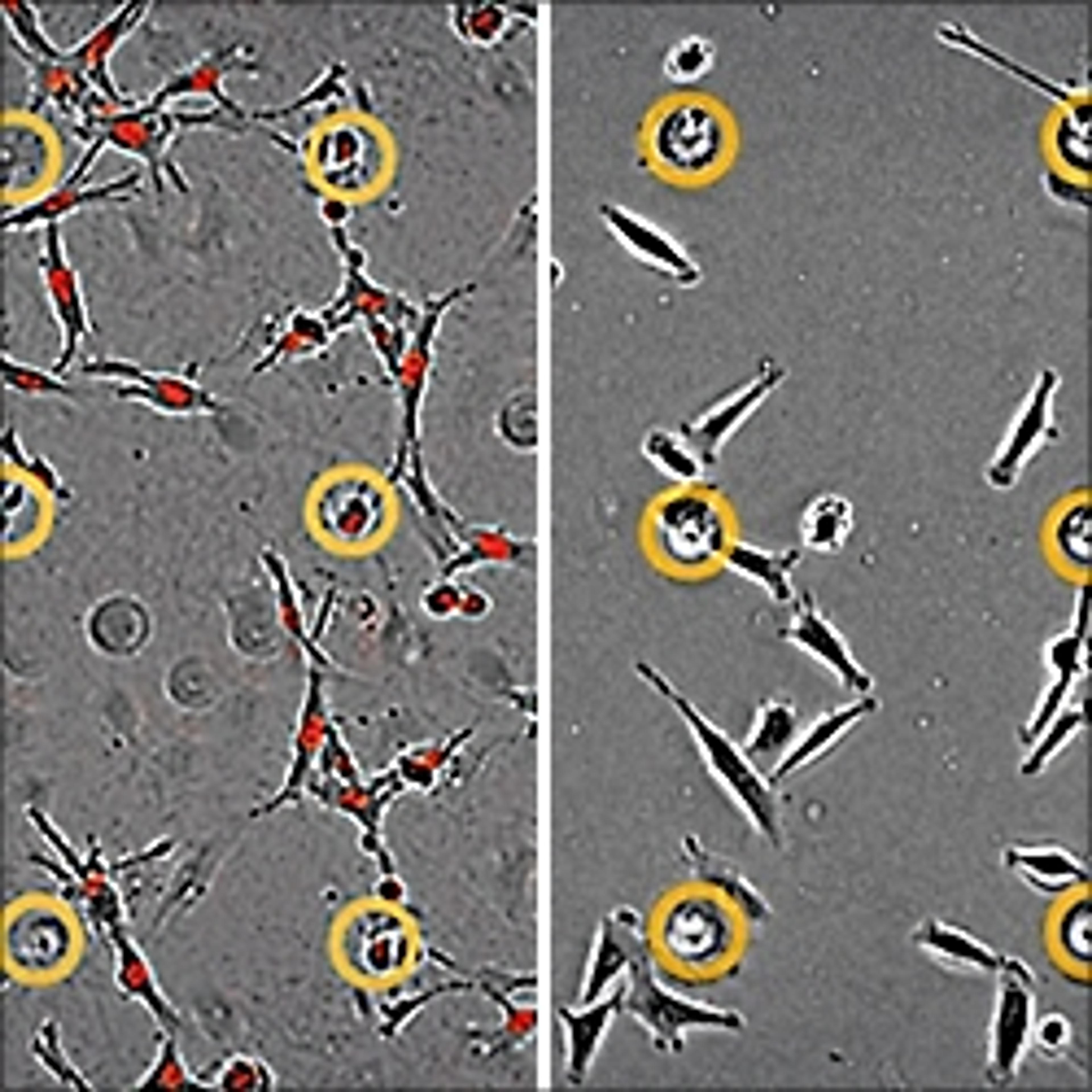
iPSC-derived motor neurons and microglia from ALS background display disease phenotype
Amyotrophic lateral sclerosis (ALS) is a neurodegenerative disorder caused by the degeneration of motor neurons, and ultimately results in death. In this whitepaper from Sartorius, explore how fibroblasts taken from a healthy individual and an ALS patient carrying a C9orf72 hexanucleotide expansion were reprogrammed to induced pluripotent stem cells, and then differentiated to motor neurons and microglia alongside controls. Results have displayed that this can be used as a robust model for C9orf72 ALS research and drug discovery.

Sartorius presents recent developments in COVID virology
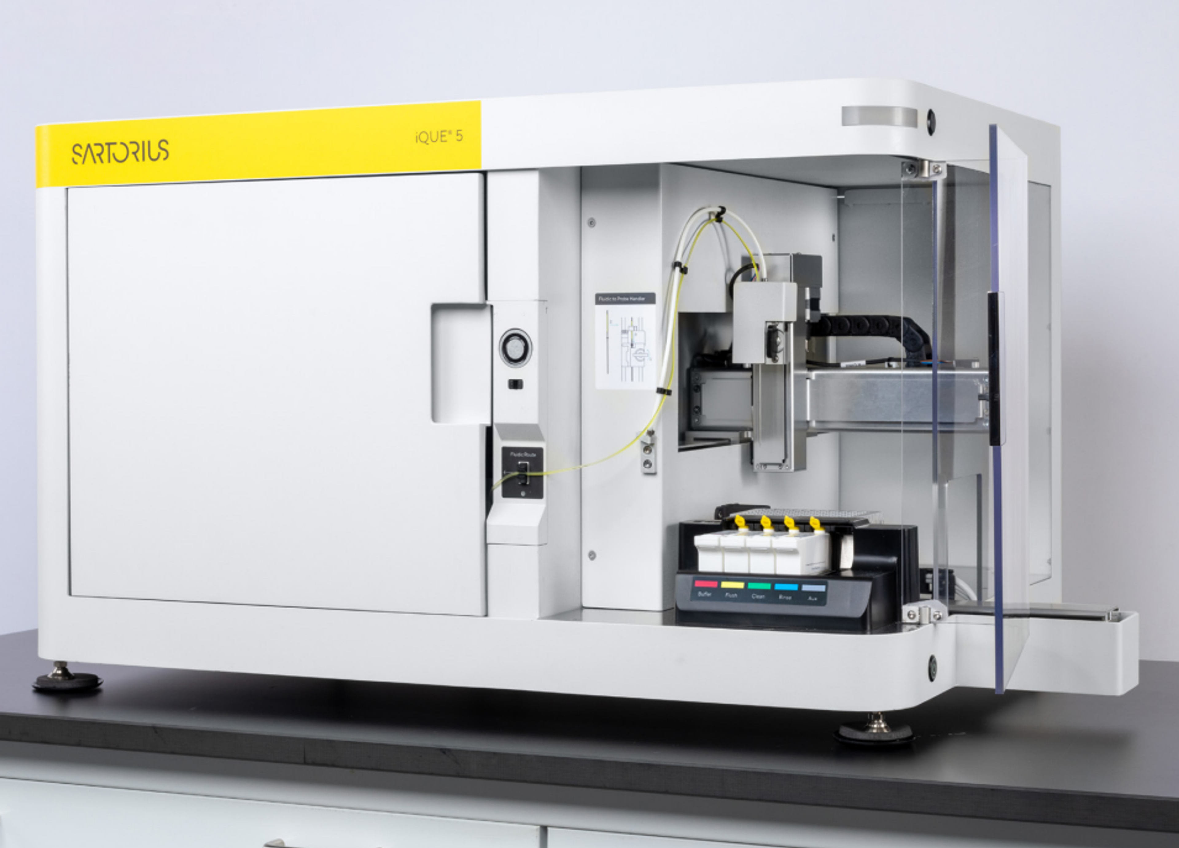
In this application note, Sartorius presents a series of four articles summarising recent developments in Covid virology. Download the application note below to discover how researchers are using the iQue Qbeads® DevSceen SAv bead kit, iQue® Screener Plus, and Incucyte® S3 Live-Cell Analysis system for their virology research.
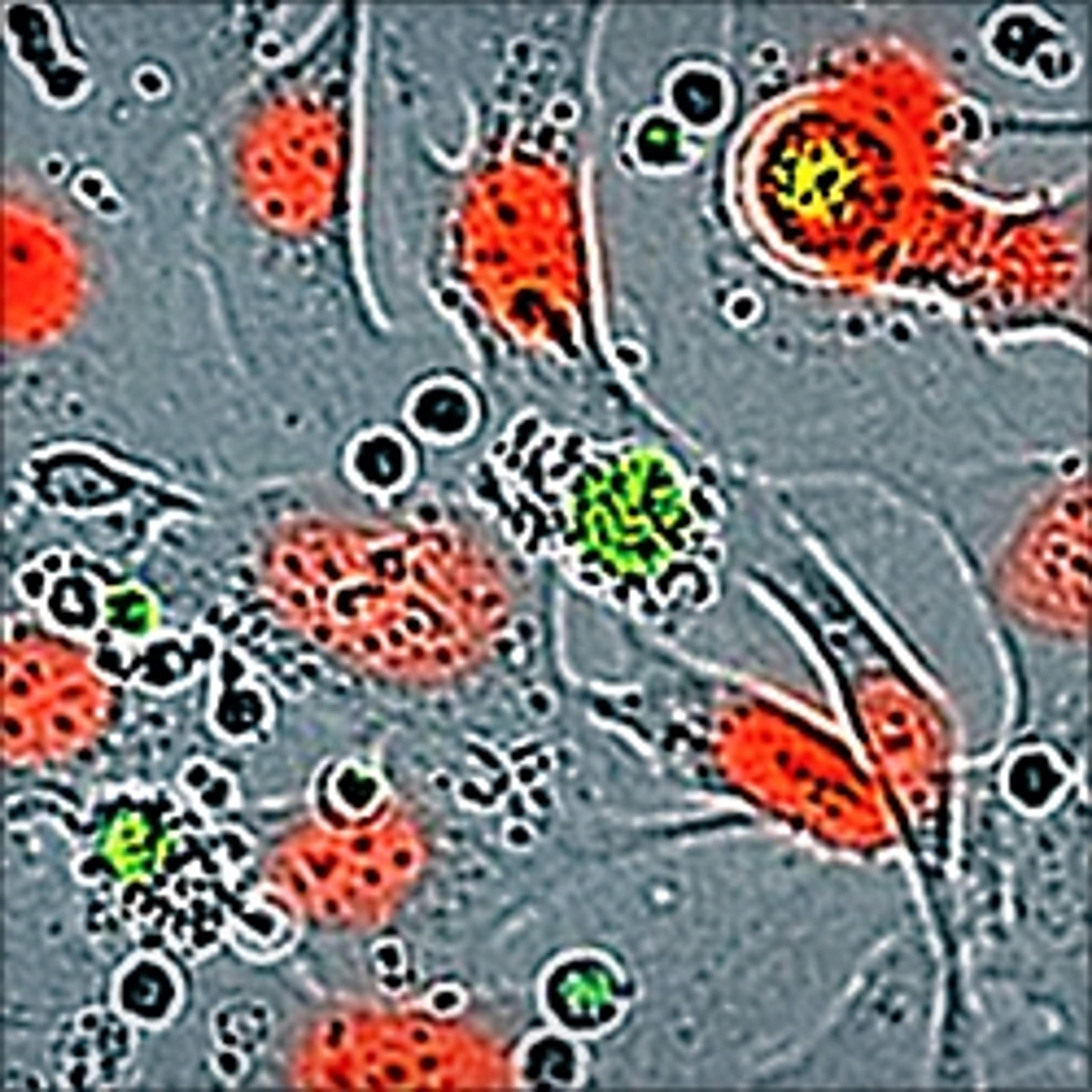
Quantitative, live-cell kinetic analysis of microglial function and morphology
In this whitepaper, Sartorius explains how to characterize Rat Primary, iPSC-derived, and immortalized microglia. Sartorius also presents data evaluating the ability of these cells to phagocytose pHrodo® labeled E. coli bioparticles and apoptotic Neuro-2A (N2A) cells using a quantitative, live-cell imaging approach with the Incucyte® S3 Live-Cell Analysis System for Neuroscience.
Long-term live cell visualization and quantification of neuronal activity
In this application note, Sartorius presents data to support the use of the the IncuCyte S3 for Neuroscience to characterize and refine different neuronal phenotypes and model their function in vitro. Here, Sartorius explains how this single live-cell imaging platform allows users to assess calcium flux kinetics and continuously monitor the morphology of neuronal populations, using non-perturbing reagents, validated protocols that are cell-sparing, and a built-in, guided interface for non-experts, as provided by the IncuCyte S3 Neuronal Activity Analysis Software Module.
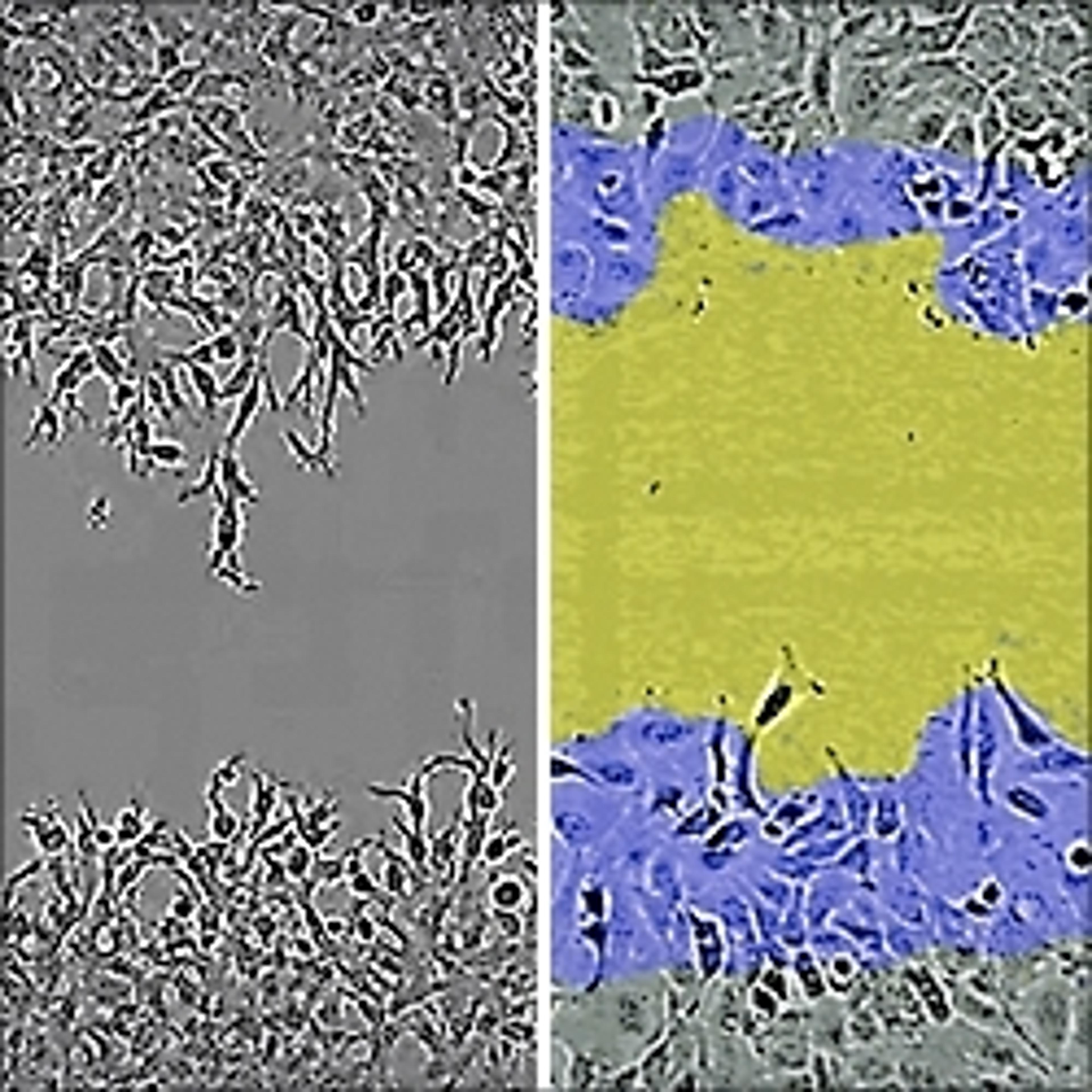
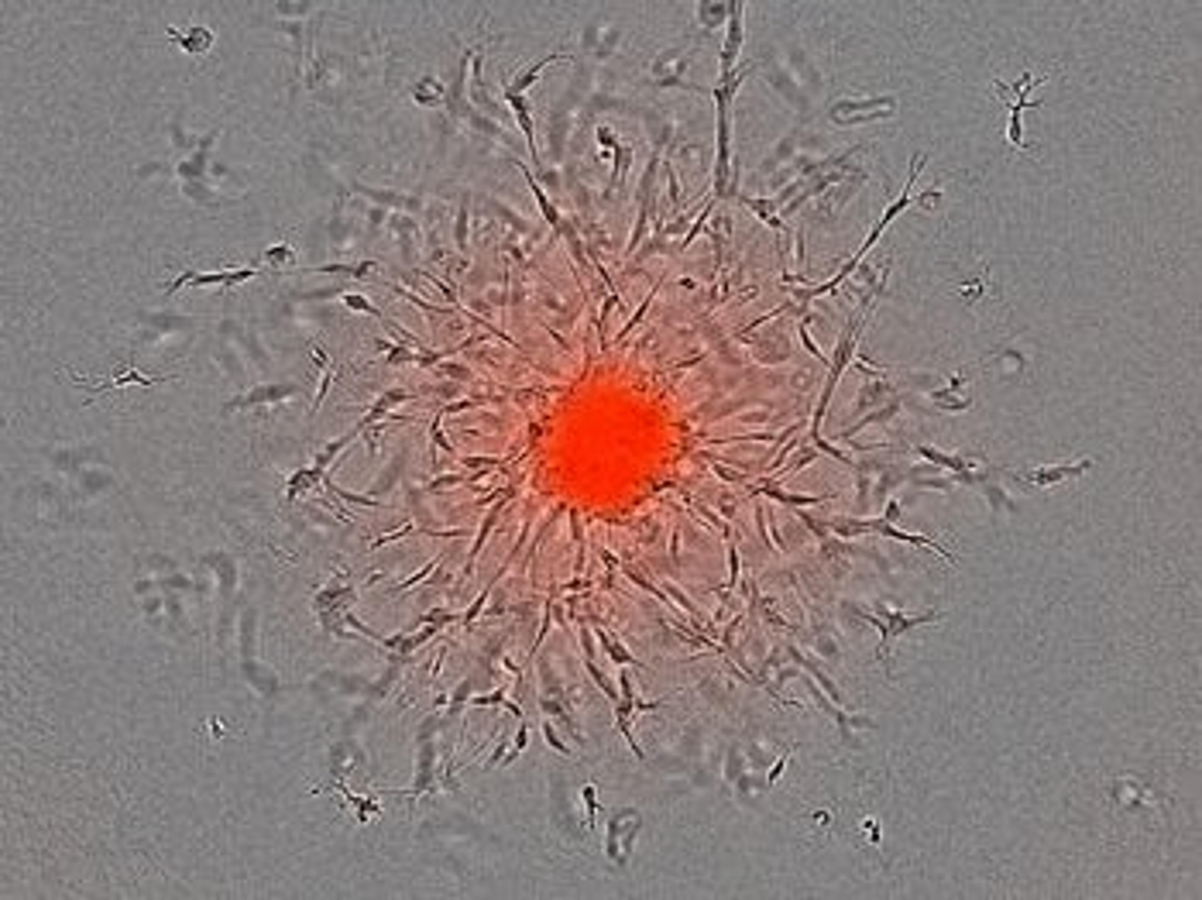
Real-time live-cell analysis of 3D tumor spheroid invasion
In this poster, Sartorius describes a simple 3D tumor spheroid invasion model to recapitulate key features of the metastatic phenotype and demonstrates use of live-cell imaging to acquire and quantify spheroid invasion.

Live-cell imaging for label-free toxicology analysis of hepatic organoids
Conventional 2D cultures of primary hepatocytes or established hepatic cell lines do not provide a true representation of the cellular mechanisms observed in vivo. In this webinar we will demonstrate an efficient and consistent protocol developed by STEMCELL Technologies to extract, grow, and differentiate liver cells in a complex 3D organoid culture system. These organoids more closely resemble the human liver and can be used in standard and high throughput assays to accommodate both academic and industry research needs.
This webinar will demonstrate the use of hepatic organoid cultures to validate a label-free live-cell toxicology screen using a powerful and flexible Incucyte® Live-Cell Analysis System.
Key learning objectives
- Organoid cultures – from tissue to mature 3D organoid culture and their downstream analysis
- Real-time kinetic analysis of organoids
- Label-free live-cell toxicology screening
- Key challenges of compound validations and data interpretation to improve the efficacy of drug discovery workflow
Who should attend?
Both academic and industry researchers using 3D in vitro models.
Certificate of attendance
All webinar participants can request a certificate of attendance, including a learning outcomes summary, for continuing education purposes.

Real-time visualization and quantification of Akt activity using live-cell imaging
Kinase signaling plays a key role in coupling extracellular stimuli with numerous downstream cellular functions including proliferation, survival, and migration. These pathways are highly interconnected, and their dysregulation has been implicated in several disease processes, including cancer initiation and metastasis, and chronic inflammation. Akt is a serine/threonine protein kinase that is upregulated across various diseases and has been extensively studied as a therapeutic target. Studying dynamic changes in kinase activity can be difficult, with standard approaches being limited to endpoint assays which cannot monitor the effects of treatment over time.
In this webinar, John Rauch, senior scientist at Sartorius, and Jasmine Trigg, scientist at Sartorius, will demonstrate the utility of the Incucyte® Kinase Akt Lentivirus Reagent, encoding a kinase translocation reporter based on a green fluorescent protein-tagged Akt substrate whose subcellular localization is phosphorylation-dependent, and a red fluorescent nuclear protein to denote the nuclear/cytoplasmic boundary. This biosensor enables Akt activity to be monitored in real-time, providing kinetic data on Akt activation and inhibition in living cells within a physiologically relevant environment.
Key learning objectives
- Gain an overview of validated assays combining the Incucyte® Kinase Akt Lentivirus Reagent and the Incucyte® Live-Cell Analysis System for image-based fluorescent readouts of Akt activity.
- Discover guidance on experimental set-up and how live-cell analysis can be built into your development workflow.
- Learn about case study data to support the use of live-cell analysis within the fields of oncology, inflammation and neuroscience.
Who should attend?
- Researchers involved in fundamental research and therapeutic development.
- Scientists interested in quantifying Akt activity, and its modulation, within the fields of oncology, inflammation, and neuroscience.
- Individuals who desire to implement live-cell imaging into their workflow.
Certificate of attendance
All webinar participants can request a certificate of attendance, including a learning outcomes summary, for continuing education purposes.
How to ensure physiological relevance in neonatal models with live-cell imaging
Dr. Sandra Leibel, assistant professor at University of California, discusses her work investigating surfactant metabolism in neonatal patients and explains the challenges that arise when attempting to model fetal lungs. Dr. Leibel highlights how the Incucyte Live-Cell Analysis System helps streamline modeling workflows by enabling the close monitoring and imaging of developing lung cells within a petri dish.
Classification of cell morphology with advanced multivariate analysis
Watch this on-demand webinar to discover a user-friendly workflow that yields quantitative analysis of a wide range of biological models
The quest for experimental cancer therapeutics: Accelerating access to assay data
Find out about the 'revolutionary' technology enabling one lab to provide higher-quality, more timely data for cancer researchers
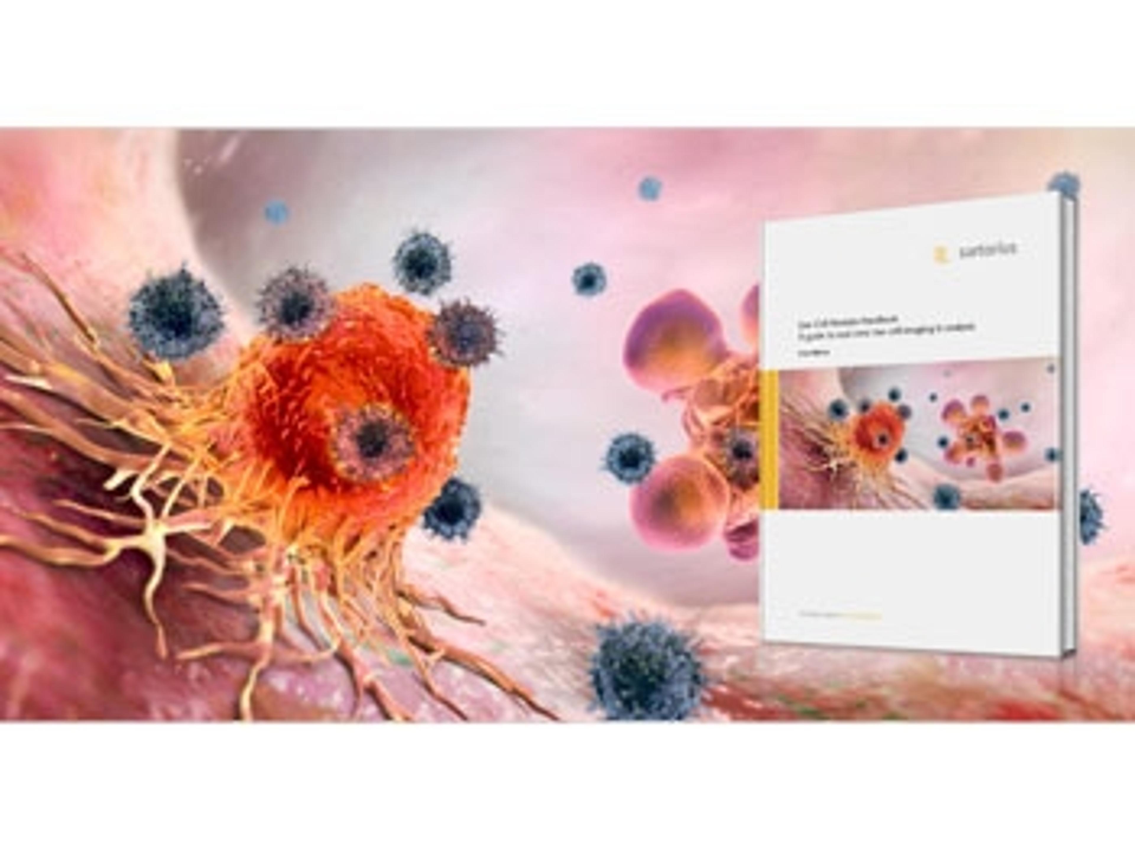
The third edition of the Live-Cell Analysis Handbook published
Latest edition includes two new chapters for scientists who perform cell biology assays
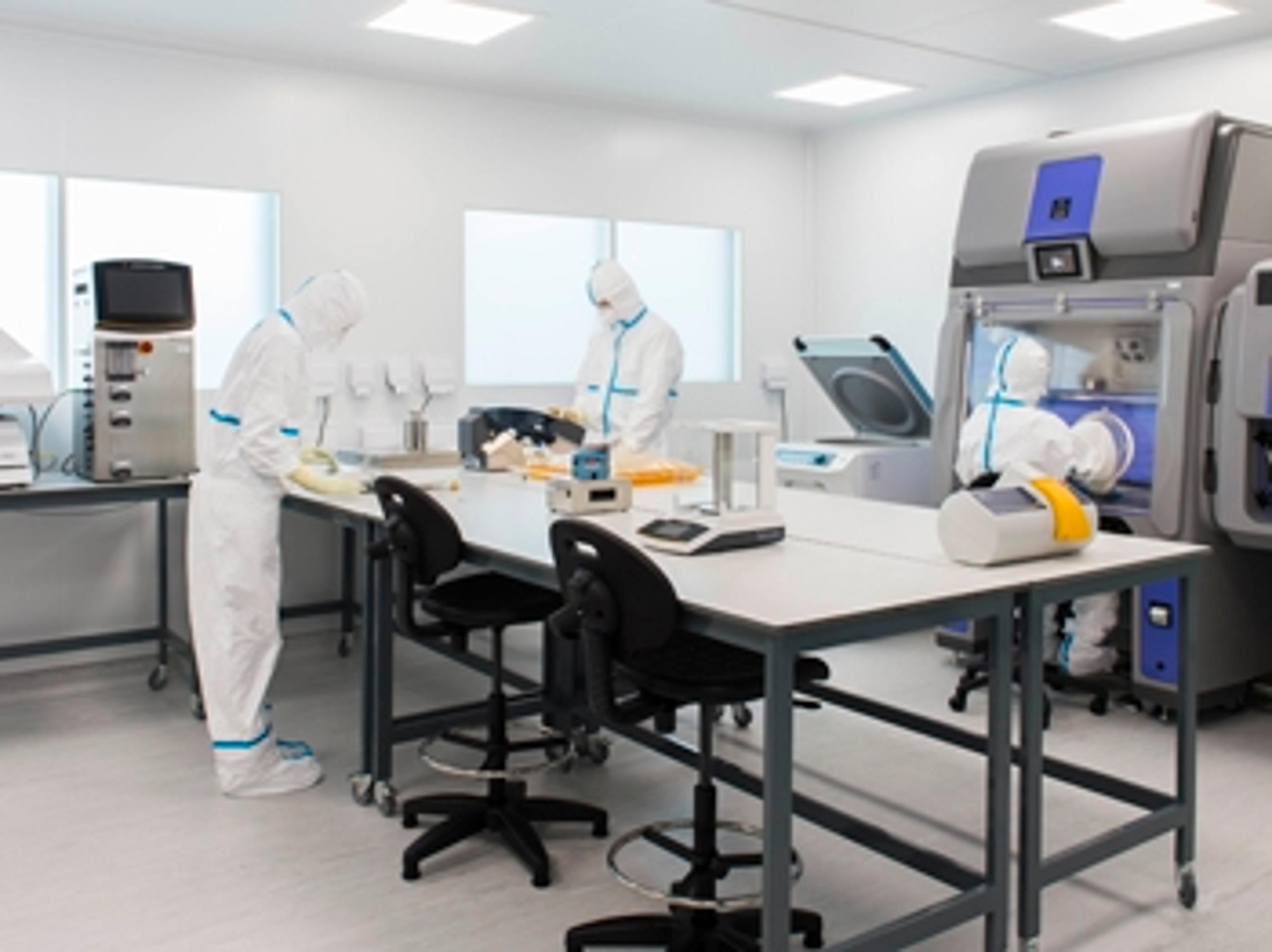
Sartorius launches new services for mammalian cell bank manufacturing
Integrated package of new and established services saves time and minimizes risks