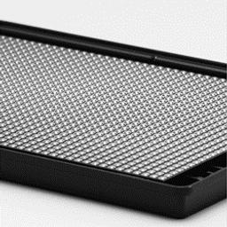
Corning 1536-well Spheroid Microplates
With their novel and proprietary design, these microplates are ideal for generating and analyzing 3D multicellular spheroids in the same microplate. The Ultra-Low Attachment (ULA) surface enables uniform and reproducible 3D multicellular spheroid formation. The black opaque microplate body shields each optically clear, round bottom well from well-to-well cross-talk.
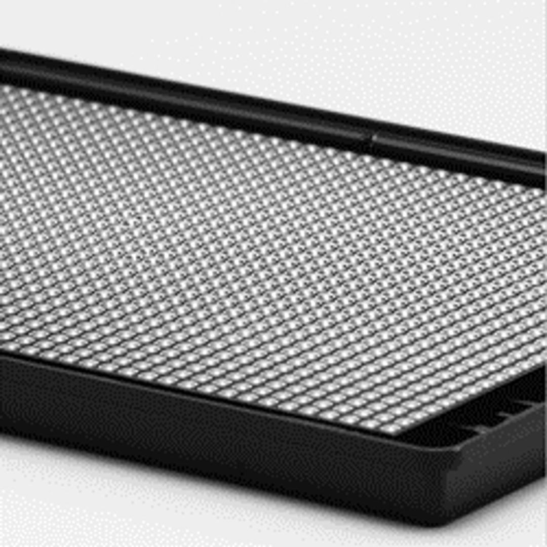
1536-well spheroid microplates

The supplier does not provide quotations for this product through SelectScience. You can search for similar products in our Product Directory.
Reliable/reproducible results, Optically clear, compatible with with HTS instruments.
ADME and Toxicology, Neurobiology and metabolic disease , High throughput drug screening
Adjusting cell concentration is an easy way to vary the size, and imaging was very straightforward, High efficiency of the products made work easier. There is reproducibility and high quality of results which make it reliable for work. Good service at a rignable price.
Review Date: 6 Oct 2021 | Corning Life Sciences
With their novel and proprietary design, these microplates are ideal for generating and analyzing 3D multicellular spheroids in the same microplate. The Ultra-Low Attachment (ULA) surface enables uniform and reproducible 3D multicellular spheroid formation. The black opaque microplate body shields each optically clear, round bottom well from well-to-well cross-talk.
- 1536-well format allows for high throughput 3D cell culture
- Optically clear round bottom with black opaque microplate body
- Covalent attachment of Ultra-Low Attachment surface to reduce cellular adhesion to well surface
- Novel well geometry aids in the generation of uniform, single spheroids across all wells, which enables automated visualization.
- Unique design shields each well to minimize well-to-well cross-talk.
- You can culture and assay spheroids in the same microplate, without the need for transfer to a new microplate.
- Compatible with the GNF ultra high throughput system

Neurosphere formation, differentiation, and migration of human neural stem cells
Having an easy-to-use, reproducible neurosphere culture and analysis method for studying hNSC proliferation, migration, and neurotoxicity greatly enables their use for drug discovery and cell therapy applications. In this study, Corning spheroid microplates were used for neurosphere formation, proliferation, and migration of hNSCs in an easy-to-use format that is amenable to high throughput screening. The spheroid microplates were used for neurosphere culture of hNSCs over the course of 96 hours, throughout which multipotency was maintained as assessed through Nestin and SOX2 marker expression, followed by subsequent harvesting of neurospheres for differentiation into neuronal, astrocytic, and oligodendrocytic lineages.
Analysis of RNA transcript levels reveals upregulation of hypoxia markers for pancreatic cancer cells cultured in 3D
Pancreatic ductal adenocarcinoma (PDAC) is a notoriously aggressive tumor type due to its high levels of metastasis and recurrence and is the fourth leading cause of cancer-related deaths in the Western world. Pancreatic tumors typically lack vasculature and are hypoxic due to oxygen diffusion limitations. In this study, the PDAC cell line PANC-1 was seeded at densities ranging from 500 to 5,000 cells in Corning spheroid microplates and the relative expression of hypoxia markers HIF-1α, GLUT-1, and CA IX were analyzed compared to 2D culture using RT-qPCR to assess the correlation between spheroid size and hypoxia marker expression.
3D Imaging of Optically Cleared Spheroids in Corning Spheroid Microplates
The use of three-dimensional (3D) cell cultures for in vitro drug discovery assays has increased dramatically in recent years because 3D cell culture models more accurately mimic the in vivo environment compared to traditional two-dimensional (2D) monolayer cultures. However, current imaging-based analysis of these 3D cultures relies upon techniques originally developed for 2D cell culture, and as such, has significant limitations. Specifically, the light scattering inherent with thick microscopy specimens prevents imaging the entirety of 3D spheroids, which are typically >100 µm in diameter. This technical limitation introduces a sampling bias in imaging analysis in which only the exterior cells of a spheroid can be imaged. To accurately survey the cellular environment and response of the spheroid, the field needs new techniques to overcome the sampling bias.

Organoid-based 3D model of human glioblastoma: Recreating brain tumors in a dish
Discover how a brain organoid core facility is applying its glioblastoma model towards developing patient mini-brains

Ultra-high-throughput screening (uHTS): How scientists at Scripps Research target global medical breakthroughs
From assay inception to dose response and medicinal chemistry, discover the cutting-edge technology used to identify small molecules with clinical potential, in this SelectScience interview