Organoid-based 3D model of human glioblastoma: Recreating brain tumors in a dish
Discover how a brain organoid core facility is applying its glioblastoma model towards developing patient mini-brains
7 Apr 2021
Glioblastoma, the deadliest of primary brain tumors in adults, has a median survival of only about 15 months, with less than 6% of patients surviving past the five-year mark. The urgent need to fully understand the cancer biology of glioblastoma is thwarted by the lack of a suitable disease model that mimics the cerebral microenvironment.

In recent times, cerebral organoids, the self-assembling three-dimensional cellular models derived from stem cells, have opened up the possibility of replicating the brain’s milieu to closely examine and study glioblastomas.
In this SelectScience® interview, we speak with Dr. Richa Singhania, Director of the Starr Foundation Glionoid Translational Core at Weill Cornell Medicine, to learn about the key considerations in successfully recreating gliomas in a dish.
3D organoid co-cultures replicate glioblastomas in vitro
“We've successfully developed a cerebral organoid-based 3D model of human glioblastoma to replicate the fundamental biology of these tumors,” says Singhania. “We work towards developing a personalized, targeted treatment for brain cancer patients by integrating cerebral organoid technology with automated high-throughput screening.”
For modeling glioblastoma in organoids, the core facility uses a co-culture model. Patient-derived glioma stem cells are co-cultured with cerebral organoids to induce tumor formation. “We call our model GLICO – glioblastoma cerebral organoids – where these glioma tumors grow within the context of the human brain microenvironment as provided by the organoids,” explains Singhania.
GLICO generation is a multi-step process. First, glioma stem cells are isolated from fresh patient tumor biopsies. They are then tagged with a fluorescent reporter to enable visualization of tumor growth within the organoid in real time. Finally, these tumor cells are co-cultured with mature cerebral organoids that are generated from human embryonic stem cells (hESCs) or induced pluripotent stem cells (iPSCs).
“Within a couple of days after co-culturing, tumor cells start infiltrating the organoid,” notes Singhania. “Following this invasion, the tumor cells proliferate and migrate throughout the organoid, ultimately forming tumors that closely resemble the glioblastoma seen in patients.” These tumors mimic original gliomas in terms of their characteristic features, such as infiltrative behavior, as well as key genetic and molecular features.
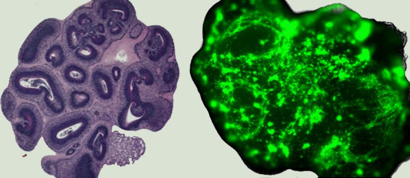
An image of the 'normal' and 'glioma' cerebral organoids Image © finelab.weill.cornell.edu
A human-specific brain microenvironment
GLICO tumors are able to closely recapitulate the phenotypic and molecular heterogeneity observed in patients' gliomas, a feat that traditional cell lines or animal-based models have repeatedly failed at.
“The first and foremost feature that we're able to capture in this GLICO model is how the tumors grow in the same manner as they would within the human brain,” notes Singhania. “This means, we can now study the behavior of the tumor and potentially find targets to stop its invasion into the brain.”
In this setting, the organoids serve as mini-brains, acting as a surrogate for the human brain. “We believe that the GLICO platform offers a unique tool to study fundamental tumor biology as it occurs in humans,” notes Singhania. “It enables us to explore tumor-host interactions in the context of a natural human tumor microenvironment and test potential drugs.”
Conventional animal models, where human cells are grown within the mouse brain, present a cross-species problem, making it impossible to capture the essential cellular crosstalk between the tumor cells and healthy cells. The organoids, on the other hand, are derived from human stem cells, making it possible to recreate a human-specific brain microenvironment. “The advantage of this model is that it can be readily manipulated, genetically and molecularly, for studying the functional consequences of gene mutations or alterations,” explains Singhania. “As such, it offers a robust platform for drug discovery and toxicology studies.”
Developing high-quality organoids: The right conditions
Due to the complexity of the human brain, cerebral organoid generation entails a four- to six-week-long process. “It is very critical to start with high-quality human pluripotent stem cells that have been cultured under the right conditions, to begin with,” Singhania says.
“Corning has a fantastic product line to support our research with brain organoids. We use the Corning® Matrigel® hESC-Qualified Matrix for culturing human pluripotent stem cells.” Extracellular matrices are an important consideration in generating 3D cellular models. They not only lend structural support for the cells to self-assemble but also provide biochemical signals for cell migration and growth.
Singhania continues: “We have a very critical step in the organoid production timeline where we have to embed the embryonic bodies into a Matrigel droplet. For this step, we utilize the Corning® Matrigel® Basement Membrane Matrix. Here, again, the composition of the Matrigel is important, and we work closely with Corning representatives to obtain good reproducibility. Both these products that we use in the pipeline play a key role in maintaining the health and the right differentiation of the organoids.”
Moreover, each lot of the Corning® Matrigel® Matrix is optimized to form a stable 3D dome structure that enables organoids to grow from both healthy and diseased cell origins. “We also use Corning's spheroid microplates, which we find extremely useful for our automated organoid-based drug screening platform,” Singhania adds.
The patients’ own mini-brains
An important project at the Weill Cornell Medicine brain organoid facility is to use this organoid-based glioblastoma model for performing clinically relevant drug screening. An automated high-throughput screening platform facilitates the screening of drug libraries in GLICO tumors.
“Going forward, we want to derive autologous brain organoids – our patients’ own mini-brains in a dish – to generate and utilize patient-specific tumor organoids as a precision medicine tool for tumor modeling and personalized drug testing,” explains Singhania. The goal of such an endeavor is to essentially predict if a patient with a particular mutation or a gene abnormality would benefit from a proposed treatment in the clinic.
“Pairing the patients' tumor cells with their own mini-brains would be pathbreaking. It will give us the opportunity to create personalized solutions for brain cancer patients,” Singhania adds.
The future of brain organoids in translational research
Brain organoids have catapulted neuroscience research in a way that was once inconceivable. A close-to-real human brain microenvironment is now feasible at a fraction of the cost, time, and resources required by traditional mouse models.
“There's been continuous interest and improvement in the organoid technology, which is causing a paradigm shift in the study of neurological disorders,” notes Singhania. “Recent organoid studies have focused on brain regional interconnectivity and neural circuit formation in a dish, providing new avenues to explore neuropsychiatric disorders.”
In light of the COVID-19 pandemic, brain organoids were used to model the viral infection and inspect its effects on the central nervous system. Singhania continues: “Research groups performed experiments where they infected the brain organoids with the novel coronavirus. They demonstrated that SARS-CoV-2 preferentially targeted the choroid plexus, breaking the brain-cerebrospinal fluid barrier, allowing leakage of immune cells and cytokines and triggering neuroinflammation.”
“Brain organoids hold a tremendous potential,” Singhania summarizes. “It’s an unprecedented opportunity for patient-centric research. With each passing day, the technology is only going to get better to make these model systems more predictive for patients.”
Do you use Corning Life Sciences products in your lab? Write a review today for your chance to win a $400 Amazon gift card>>
Related products
Request Quote for All Products
Corning 1536-well Spheroid Microplates
Corning Life SciencesWith their novel and proprietary design, these microplates are ideal for generating and analyzing 3D multicellular spheroids in the same microplate. The Ultra-Low Attachment (ULA) surface enables uniform and reproducible 3D multicellular spheroid formation. The black opaque microplate body shields each optically clear, round bottom well from well-to-well cross-talk.
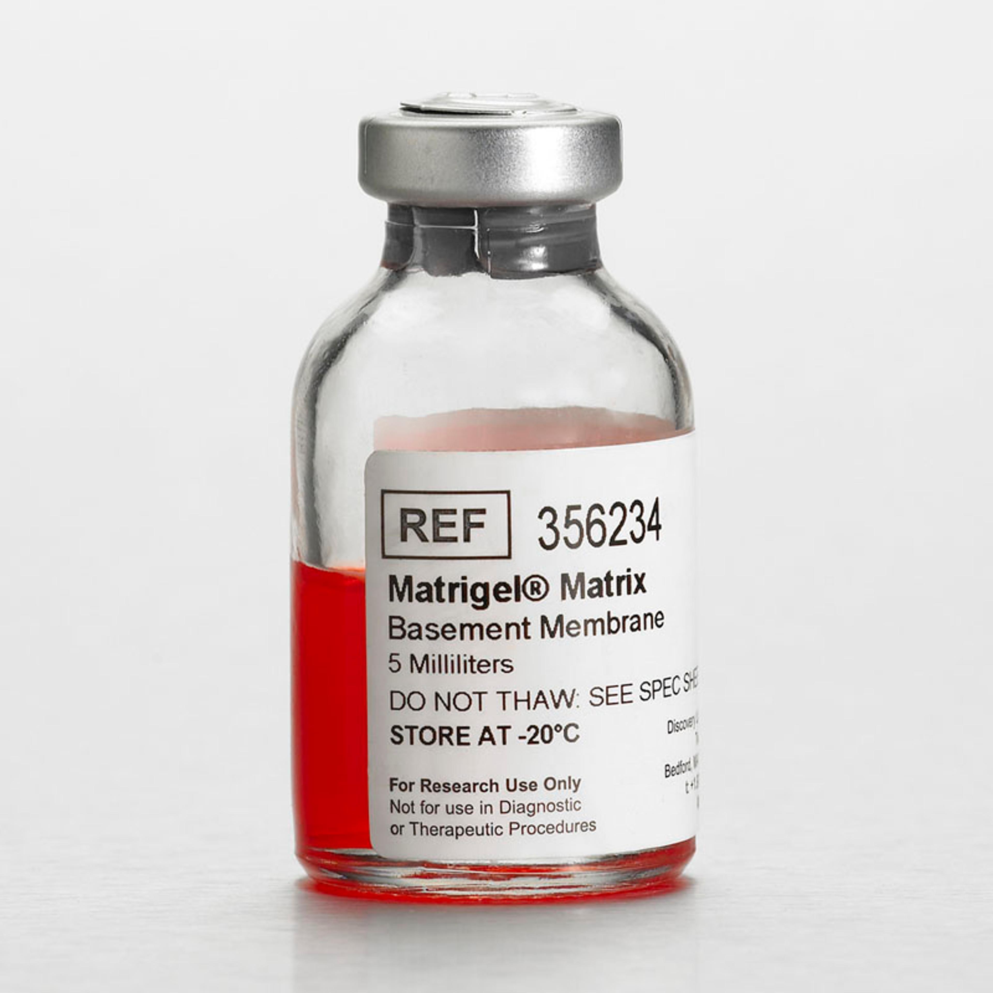
Corning® Matrigel® Basement Membrane Matrix, LDEV-free, 5 mL
Corning Life SciencesCorning Matrigel matrix is a solubilized basement membrane preparation extracted from the Engelbreth-Holm-Swarm (EHS) mouse sarcoma, a tumor rich in extracellular matrix (ECM) proteins, including Laminin (a major component), Collagen IV, heparan sulfate proteoglycans, entactin/nidogen, and a number of growth factors.Quality Mouse colonies are routinely screened for pathogens via Mouse Antibody Production (MAP) testing Extensive PCR testing is performed to screen for a number of pathogens, including LDEV, to ensure strict control of raw materials used during the manufacturing process Tested and found negative for bacteria, fungi, and mycoplasma Protein concentrations are determined by Lowry method Endotoxin units are measured by Limulus Amoebocyte Lysate assay Corning Matrigel Matrix gel stability is tested for a period of 14 days at 37°C Biological activity is determined for each lot using a neurite outgrowth assay. Chick dorsal root ganglia are plated on a 1.0mm layer of Corning Matrigel Matrix and must generate positive neurite outgrowth response after 48 hours without addition of nerve growth factor. Source Engelbreth-Holm-Swarm Mouse Tumor Preparation and Storage Store at -20°C. Avoid multiple freeze-thaws. Do not store in frost-free freezer. KEEP FROZEN. Application Cell Growth and Differentiation Metabolism/Toxicology Studies Invasion Assays in vitro and in vivo Angiogenesis Assays in vivo Angiogenesis Studies and Augmentation of Tumors in Immunosuppressed Mice For Customers in California only: WARNING Please refer to Safety Data Sheet (SDS) Section 15 for more information regarding California Proposition 65.
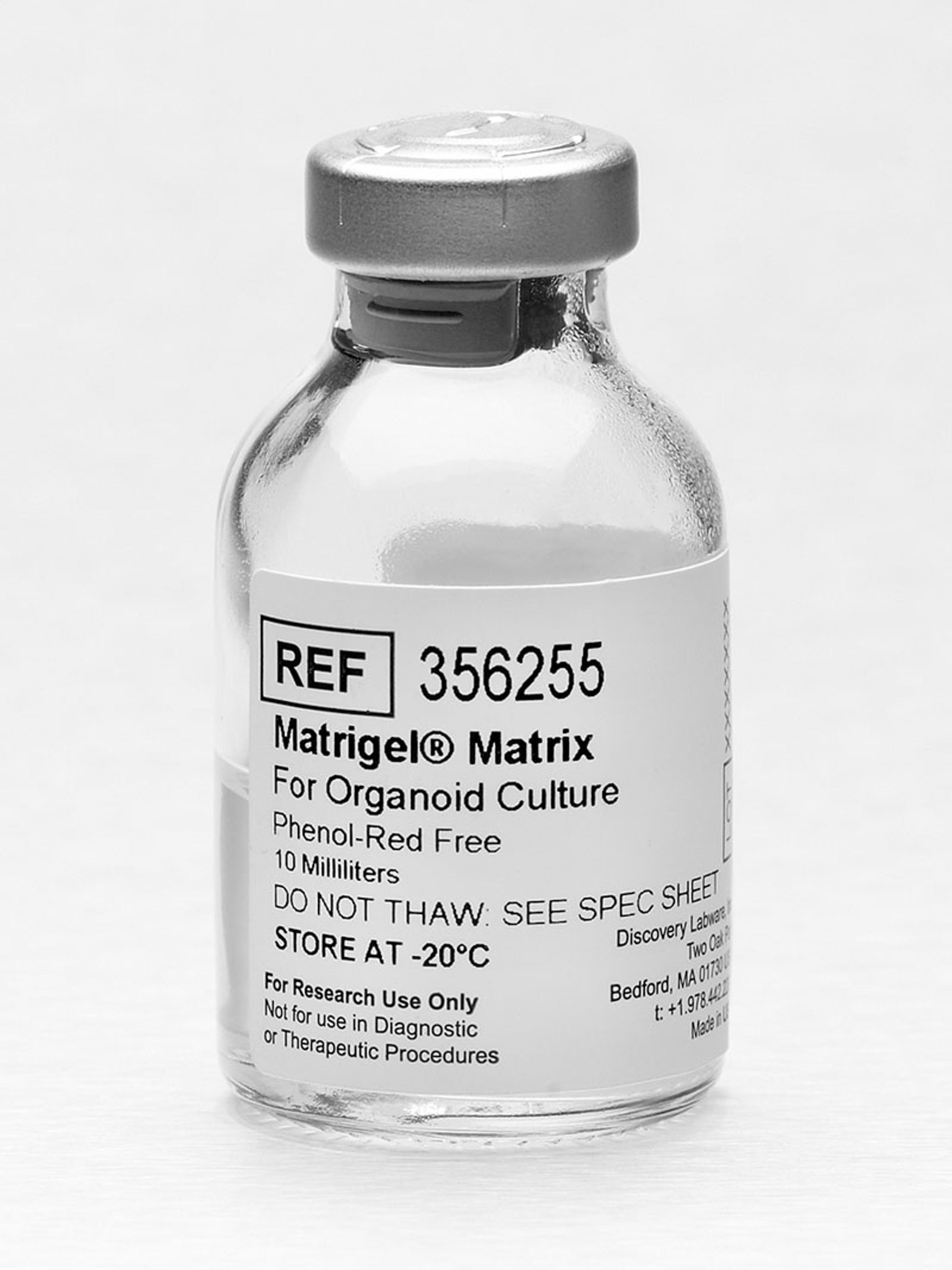
Corning® Matrigel® Matrix for Organoid Culture, Phenol Red-free, LDEV-free, 10 mL
Corning Life SciencesCorning Matrigel Matrix for Organoid Culture is a solubilized basement membrane preparation that has been optimized for organoid culture. Verified to support both mouse and human organoid growth and differentiation from healthy and/or diseased tissue, each lot has a specific elastic modulus value indicating the stiffness of the gel formed and is qualified to form stable 3D domes commonly used in organoid culture protocols. Available in 10mL LDEV-free, Phenol Red-free format.Corning Matrigel Matrix for Organoid culture is extracted from the Engelbreth-Holm-Swarm (EHS) mouse sarcoma, a tumor rich in extracellular matrix (ECM) proteins, including Laminin (a major component), Collagen IV, heparan sulfate proteoglycans, entactin/nidogen, and a number of growth factors. Quality Tested for ability to form and maintain stable 3D domes/droplets on multi-well plates. Elastic Modulus (matrix stiffness) measured using a rheometer. Protein concentrations determined by Lowry method Mouse colonies routinely screened for pathogens via Mouse Antibody Production (MAP) testing Extensive PCR testing is performed to screen for a number of pathogens Tested and found negative for bacteria, fungi, and mycoplasma Endotoxin units measured by Limulus Amoebocyte Lysate assay Matrigel Matrix gel stability is tested for a period of 14 days at 37°C