COMET
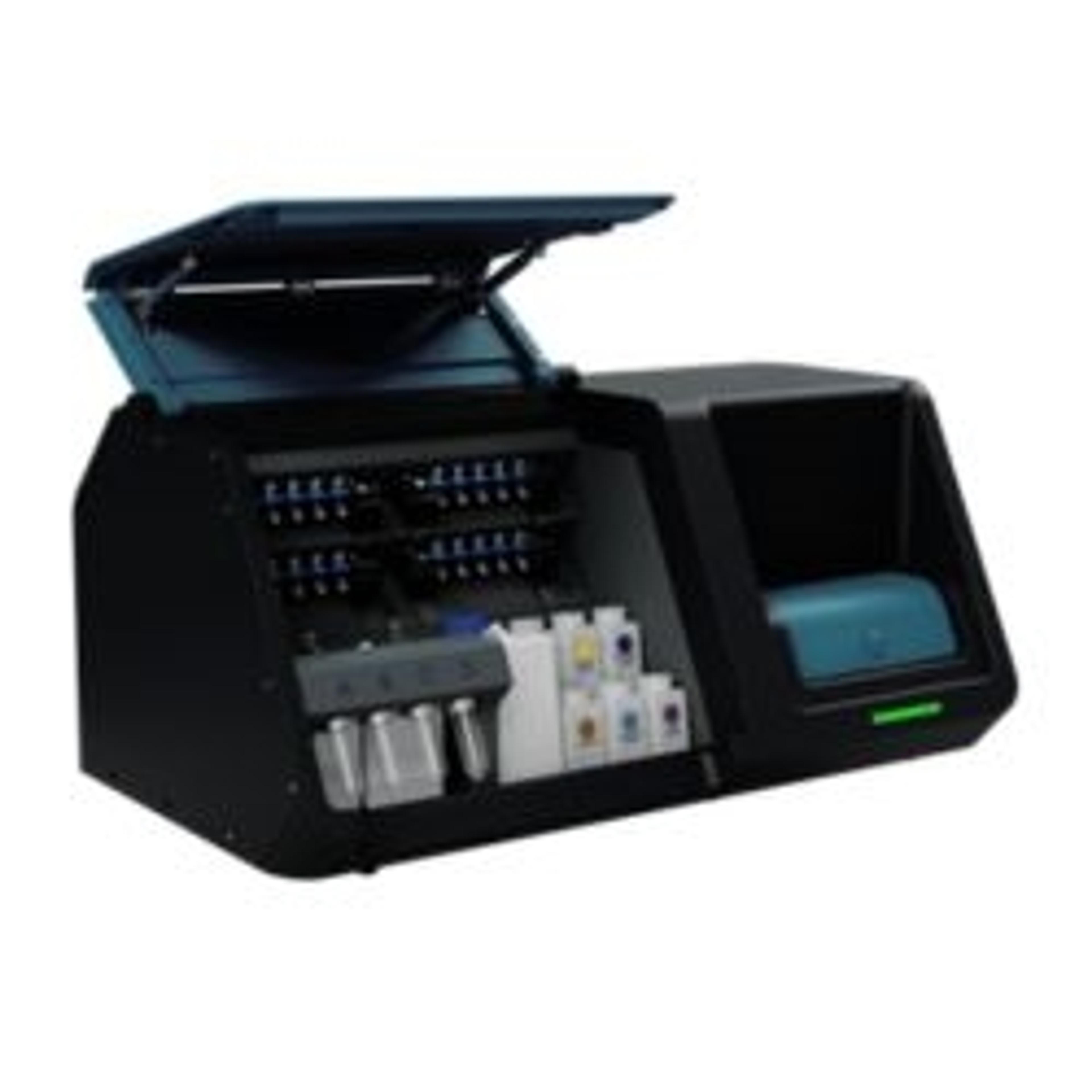
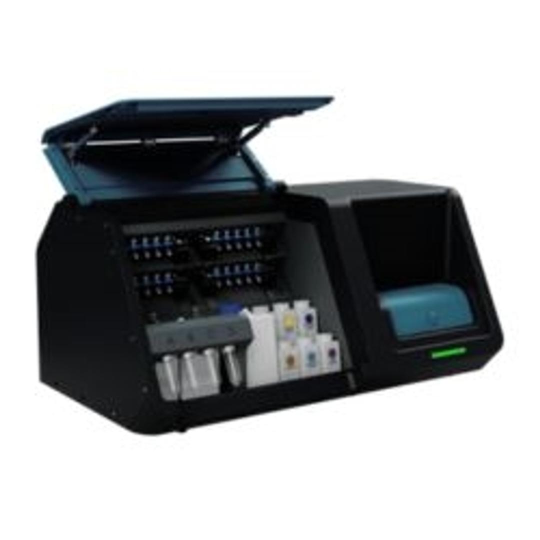
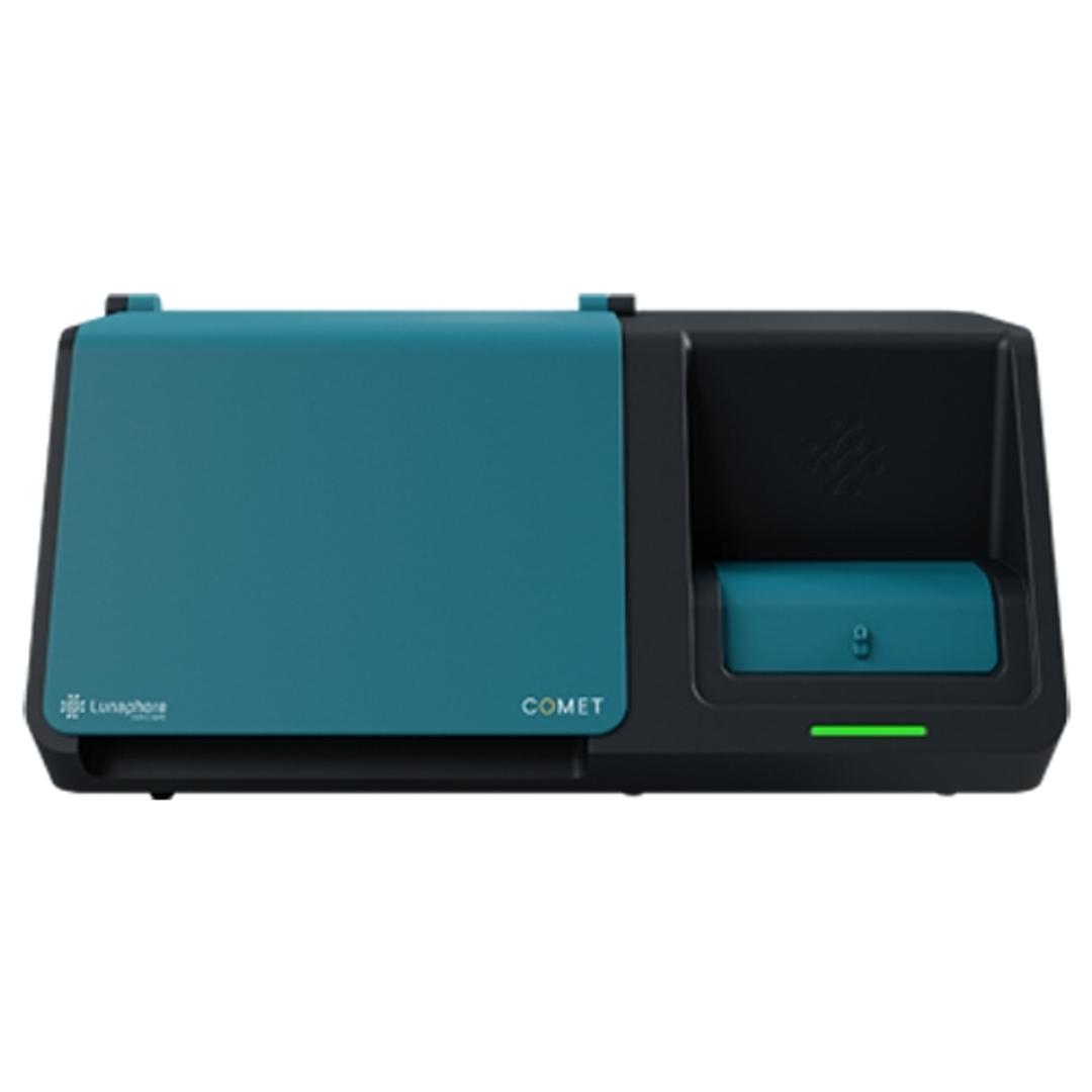
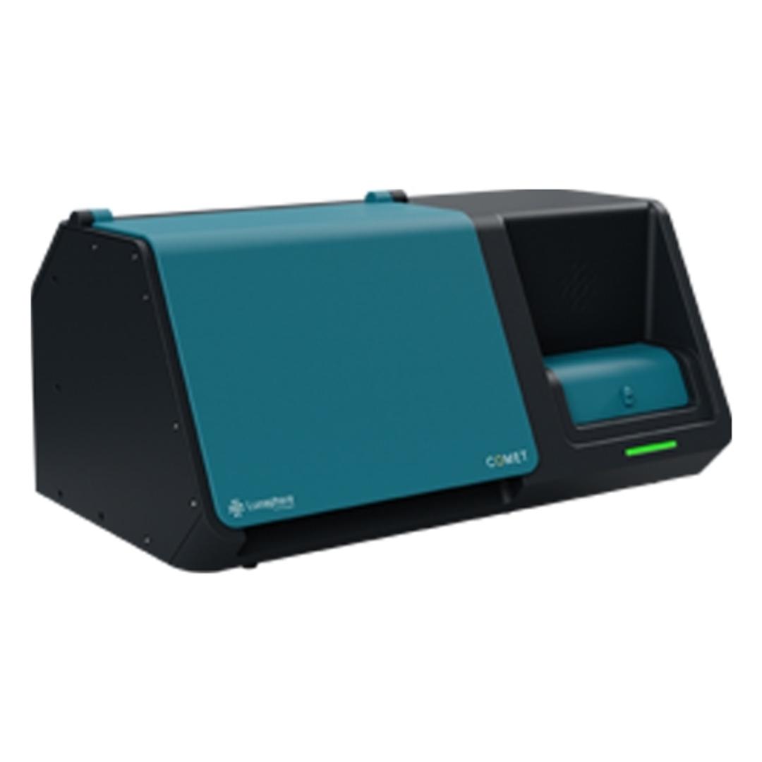
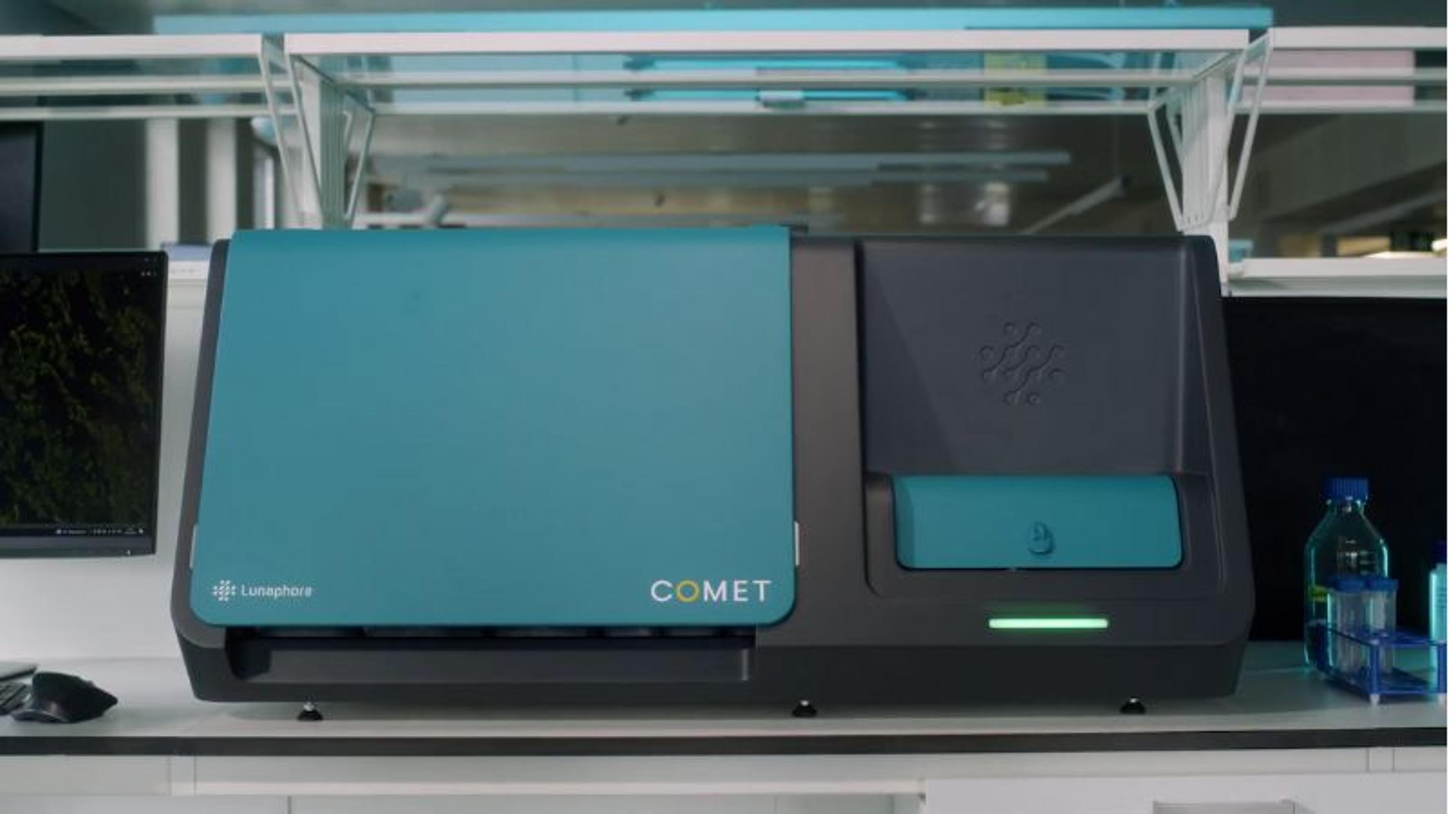
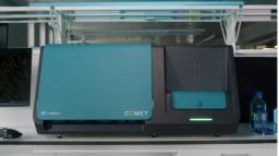
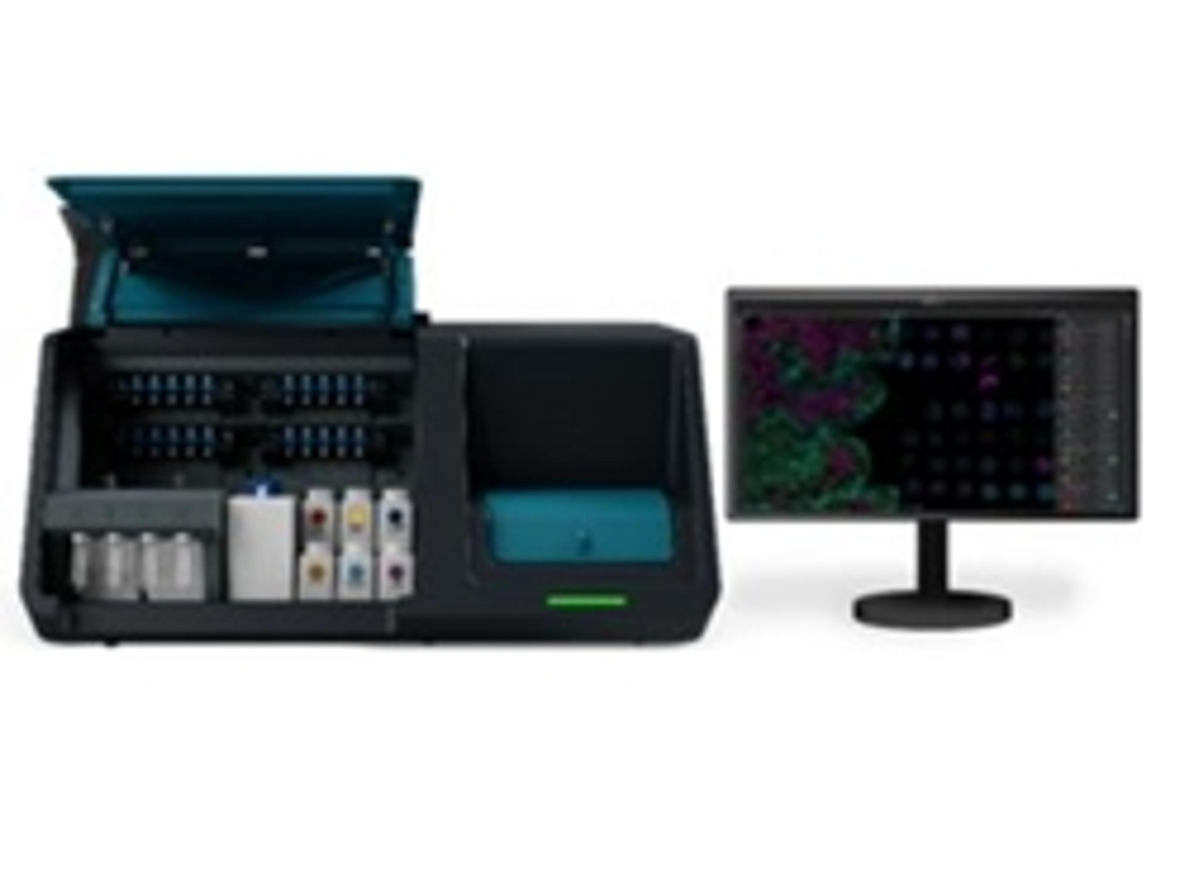
COMET™ is a fully automated, high-throughput, hyperplex platform for the analysis of Formalin-Fixed, Paraffin-Embedded (FFPE), and frozen tissues.



Receive your quote directly from the manufacturer.
The COMET™ is a walk-away automation system to scale-up spatial biology assays with unmatched throughput. It can perform proteomics, transcriptomics and same-section multiomics applications. For protein detection, COMET™ works with standard, off-the-shelf, label-free primary antibodies and performs sequential immunofluorescence (seqIF™) assays that consist of sequential cycles of staining, imaging, and elution. In each cycle, protocol conditions can be finetuned to stain and image up to 4 antibodies. Runs of 40 markers can be performed fully automated in just a few hours on the same tissue sample and without user intervention.
For multiomics application, detection of RNA and protein is performed simultaneously on the same tissue section at single-cell level. The system integrates the entire staining or target probe hybridization, imaging and image pre-processing workflow without the need for any user intervention. The workflow includes an optimized target retrieval step and a protease-free pretreatment to maximize RNA and protein target detection as well as epitope unmasking while preserving tissue morphology. The use of RNAscope™ HiPlex and seqIF™ allows fully flexible RNA and protein target selection to build your panel.
Brochures

Automated multiomics on COMET
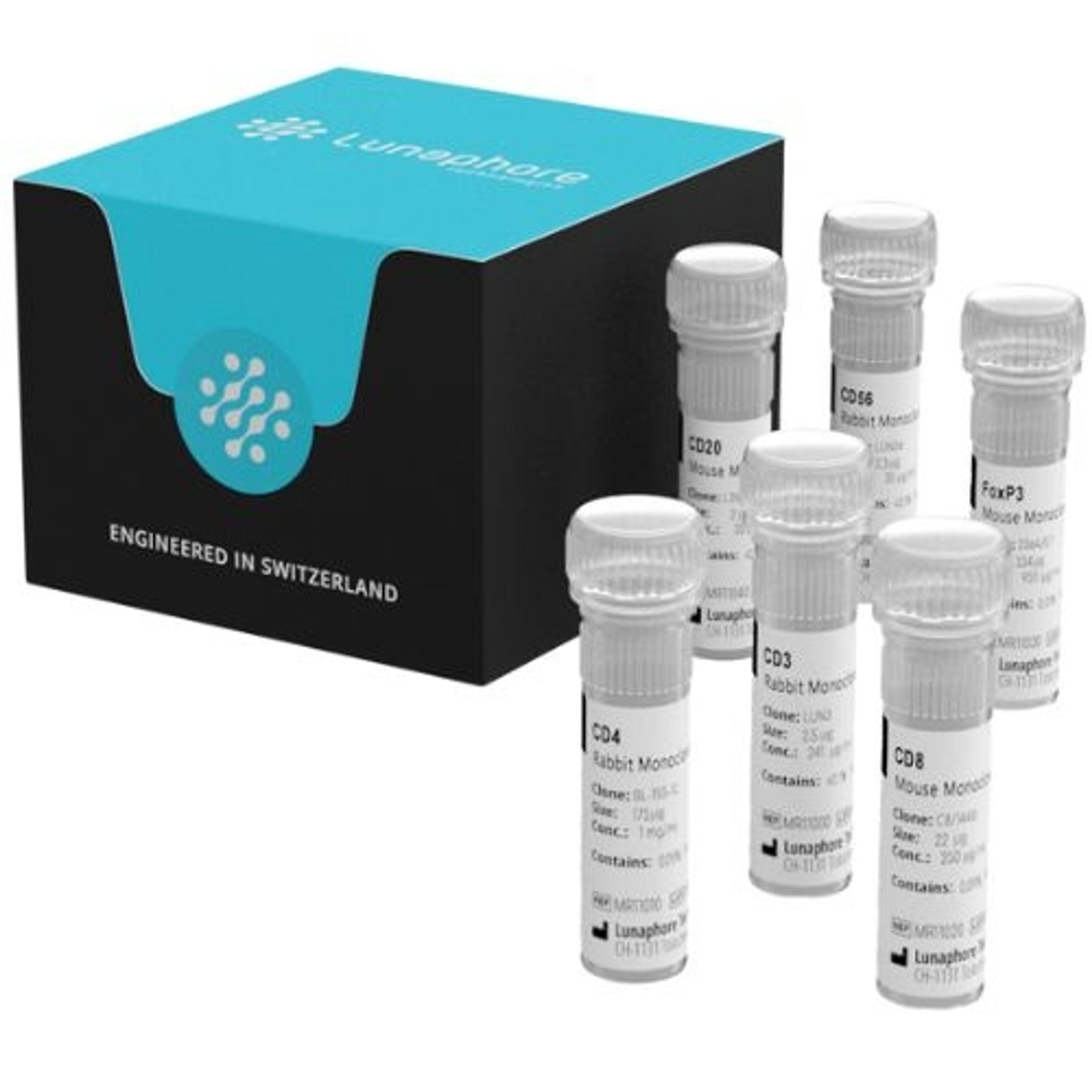
In this product brochure, Lunaphore describes its COMET™ platform, which performs automated cycles of RNA detection using RNAscope™ and protein detection using sequential immunofluorescence (seqIF™), enabling the use of clinically proven immunohistochemistry (IHC) antibodies. The brochure also includes the RNAscope™ HiPlex Pro for COMET™ RNA detection kit, which has a large and widely established library of probes. Lunaphore also details how for protein detection, scientists can use standard, non-conjugated primary antibodies as their markers, and have the ability to characterize 10 new antibodies per week and add them to their marker library.
Multiomics adds new dimension to biological research
A comprehensive understanding of biology and disease requires an integrative approach that combines information from different biological inputs. Multiomics methodologies enable researchers to achieve a comprehensive understanding of molecular changes contributing to tissue development, cellular response, and disease. In this free infographic:
- Explore the omics landscape
- Discover multiomics applications in translational research
- Learn about a platform designed to revolutionize your spatial multiomics studies
COMET: Streamline hyperplex immunofluorescence at single-cell resolution
In this application note, Lunaphore Technologies demonstrates how to build an inclusive hyperplex panel to streamline IO research using COMET™. Understanding the tumor microenvironment (TME) can aid in tumor classification, identification of biomarkers, and development of targeted therapies. One approach to studying multiple proteins in the TME is multiplex immunofluorescence (mIF), but current assays struggle with long turn-around times and tissue degradation. Lunaphore has developed the COMET, a revolutionary microfluidic instrument that performs hyperplex assays using a sequential immunofluorescence (seqIF™) approach. COMET allows researchers to create and customize panels with their off-the-shelf antibodies.

Single-section spatial proteomics enables comprehensive lung cancer diagnosis
Monday, November 30 at 14:00 GMT | 15:00 CET | 09:00 EST | 06:00 PST
Lung cancer diagnosis increasingly depends on small biopsy samples, placing growing pressure on laboratories to extract as much clinically relevant information as possible from limited tissue. Conventional sequential testing for histopathology, PD-L1 expression, molecular biomarkers, and immune profiling can rapidly exhaust precious samples and extend the time to treatment decisions.
In this SelectScience® webinar, discover a single-section multiplexed imaging approach designed to bring these analyses together in one tissue-preserving workflow. The method enables simultaneous tumor classification, PD-L1 scoring, assessment of actionable biomarkers, and detailed immune profiling, while achieving 96% concordance with standard pathology.
Explore how multiplexed imaging could help streamline lung cancer diagnostic workflows by reducing the need for multiple sequential assays and enabling quantitative, automated analysis from a single tissue section. You will also learn how the resulting high-dimensional spatial data can provide new insights into the tumor microenvironment, creating opportunities for both clinical assessment and translational research.
Certificate of attendance
If you attend the live webinar, you will automatically receive a certificate of attendance, including a learning outcomes summary, for continuing education purposes.
If you view the on-demand webinar, you can request a certificate of attendance by emailing editor@selectscience.net.
Webinar details
Cost: Free to attend
Location: Online
Duration: 60 minutes
Registration is required to secure your place. If you register but can’t attend live, you will receive a link to the on‑demand recording once it becomes available.

In situ Hyperplex Techniques to Study the Cytotoxicity of Immunotherapies in Non-small Cell Lung Cancer
In this webinar, Dr. Amine Majdi, postdoctoral fellow at the Centre de Recherche des Cordeliers, will share his research focusing on the emerging role of immunogenic cell deaths in non-small cell lung cancer (NSCLC) and its correlation with prognosis based on spatial cell distribution.
Tissue architecture not only establishes the groundwork but also influences cell interactions and phenotypes significantly. Dr. Majdi will discuss his recent investigations utilizing the COMET™ platform that his team uses to analyze and map tissues effectively. Simultaneous visualization of multiple biomarkers is essential, facilitated by multiplex immunofluorescence (mIF). This technique yields comprehensive insights into cell phenotypes while preserving spatial context and neighboring information.
Key learning objectives
- Learn about the latest research regarding NSCLC and immunotherapy-associated cytotoxicity with the COMET™ platform
- Discover how to obtain highly reproducible, hyperplex data with a fully automated workflow on COMET™
- Explore how to use spatial biology to identify and validate biomarkers
Who should attend?
Researchers from academics to the industry settings interested in immunology and immuno-oncology.
Certificate of attendance
All webinar participants can request a certificate of attendance, including a learning outcomes summary, for continuing education purposes.
Scalable hyperplexing with COMET
In this video, Lunaphore Technologies introduces COMET™, a fully automated, high-throughput, hyperplex platform that ensures scalability and reproducibility without the need to conjugate primary antibodies. It provides walk-away automation for staining, imaging, and image preprocessing, generating highly robust and reproducible data with full tissue preservation.

How spatial biology is driving tumor microenvironment research
Learn how a leading cancer researcher is utilizing the COMET™ technology in his research

Spatial biology techniques in tumor microenvironment analysis
Researchers in the US are using multiplex immunofluorescence to unravel the complexities of the spatial biology within the microenvironment of brain tumors
Unraveling the tumor microenvironment of pancreatic cancer
Dr. Julienne Carstens highlights the critical role of spatial biology in understanding the metastatic progression of PDAC and advancing personalized immunotherapy