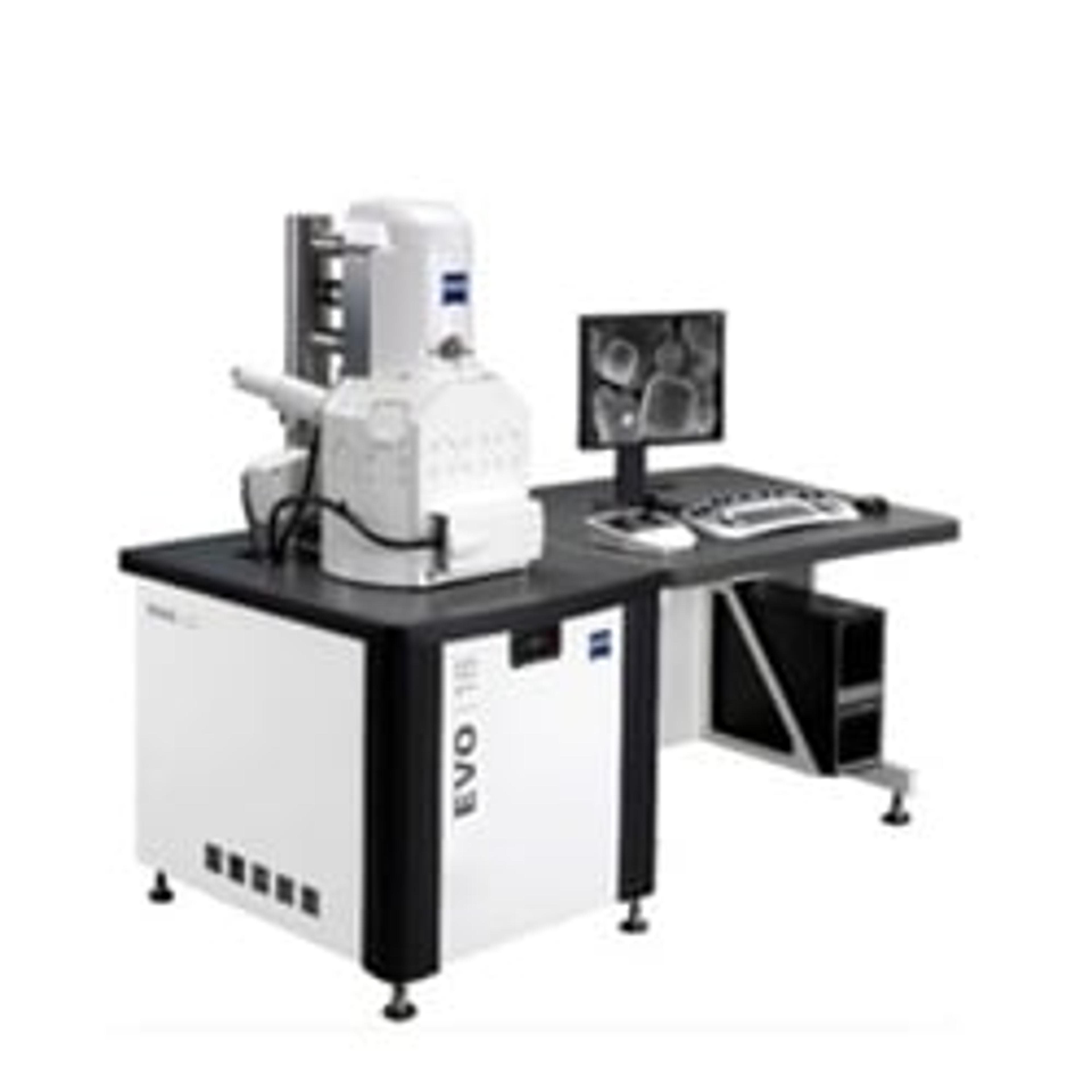
ZEISS EVO Family
Your Interactive SEM for Multi-User Environments


Receive your quote directly from the manufacturer.
Highly recommend Zeiss for SEM/EDX.
Particle identification
SEM/EDX 15 is an excellent addition to our lab. It is used every day. Sales and support staff are excellent. Highly recommend Zeiss for SEM/EDX.
Review Date: 12 Mar 2020 | ZEISS Research Microscopy Solutions
I totally recommend this instrument for everyone.
Polymeric sciences
This system is easy to use and the service is always great.
Review Date: 26 Mar 2019 | ZEISS Research Microscopy Solutions
Material both metallic as well as non-metallic samples can be analyzed
Highly precise analytical equipment and it gives images with high resolution. Shows fast scanning electron micrograph and phase information/details. It is worth for price as it gives detail information for both solid as well as powder samples. All analytical labs should have this SEM.
Review Date: 14 Oct 2015 | ZEISS Research Microscopy Solutions
Forensic / Material Analysis
The SEM from ZEISS is very easy to use and I can strongly recommend it. Use the long-life-cathodes and you'll be happy.
Review Date: 26 Aug 2015 | ZEISS Research Microscopy Solutions
Environmental
I rated its ease of use to be above average because the system can be unstable, especially with back-scattered electron detector. After sales support has been excellent, though with a not-cheap service agreement. All in all, I think the product is worth the money we paid.
Review Date: 26 Aug 2015 | ZEISS Research Microscopy Solutions
The ZEISS EVO series combines high definition scanning electron microscopy with an intuitive user-friendly experience.
Discover substantial improvements in productivity and dramatically reduced training costs with SmartSEM touch user interface. Empower all users to generate excellent images and achieve highest throughput for visual inspection workflows.
SmartSEM touch is a welcome addition in the multi-user environment. Experience excellence in high vacuum (HV), variable pressure (VP) and extended pressure (EP) imaging, thanks to the latest detector technology. Significant improvements in signal yield and contrast ratio herald a step change in imaging efficiency.
EVO is a highly flexible, easy-to-use, high definition imaging and analysis tool delivering fast, accurate, repeatable results across all samples.

Python Blood Analysis by STEM
Understanding veterinary pathologies enables better conservation. This case study describes the analysis of python blood using cells prepared conventionally for electron microscopy, and thin sections subsequently imaged using an EVO15 HD SEM (scanning electron microscope) fitted with a transmission electron microscope (STEM) attachment.
ZEISS Microscopy Solutions for Steel and Other Metals
ZEISS Microscopy offers multi-modal characterizations and advanced analysis options for industry and research. This application note provides a variety of applications of ZEISS products to analyze steel and other metals.
Multiscale Analysis of Bacteria Population in Legume Root Nodules with "Shuttle & Find"
Correlative light and electron microscopy (CLEM) combines the overview information of fluorescence light microscopy (FLM) with structural details in scanning electron microscope (SEM) for high-content imaging. Hence, CLEM is very useful for investigating infection and colonization of the legume hosts by their rhizobial symbionts. This white paper illustrates a methodology involving the shuttle & find interface of CLEM to enable fast and reliable analysis of the bacterial infection of legume root nodule cells.
Stubscope Infrared Camera: High Resolution View of an Individual Stub with ZEISS EVO
Throughput of the scanning electron microscope can be limited by the need to find small features on a large specimen. This application note showcases the capabilities of the Stubscope for navigation assistance in SEM.
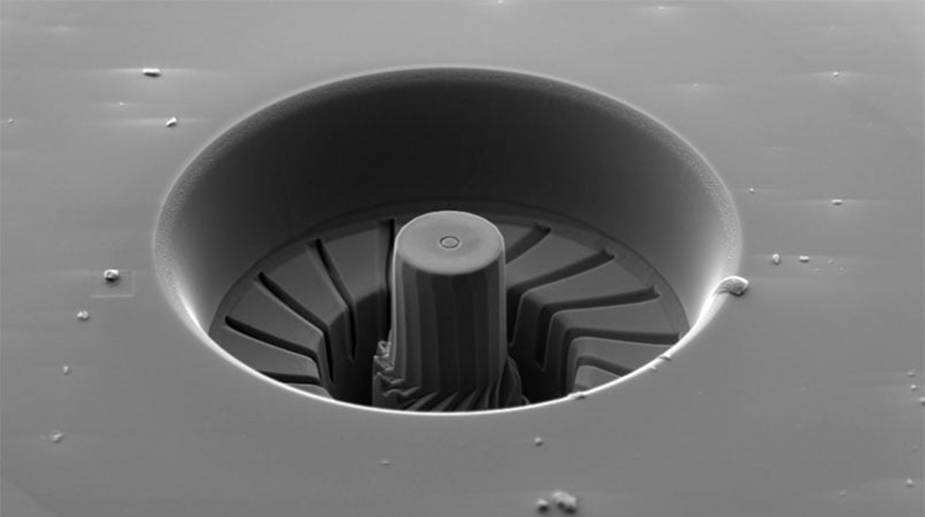
Beam Deceleration Imaging with ZEISS EVO Beam
EVO conventional scanning electron microscope has a new sample bias module that enables beam deceleration imaging. This, combined with the ZEISS HD BSE detector, produces high quality images with enhanced surface contrast and topographical detail.
Microscopy in the Field of Tool Engineering - the Microstructure of Hard Metal Drills
The combination of microscopic and quantitative micro-structural analysis is a powerful method of obtaining information on geometry, usage properties and wear conditions of tools. The mechanical and usage properties of hard metal tools are crucially affected by their microstructure; therefore microscopic investigation of the micro- and macro-structure of the tools is a powerful and fast technique for checking the quality and corresponding usage properties of tools such as drills. This application notes presents possible applications of different microscopy systems from optical light to electron microscopy.

Microscopy with ZEN core: A unified approach to imaging and analysis
April 29, 2025 - 09:00 BST / 10:00 CEST / 16:00 CST / 17:00 JST
Join us for an informative webinar sponsored by ZEISS, where we will introduce ZEN core, a sophisticated software solution designed to enhance your microscopy workflows. This session will focus on how ZEN core provides a streamlined user interface that accommodates both light and electron microscopy, allowing users to transition seamlessly between modalities.
During the session, you will discover how ZEN core's intuitive user interface adapts to the unique needs of your laboratory allowing users of all experience levels to efficiently image, analyze, and manage data. We will explore the advanced imaging capabilities, automated workflows, and powerful AI features that make ZEN core the command center for both light and electron microscopes.
Join us for a live demonstration of ZEN core and take part in our Q&A session to address your specific inquiries. Register today to learn how ZEN core can support your microscopy research and analysis needs.
Key learning objectives:
- Understand how ZEN core fosters seamless collaboration in connected laboratory environments
- Explore the integration of multiple microscopy techniques within a unified platform
- Learn how ZEN core ensures data consistency and standardization across instruments and locations
Who should attend?
This webinar is ideal for researchers in both academia and industry looking to improve their microscopy practices and enhance productivity.
Certificate of attendance
All webinar participants can request a certificate of attendance, including a learning outcomes summary, for continuing education purposes.
If you view the on-demand webinar, you can request a certificate of attendance by emailing editor@selectscience.net.
How ZEISS Microscopes are Shaping the Future
In this video, learn how ZEISS is developing technologies for use at the cutting edge of science, consumer electronics, energy, construction, and more.
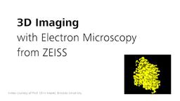
ZEISS 3D Electron Microscopy for Life Sciences
Cut your samples with an integrated ultramicrotome, slice them with a focused ion beam (FIB) gun, or image nondestructively with X-ray. Get a maximum of information from your samples in 3D and correlate with light microscopy. Experience the largest portfolio of 3D imaging for light, electron and X-ray microscopy in this video from ZEISS Microscopy.
New Generation of ZEISS EVO Scanning Electron Microscopes Introduced
Modular platform for intuitive operation, routine investigations and research applications
ZEISS Opens New Microscopy Customer Center
ZEISS Announces Partnership with ECR Engines
Providing analytical microscopes for NASCAR Engine Producer