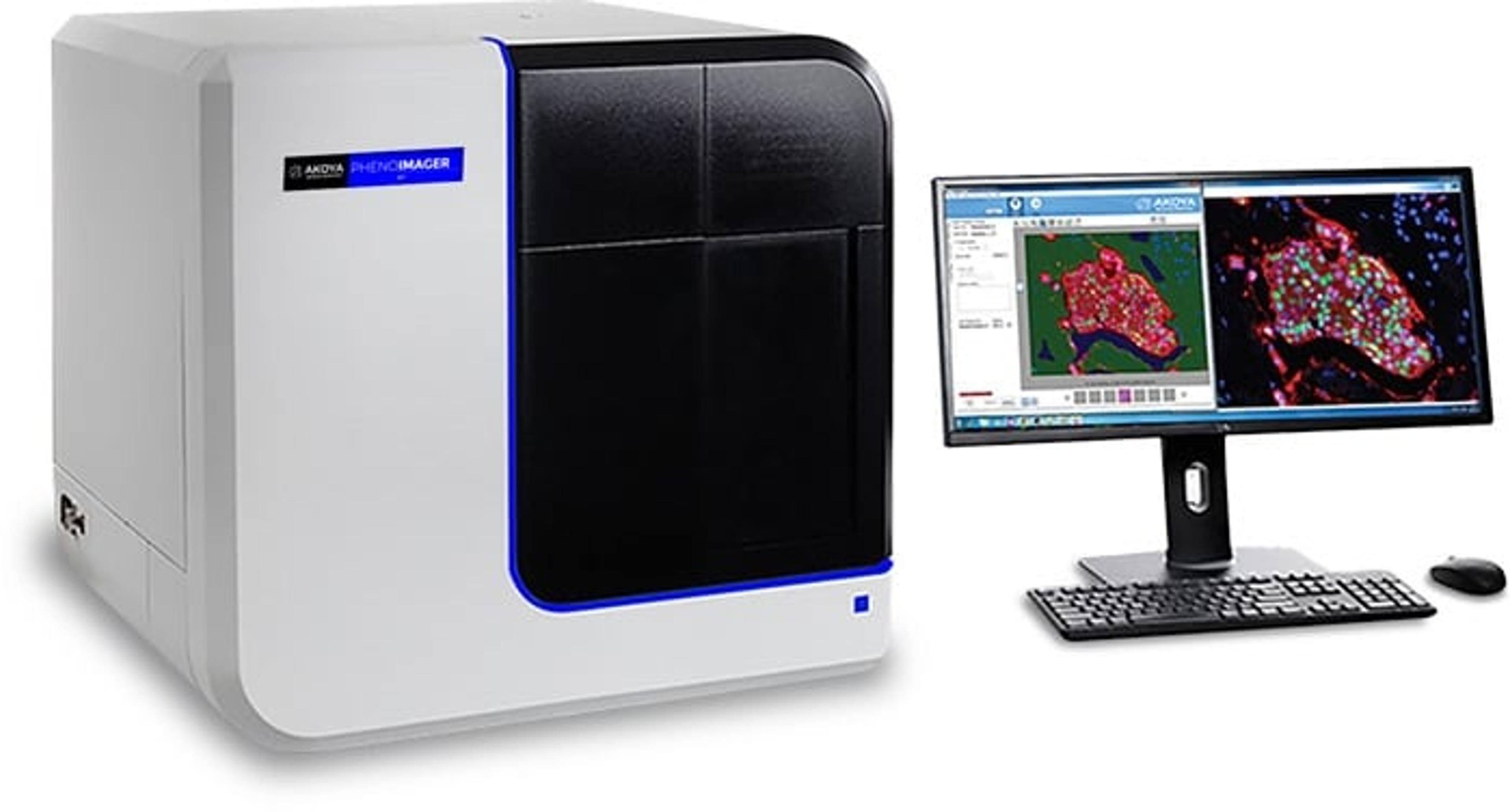
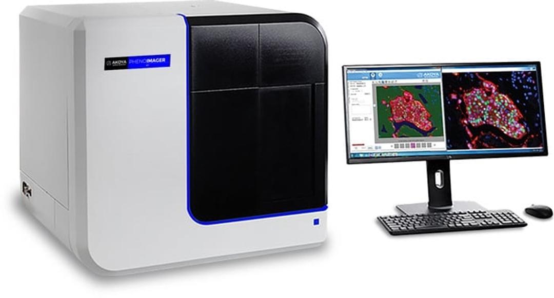
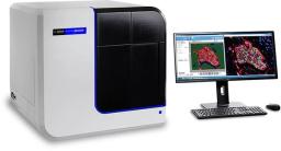
PhenoImager® HT
PhenoImager ® HT (formerly Vectra ® Polaris™) is the fastest and most highly cited whole-slide, single-cell resolution imaging platform for spatial phenotyping and the development of spatial signatures. Featuring Akoya’s patented Multispectral Imaging (MSI) and spectral unmixing technologies, this platform can be easily integrated into high-throughput workflows to accommodate projects regardless of your scale.
PhenoImager™ HT


The supplier does not provide quotations for this product through SelectScience. You can search for similar products in our Product Directory.
Key to complete studies successfully
In the field of cancer research studies
The product is quiet good and user friendly. It helps imaging and also analysis as it has built in image analysis facility. Cool machine
Review Date: 20 Apr 2023 | Akoya Biosciences
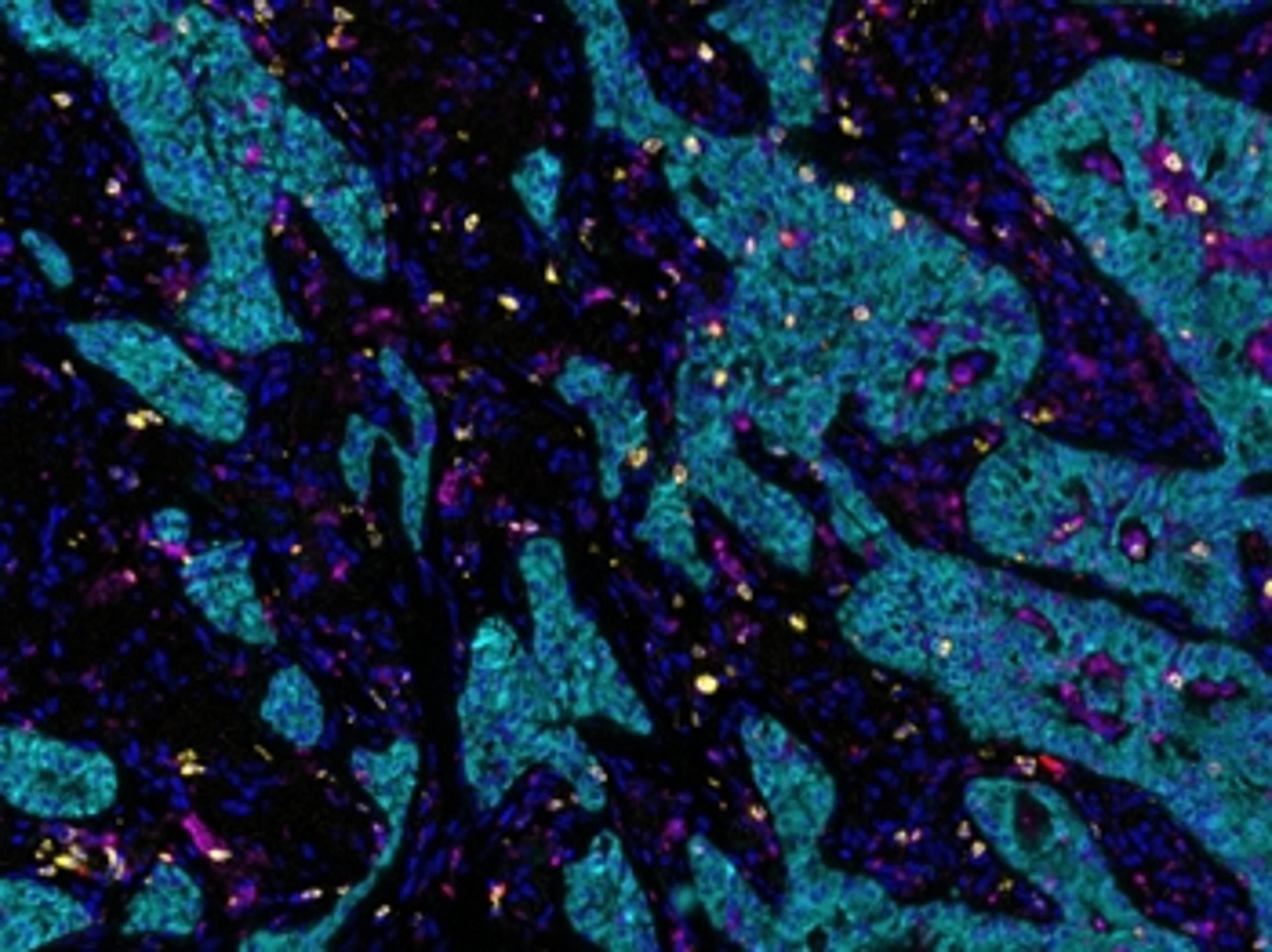
A game-changer in immunohistochemistry-based research of tumor microenvironments.
Analysis of immune markers in tissue - multiplex IHC
Having the Vectra Polaris in our lab has transformed our ability to analyze and understand the tumor immune microenvironment. Within a year of having this technology we have developed novel 9-plex immune marker panels and applied them to endometrial, bladder and esophageal cancer tissues. PhD students with little to no experience of IHC have quickly mastered the ability to perform multiplex IHC and are also now fully competent at using the software needed to analyze their samples. Before obtaining this technology, we were unable to carry out equivalent immunohistochemistry analyses, both in terms of multiplex staining capability and also quantitative regional analysis of multiplex-stained tissue sections. The increased information and thus understanding of the tumor microenvironments this technology is revealing to us will greatly help inform us in our development of more effective combination immunotherapies.
Review Date: 3 Dec 2020 | Akoya Biosciences
PhenoImager HT running the newly released 2.0 software provides researchers with a unique technology stack combining onboard spectral unmixing, rapid imaging and manageable data outputs.
Key Benefits:
- Speed: The fastest imager for spatial phenotyping and signature development, PhenoImager HT supports walk-away automation with a straightforward and user-friendly workflow enabling you to scale projects effortlessly.
- Accuracy: A new onboard spectral unmixing process that runs seamlessly in parallel with imaging means never having to compromise on data quality, regardless of your project size.
- Throughput: PhenoImager HT is ideally suited for an automated mIF staining and imaging workflow, providing the capacity to image over 400+ multiplex stained slides per week to fit any project of your scale.
- Proven: Akoya’s technologies have been cited more than a 1,000 times. With the installation of our 1000th instrument, we are proud to be one of the most trusted partner in the field of spatial biology.
Key Features:
- Whole-slide scanning: 10X-40X in brightfield or fluorescence
- Multispectral Range: 440 – 780 nm
- Multiplexing Capability: Separates up to 9 colors, even if overlapping (integrated spectral unmixing)
- Slide Capacity: 80 slides with continuous loading technology
- Automation: Touchless, with walk-away image acquisition
- Scan Speeds: Generate whole slide scans of up to 7 colors in less than 12 minutes (15mm x 15mm)
- Sample Types: Tissue microarrays and tissue sections
- Data Output: Akoya Biosciences’ whole slide scan image compatible with 3rd party analysis software packages (.qptiff); Multispectral field format (.iM3); monochrome or color images (JPEG, single-layer TIFF, BMP, or PNG)
- Analysis Software: Integrated inForm and phenoptr tissue analysis software packages support configurable projects for biomarker quantification and spatial analysis.
Transform your discovery research with spatial phenotyping
Spatial phenotyping of whole tissue sections enables the study of different cell phenotypes and how they interact in situ to impact disease pathology and progression. In areas such as oncology, immunology, and neurodegenerative disease, this is rapidly increasing our understanding of underlying cell-level biology and holds the potential to drive a new wave of therapeutic discovery.
In this new eBook, we provide a user guide to spatial phenotyping and highlight a range of its benefits and applications, including how it can enable researchers to:
- Unlock single-cell discoveries in FFPE tissues
- Explore tumor heterogeneity
- Visualize tumor-infiltrating lymphocytes
- Perform highly multiplexed hypothesis-free biomarker discovery
- Conduct hypothesis-driven biomarker discovery and high-throughput translational research

Using spatial phenotypic signatures to predict immunotherapy response
In this infographic, discover how spatial phenotypic signatures can act as invaluable whole slide image-based quantitative biomarkers to predict immunotherapy response, and learn more about Akoya Biosciences’ comprehensive multiplex immunofluorescence solutions for spatial phenotyping.
Optimized immunofluorescence staining using Opal Polaris 7-Color PD1/PD-L1 Panel Kits for lung cancer and melanoma
Using lung cancer and melanoma, this study shows a complete workflow for spatially resolved, tissue-based biomarker discovery to elucidate cellular heterogeneity in the tumor microenvironment.
Spatial biology: Context matters
In this video, Akoya Biosciences explains the importance and potential of spatial phenotyping in fields such as cancer, immunology and infectious disease research and how imaging solutions can enable you to phenotype the full cellular diversity of a tissue sample, with spatial context, just like a single cell GPS.
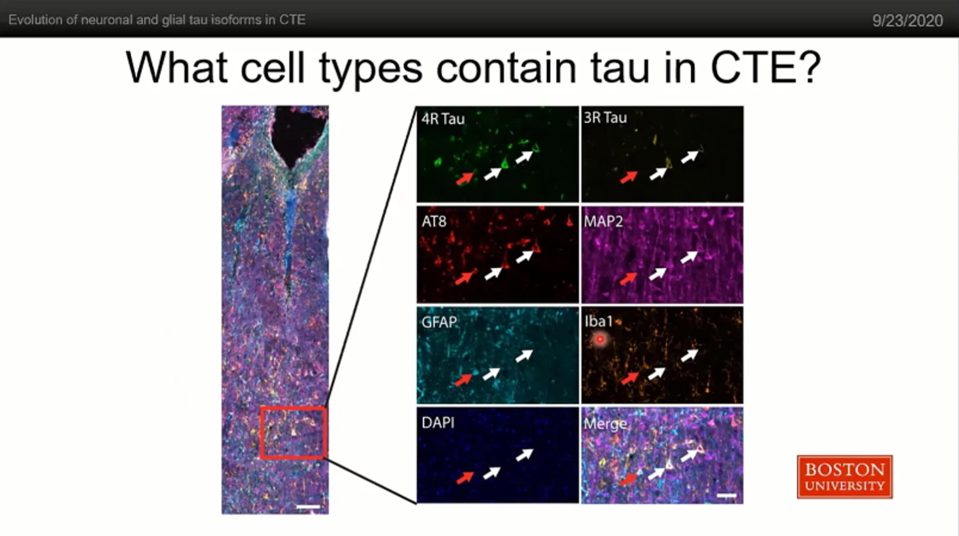
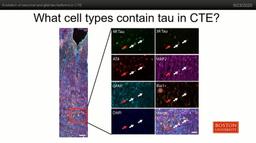
Evolution of neuronal and glial tau isoforms in Chronic Traumatic Encephalopathy (CTE)
In this video, Dr. Jonathan Cherry, Assistant Professor of Pathology and Laboratory Medicine at Boston University, speaks about how the opal staining platform and the Vectra Polaris imaging system enabled him to study the immune profile of diseased brain tissue, a historically challenging tissue type in terms of autofluorescence. Specifically, Dr. Jon Cherry discusses his work studying the evolution of neuronal and glial tau isoforms in Chronic Traumatic Encephalopathy.
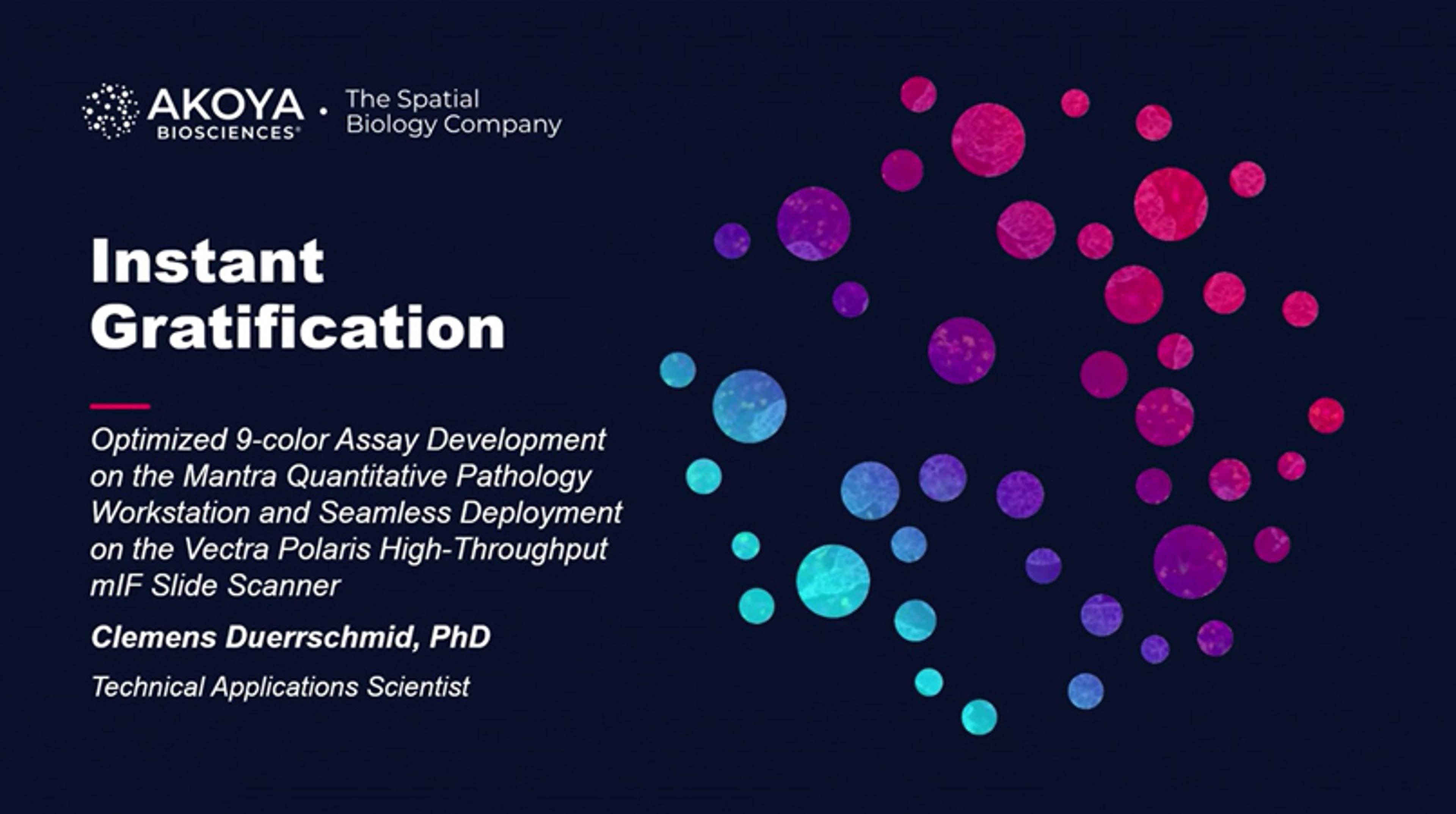
Instant gratification: Multiplexing immunofluorescence in a snap with Mantra
Here, Dr. Clemens Duerrschmid, technical applications scientist at Akoya Biosciences, discusses the ease and speed at which the Mantra™ 2 Quantitative Pathology Workstation allows Akoya to develop multiplexing immunofluorescence assays that are directly compatible with Akoya's larger high-throughput Vectra® Polaris™ scanner.
Next-generation cancer treatment: Patient stratification with immuno-oncology
This video explains how using biomarkers on tissue samples not only provides new insights into the biology of cancer but ultimately enables a new level of patient stratification. This allows the most effective treatment to be used for any given cancer leading to significantly improved outcomes for patients.

Multiplex immunofluorescence drives translational research
Find out why building expertise in multiplex immunofluorescence is critical in a translational clinical research setting

Breakthroughs in spatial biology
This special feature explores how researchers are examining the microenvironment of complex tumors, and highlights the recent advancements in microscopy and the latest discoveries driving progress in spatial biology research

Akoya Biosciences launches PhenoCode Signature Panels to accelerate development of predictive biomarkers for cancer immunotherapy
Each panel focuses on distinct areas of tumor biology and response to therapy which are of greatest interest to translational and clinical researchers
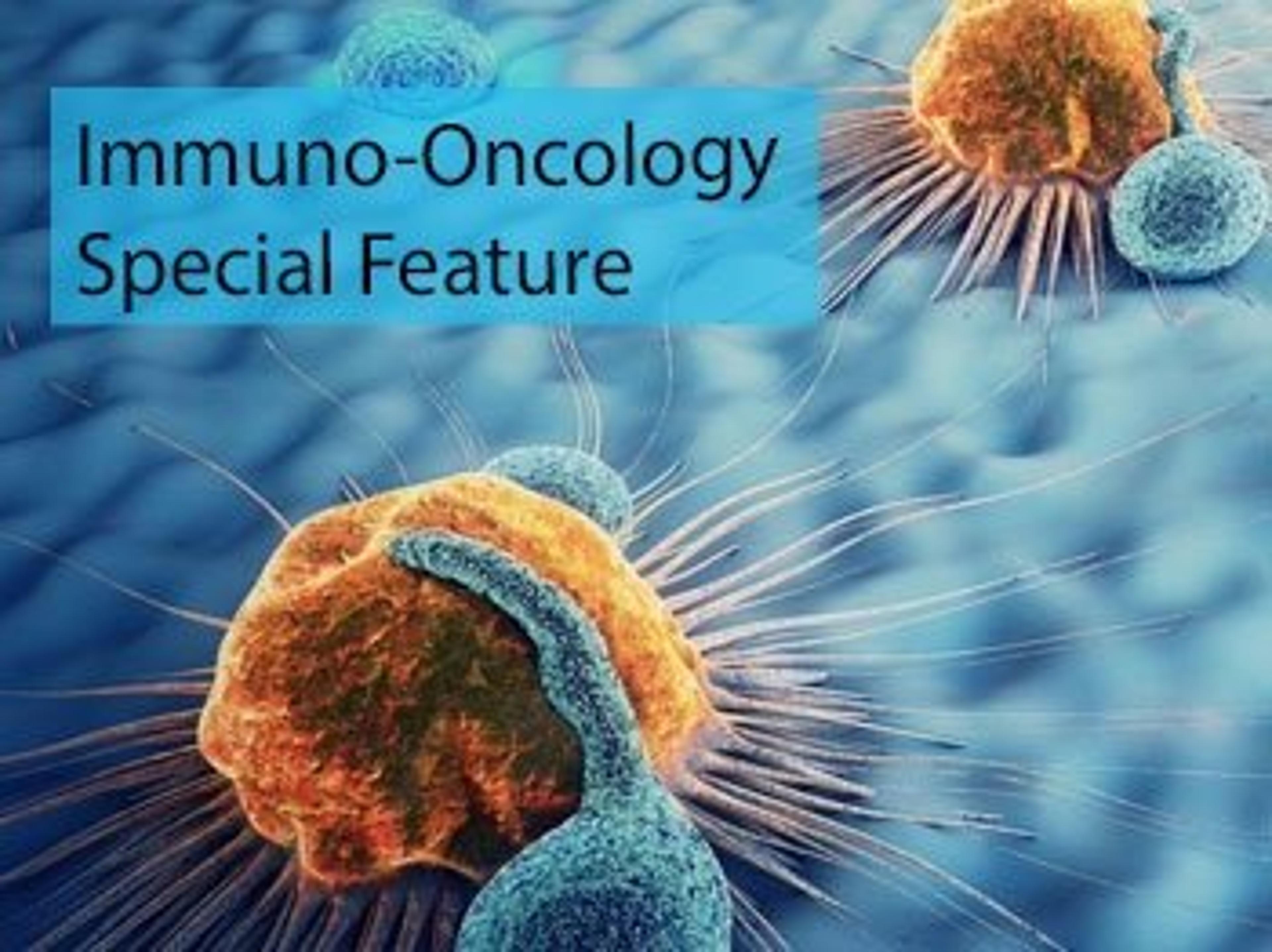
Immuno-oncology: Explore the future of cancer therapy
Discover the latest resources and innovative tools driving immuno-oncology research forward

Akoya and Johns Hopkins’ collaborative agreement supports pioneering new approach to immunotherapy biomarker discovery and validation
For the first time Astronomy and Pathology (AstroPath™) come together to discover and clinically validate new spatial phenotypic signatures using multiplex immunofluorescence
Top new resources to advance your immuno-oncology research
Exclusive interviews, free downloads, the latest methods and much more to help support your immuno-oncology research

9 top new resources for your biopharmaceuticals research
Exclusive interviews, new methods, free downloads and much more to help advance your biopharmaceutical research
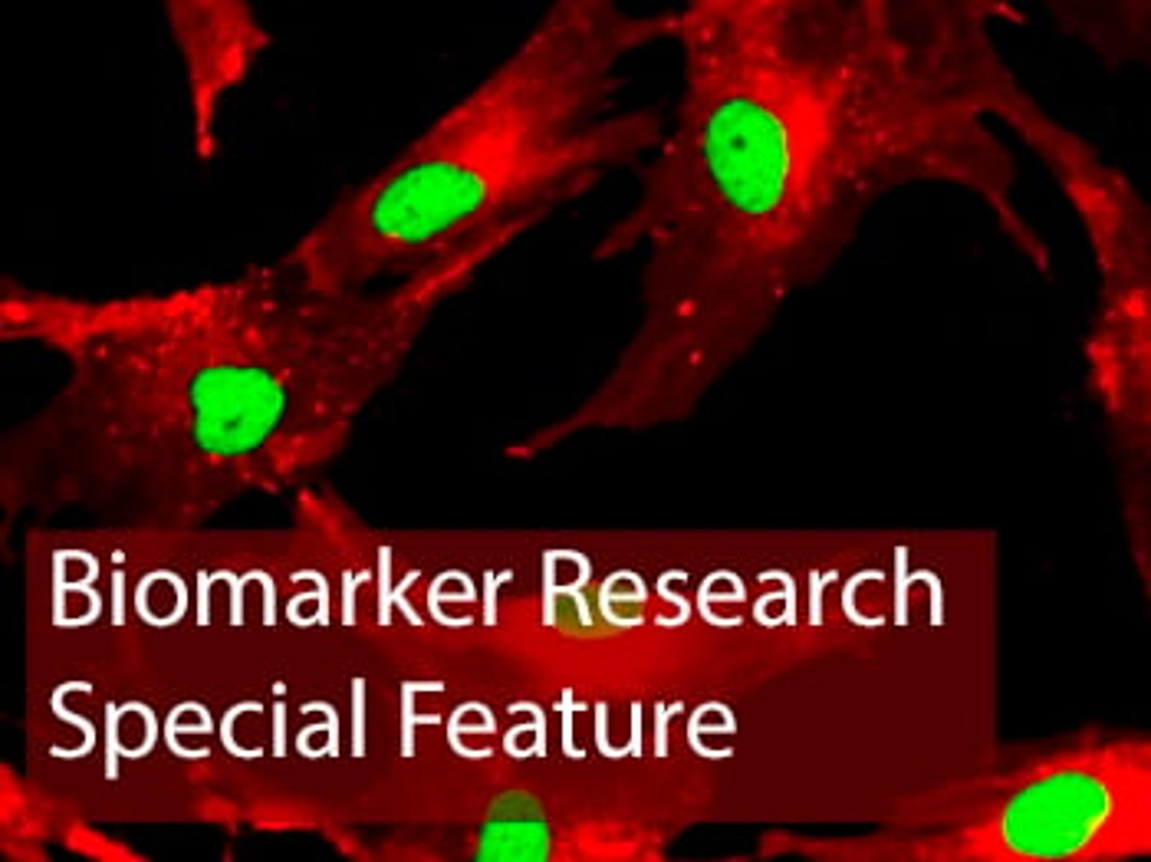
Latest advances in biomarker research: SelectScience special feature
The latest research and new method downloads: multiplex assays, ELISA, proteomics, genomics, imaging and more