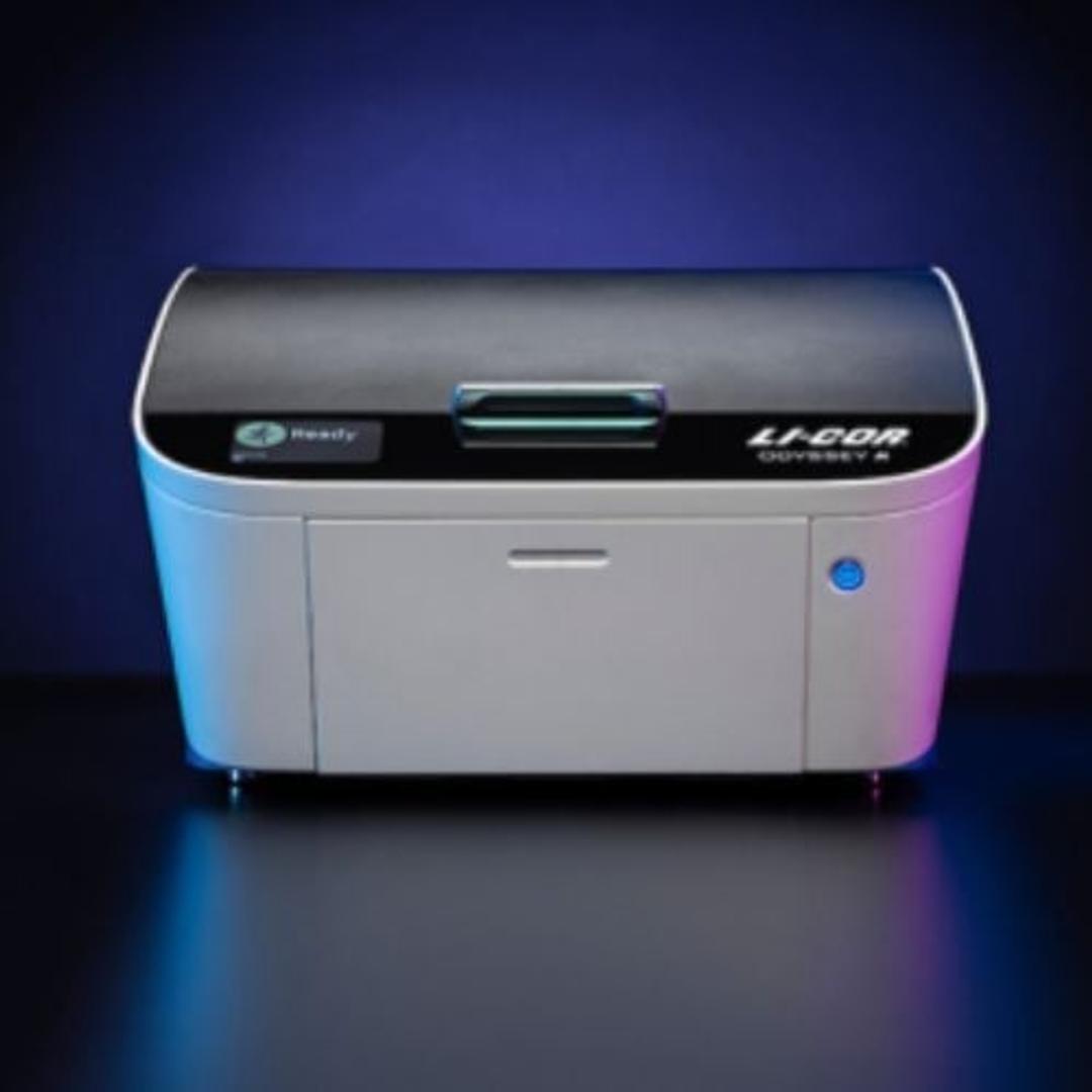
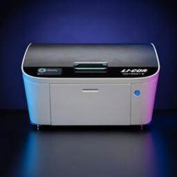
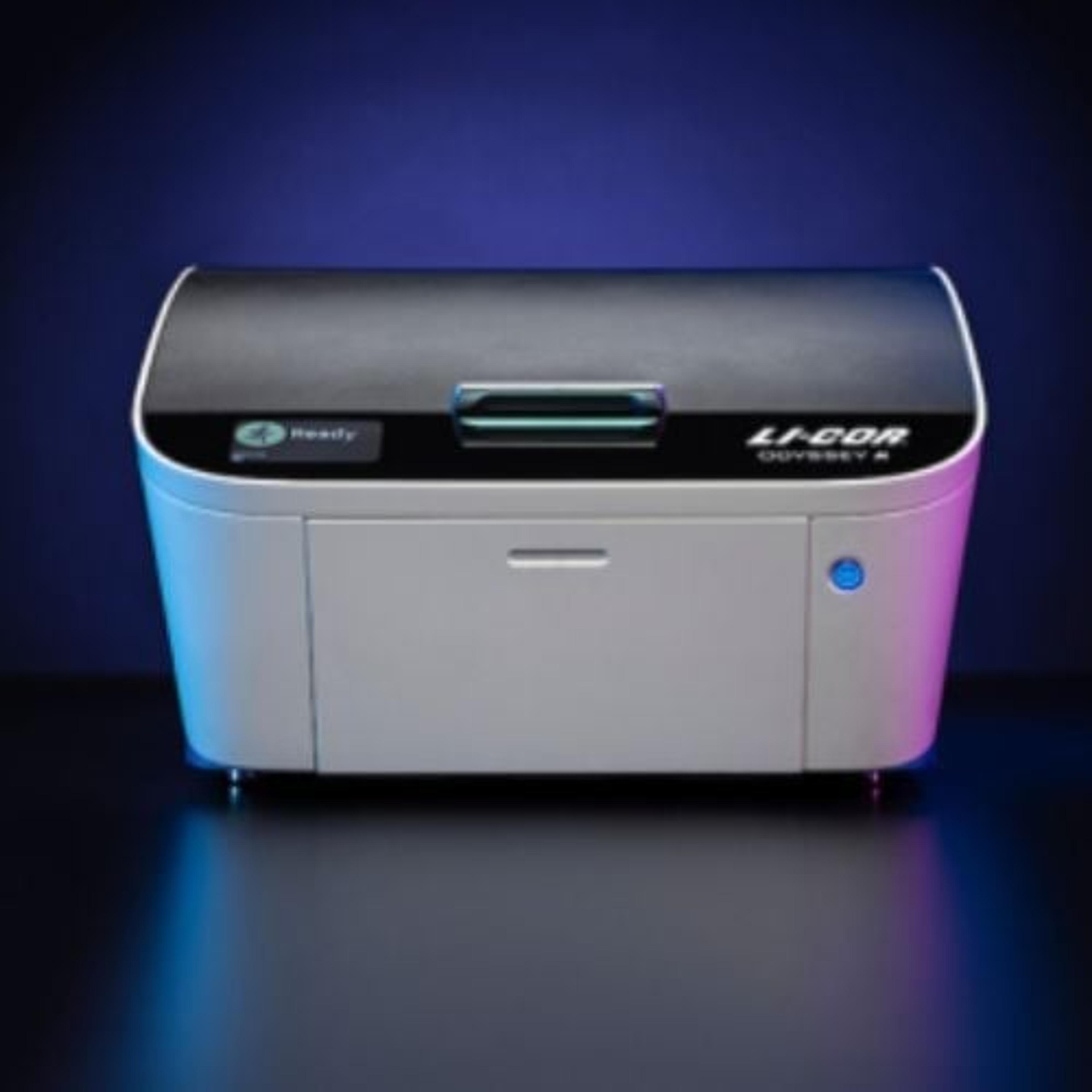
Odyssey M Imaging System
The Odyssey M is the most versatile and sophisticated biomolecular imager on the market. With 19 available channels, industry-leading 5 µm resolution, >6 logs of dynamic range, and an onboard luminescence module, the Odyssey M gives researchers a multimodal imaging solution with unparalleled capabilities and performance. Discover more with the groundbreaking Odyssey technology cited in over 40,000 peer-reviewed publications.

Receive your quote directly from the manufacturer.
Great results, amazing resolution, and low background. 2 channel scanning rocks.
Analyze protein expression in Western Blot
The Licor systems are great to use since they allow you to image at the same time your housekeeping protein and protein of interest by using the ability to scan in two different channels without having to deal with the background noise of one channel in the other. For this reason, you get a much better signal, and also, the resolution of the IR is much better than using the normal chemiluminescence machines, which allows you to quantify your proteins of interest with much better resolution. The ease of use is super simple since after the secondary you just have to place your membrane on the machine without having to use ECL or other detectors that if not evenly spread can affect your signal. With licor you just place the membrane and leave it there scanning and you can go and do something else and come back to a perfect scanned membrane. This saves you time in the long run and gives you perfect signal at the first try. Overall I am very happy with the Odyssey M and the Licor brand in general.
Review Date: 28 May 2024 | LICORbio
A superior multi-channel scanning system that delivers high quality images!
Fluorescence guided surgery
A superior multi-channel scanning system that delivers high quality images, is easy to use and saves time. If you need a high-quality, reproducible and at the same time easy to use imaging system, the Odyssey M is an excellent choice for your lab. Our lab was one of the Beta testing labs where we evaluated everything that this system had to offer. This scanner can scan 4 multi-well plates for in-cell western analysis at the same time using 4 fluorescent channels, twelve fluorescent or non-fluorescent IHC slides as well as other stained slides. A large scanning area would allow imaging of multiple Western blot memberanes or gels. The imaging time on this instrument can be reduced by selecting the region of interest. There are two highly user-friendly softwares for the acquisition and analysis of the data. I personally have used this system and can recommend it to any lab which requires imaging of above-mentioned items. At the end, I would like to thank LI-COR for their excellent support throughout the evaluation period.
Review Date: 29 Sept 2021 | LICORbio
The scans are high quality, clear, multi-channel (if desired), and easy to use.
Scanning slides, western blots, membranes, gels, and blocks of FFPE tissues at multiple channels
The Odyssey M is a high-quality image scanning system. Compared to the Odyssey CLx, the images are of higher quality and are acquired much faster. I am able to scan fluorescent IHC slides in multiple channels and detect colocalization. Scanning ~10 slides in two channels takes an hour, a very reasonable amount of time. The results are consistent when confirmed with our fluorescent microscope. The scans are reproducible. One of my favorite features is the pre-scan feature. The instrument will show you a preview of the area you have selected to scan so you can edit the area to ensure the sample is seen. Many of the other members of my lab have used this instrument for western blots and found it produced high-quality images. If you purchase this instrument, I highly recommend the empiria studio analysis software. This software has the same ease of use as the acquisition software. It guides you step-wise through image analysis. The instrument is very easy to use. It clearly prompts you through the workflow when acquiring images and analyzing them after. In order to critique the ease of use, my lab recommended that I not attend the training session and "blindly" attempt to use the machine. I found it quite easy to use. Our collaborators have had this same experience. We have been participating in Beta testing for this instrument, and we have had fantastic customer service from LI-COR. They respond in a timely manner and are always willing to help. I highly recommend working with them. This imaging system has the capacity to scan blocks of formalin-fixed paraffin-embedded tissues, stained slides, western blots, gels, membranes, and custom areas. It can image in 4 fluorescent channels as well as color. This instrument is of great value for any lab, as it can be used in many projects.
Review Date: 23 Sept 2021 | LICORbio
The Odyssey M is more than a biological assay imager. It’s a solution with innovative capabilities and performance you can’t find anywhere else. It’s a doorway to discoveries. It’s new research opportunities. It’s how you’ll empower your entire lab to follow the science wherever it leads.
The 19 channels of the Odyssey M can be imaged in dozens of combinations, while its uniform illumination and optimal sensitivity reveal robust data with every scan. Using the Odyssey M’s dynamic range of more than 6 logs, you’ll capture your full range of data in a single acquisition, and its unique line scanning technology enables complete data acquisition in minutes, not hours.
For more in-depth data from every sample scanned, we gave the Odyssey M a sophisticated sCMOS image sensor and industry-leading 5-micron resolution. No other imager can give you this level of sensitivity and dynamic range. With NIR and visible fluorescence, RGB, and optional luminescence capabilities—all 19 channels calibrated and integrated for consistent performance—you can easily perform a vast variety of supported and adjacent assays with confidence.
Perform both established and emerging gel-, membrane-, slide-, and plate-based applications, all on a single reliable instrument. With access to emerging applications, such as whole slide triage imaging, as well as already essential applications, the Odyssey M has the versatility to take your research wherever discover leads you.
Seamlessly image near-infrared and chemiluminescent Western blots, a wide variety of gel- and plate-based assays, and up to 12 slides or protein arrays with industry-leading clarity in an abundance of channels, including fluorescence and H&E. Plus, all LICORbio imaging systems and products give you access to LICORbio’s exceptional team of PhD support scientists.
LICORbio offers unparalleled support from real scientists who understand the high stakes of research. From the first demonstration to post-sale training and support, you’ll have a team you can trust every step of the way. LICORbio instruments are a lasting investment, delivering multi-functional capabilities for decades despite daily use and minimal maintenance.
LICORbio is who scientists turn to when data integrity and clarity of answers matter, because our imagers deliver unmatched dynamic range, sensitivity, and resolution for the highest data fidelity. That’s why our customers choose us again and again.

Near-Infrared (NIR) Western Blot Detection with the Odyssey Family Imaging Systems
Infrared fluorescence detection with Odyssey Family Imaging Systems provides a quantitative two-color detection method for Western blots. This application note is designed to help you achieve success with NIR Western blot detection methods.
IRDye® Peptide Labeling with Odyssey® Imaging Systems
Peptides labeled with fluorescent dyes are important as probes for in vivo imaging and as substrates for enzyme activity assays. Near-infrared (NIR) fluorophores such as IRDye® 800CW (excitation 780 nm; emission 794 nm) can offer improved sensitivity because of low NIR autofluorescence from tissues, cells, biological materials, or drug compounds. Fluorophores can also be combined with appropriate quencher dyes to create fluorogenic peptide probes, such as those used for protease activity detection. This application note provides an overview of IRDye® Peptide Labeling with LI-COR’s Odyssey® Classic, Odyssey CLx, Odyssey Sa, and Aerius Imaging Systems.
Scanning a Mouse on the Odyssey® System: Hints and Tips
This article presents several useful factors to consider when imaging mice using the Odyssey CLx infrared imaging systems.
Odyssey Western Blot Blocker Optimization for Near-Infrared (NIR) Detection
This document describes a method to determine optimal blocking conditions for NIR Western blot detection with the Odyssey family of Imagers. The specific lysate and antibodies used in your system will be evaluated in four different blocking buffer systems. The buffering system will also be evaluated in this experiment.
Maximizing the Performance of Your Western Blot
In this note find tips to make the most of your near-infrared (NIR) Western Blot. Some of the factors, including antibody quality, blocking agent selection, contamination and membrane variability that may alter the performance and imaging of a NIR Western blot are discussed. Methods of maximizing performance using the Odyssey Infrared Imaging System and Odyssey software are also described.
Seeing Beyond the Visible With IRDye<sup>®</sup> Infrared Dyes
This application note outlines IRDye infrared dyes, a useful tool for fluorescent labelling of protein and cellular assays, biochemical assays,microscopy, and in vivo molecular imaging. The dyes are bright, have excellent water solubility,and exhibit low non-specific binding. Background fluorescence is reduced at NIR wavelengths, enabling a variety of fluorescent applications that were previously impractical.
The Next Step in Near Infrared Fluorescence:IRDye<sup>®</sup> QC-1 Dark Quencher
This application note introduces the quencher dye, IRDye QC-1 for use with near-infrared (NIR) applications. IRDye QC-1 is an excellent quencher with essentially no inherent fluorescence and good water solubility, for use in many biological applications. This quencher dye also has the widest available quenching range, with the ability to quench both visible and near infrared fluorophores (~500 nm to ~900 nm).
2DE Gel Detection on the Odyssey Infrared Imaging System
The Odyssey Infrared Imaging System provides a sensitive and flexible choice for imaging two-dimensional gel electrophoresis (2DE) gels and blots. Here, LI-COR presents an overview of the 2DE process and viable options for Odyssey detection of proteins separated by 2DE.
Diet Considerations for In Vivo Animal Imaging Investigations
Diet considerations in 'in vivo' animal imaging are investigated in this application note from LI-COR. Near-infrared (NIR) optical imaging is a useful tool for in vivo study of animal samples, and diet prior to NIR has been shown to affect the quality of the result. A variety of diets are discussed with some recommendations as to various diets to avoid for optimum in vivo imaging outcome.
Odyssey® In-Gel Western Detection Using NIR Fluorescence
The Odyssey® Infrared Imaging system allows you to detect target proteins while still embedded in the gel using near-infrared secondary antibodies. Using near-infrared fluorescence detection methods for In-Gel Westerns makes this a powerful technique. Here, a protocol for In Gel Western analysis and tips for its optimisation is presented.
Find out why scientist's trust LI-COR's Odyssey imaging systems
In this video, LI-COR explains how its Odyssey imaging systems promise to provide excellent data quality including three colour western blots, in-cell western assays, and cell analysis.
Product Overview
Links
Featured products
Request Quote for All Products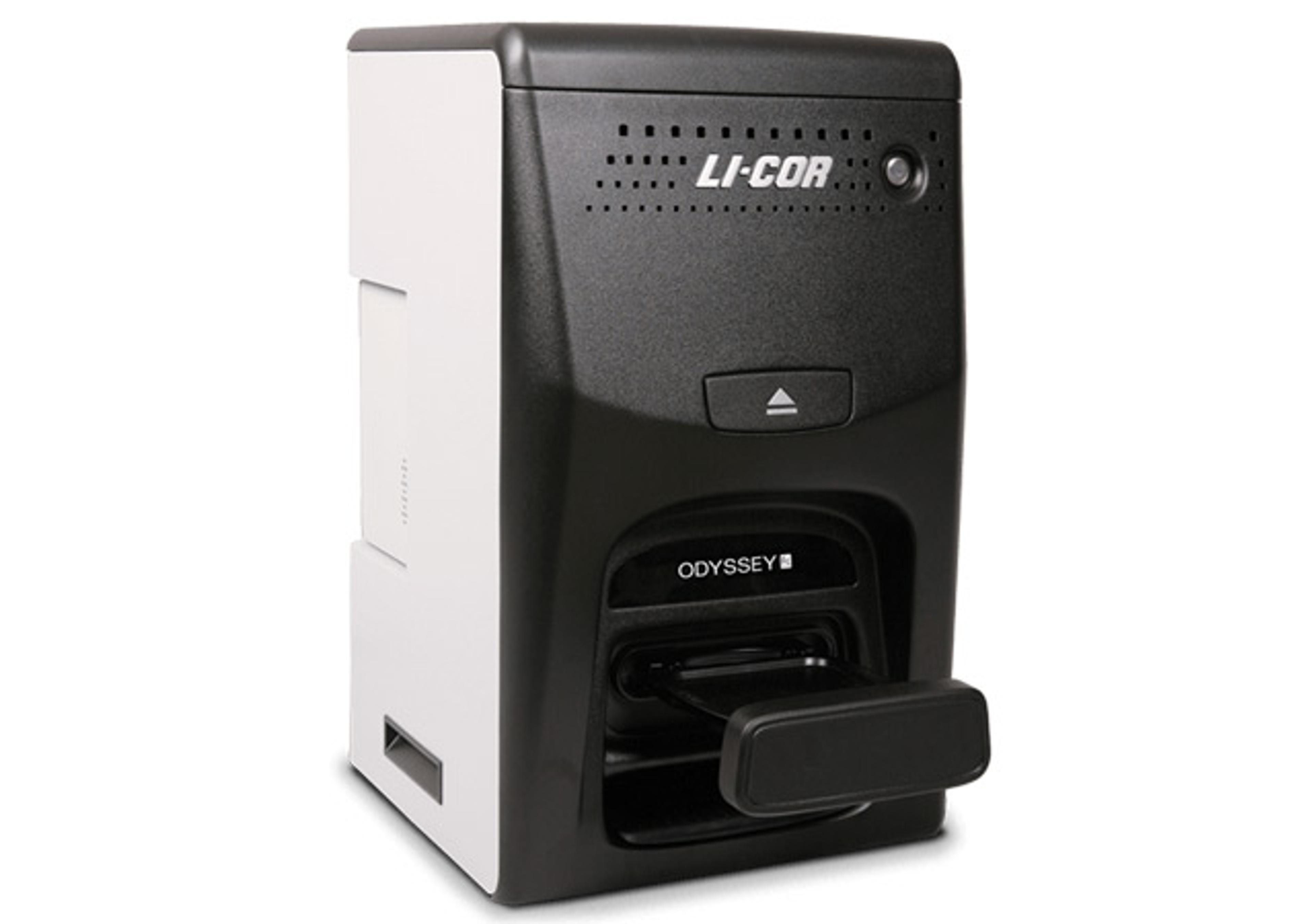
Odyssey Fc Imaging System
LICORbioThe Odyssey Fc System is the only imaging system that offers superior chemiluminescent detection and the benefits of quantitative infrared Western blot detection as well as the ability to document DNA gels using the 600nm channel.
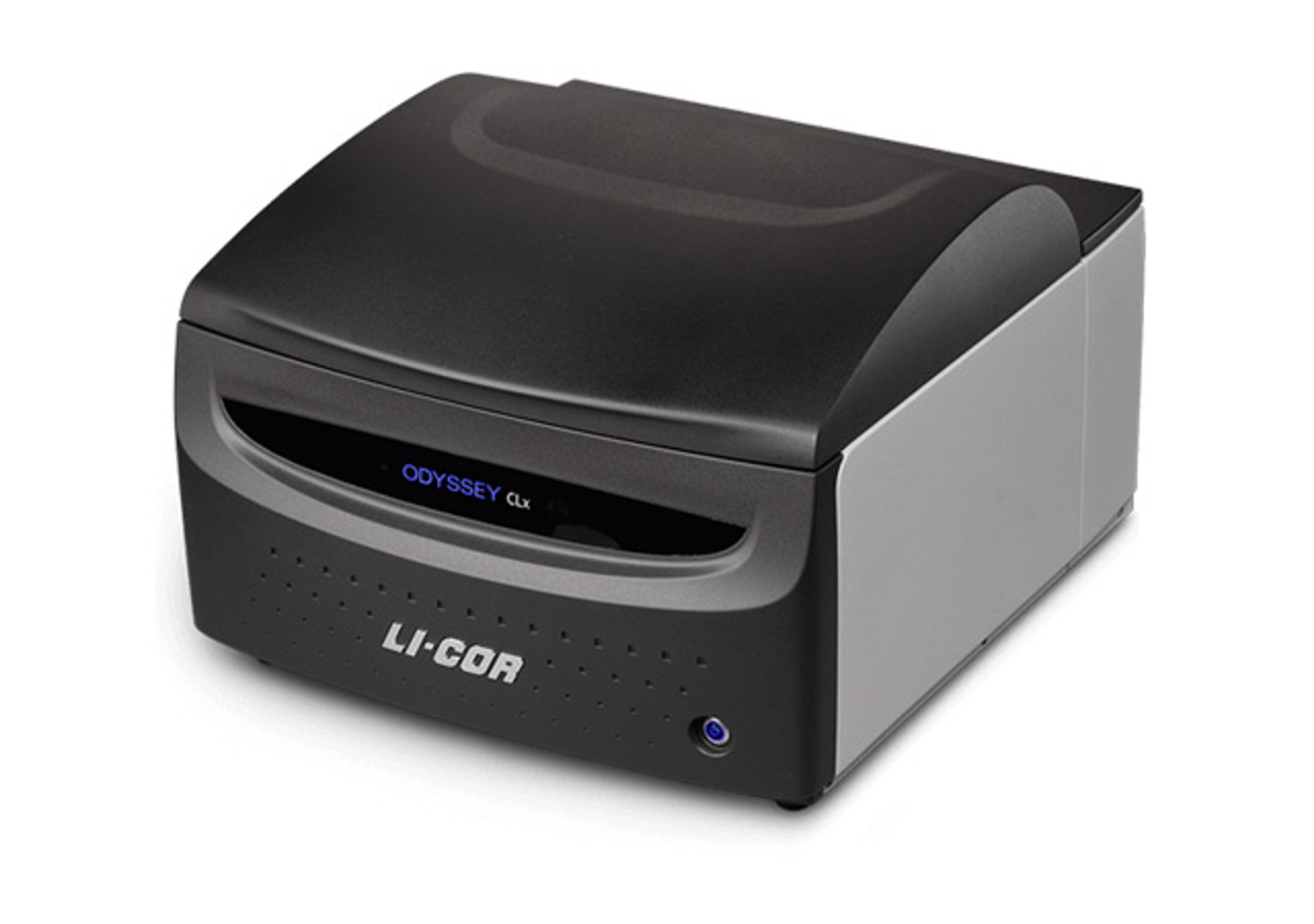
Odyssey CLx Infrared Imaging System
LICORbioThe Odyssey® CLx Infrared Imaging System gives you accurate, reproducible quantitative Western blotting results with near-infrared fluorescence.