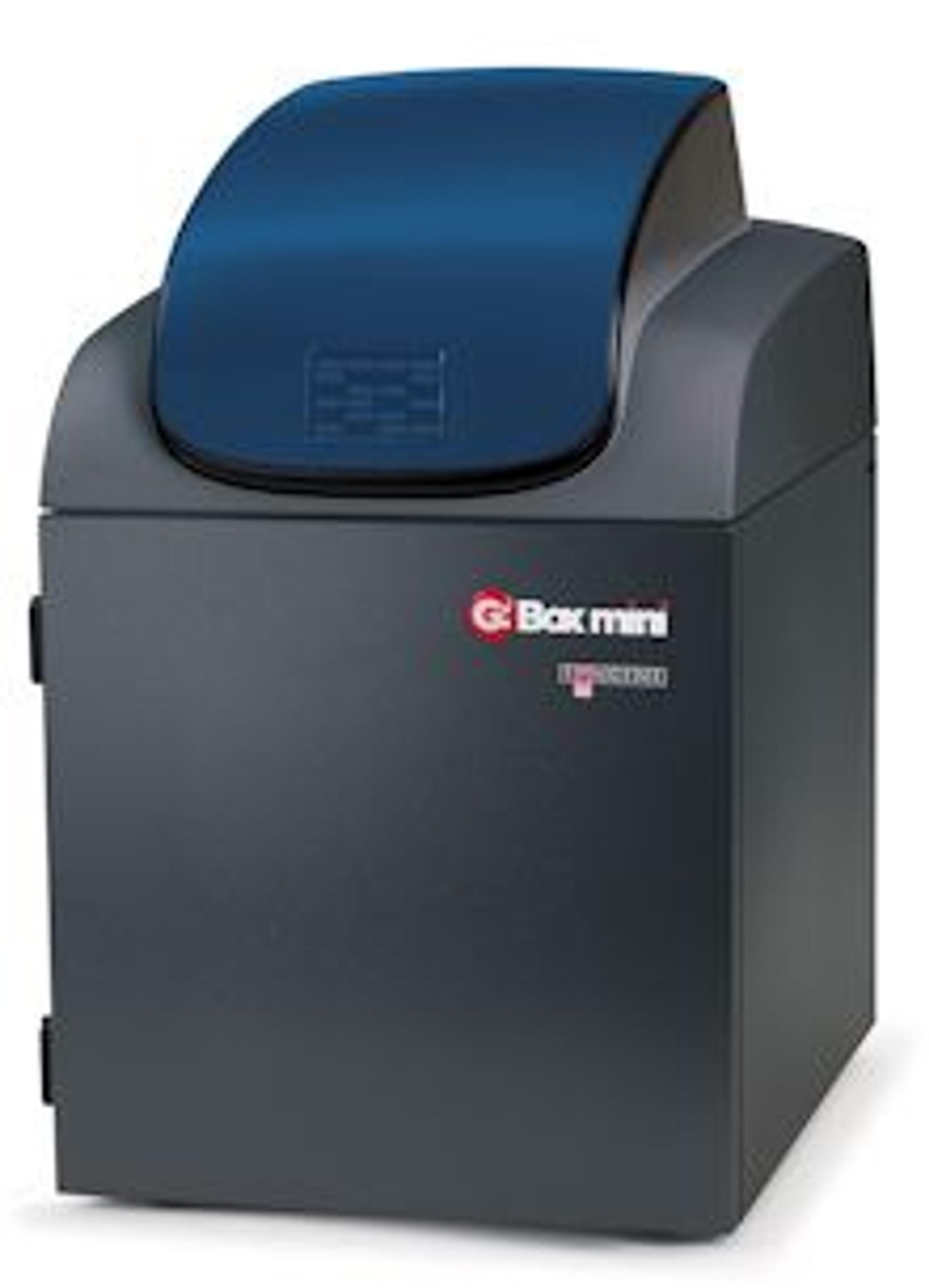
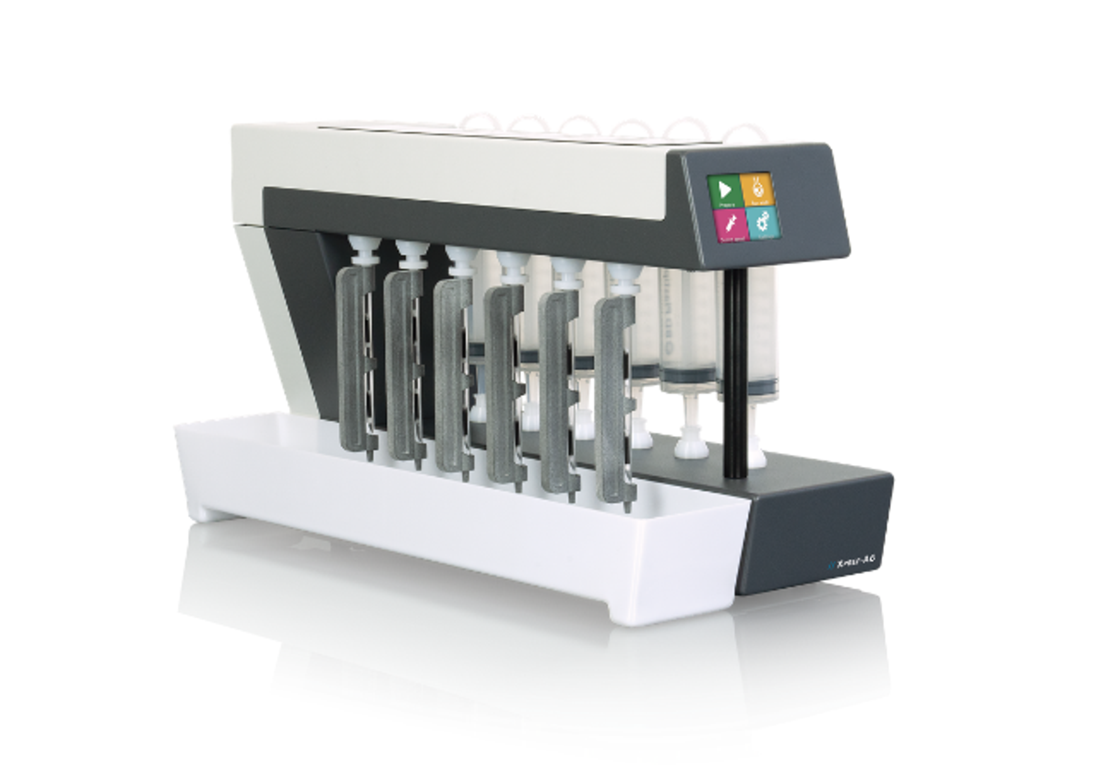
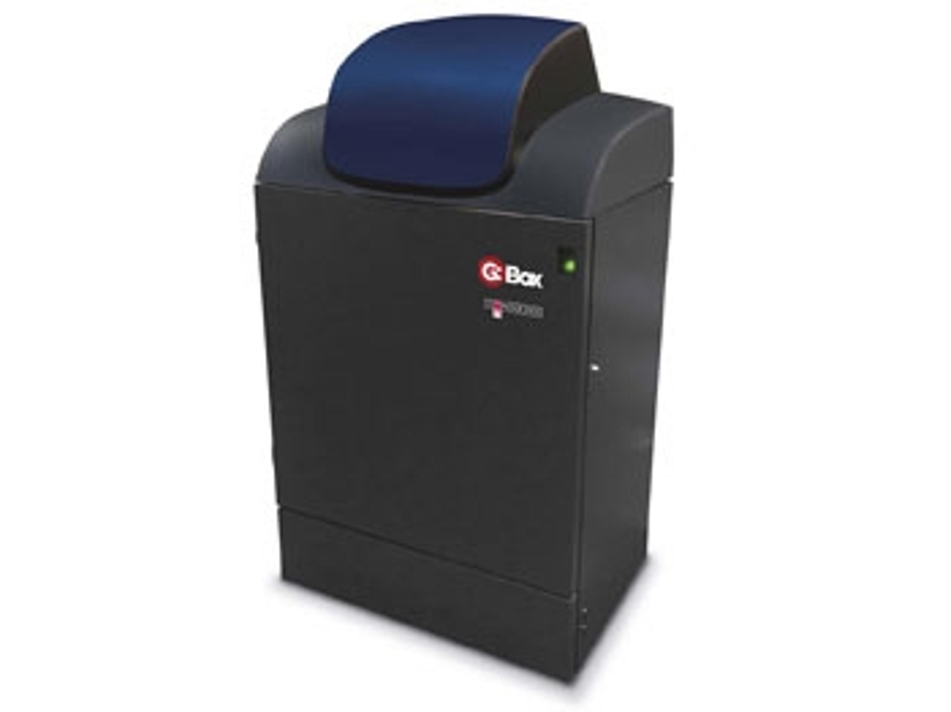
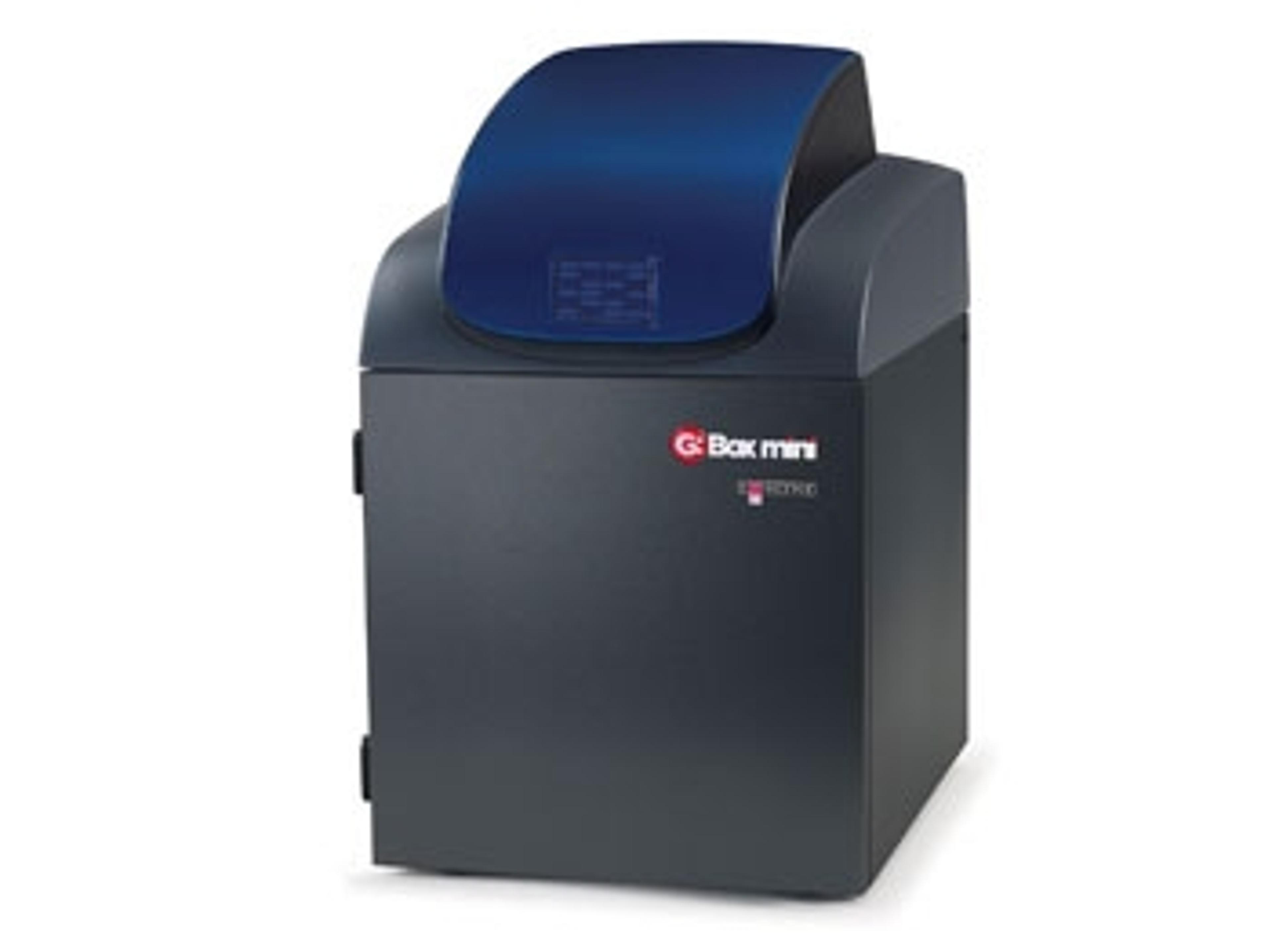
G:BOX mini
G:BOX mini is a compact, multi application imaging system for accurately imaging mini and midi fluorescence and visible gels, multiplex fluorescence westerns, stain-free gels and chemiluminescent blots.


The supplier does not provide quotations for this product through SelectScience. You can search for similar products in our Product Directory.
Great investment.
Western blots on mouse tissue samples
Very user-friendly software, that is easy to learn. The imager occupies a small lab space that is a huge advantage. There is a possibility to upgrade to fluorescence, that might worth considering.
Review Date: 13 Dec 2017 | Syngene
G:BOX mini is a compact, multi application imaging system for accurately imaging mini and midi fluorescence and visible gels, multiplex fluorescence westerns, stain-free gels and chemiluminescent blots.
Need to quantify multiple different fluorescent proteins on one blot and capture images of chemi blots, but your lab is limited on space? Then the G:BOX mini is just the compact imaging powerhouse you’ve been looking for. With stain-free, RGB and IR excitation options for Alexa Fluor® 488, 546, 647 and IR dyes, you’re just a click away from brilliant multiplex images from one single Western.
Using the G:BOX mini you’ll also get perfectly exposed images of your chemi labelled proteins without the bother of film. Simply place your blot in the darkroom, click on GeneSys capture software and your imager will do the rest with no fuss and no messy chemicals.
G:BOX mini is a compact, multi-application imaging system combining a unique motor driven stage and cooled high resolution 6 or 9 megapixel camera enabling you to generate accurate optical images, not just digitally enhanced ones. With a G:BOX mini you’ll see separate close chemi and fluorescent bands or spots even on complex gels and know they’re really part of your data.
HI-LED lighting options cover the full spectrum of high intensity blue, green, red and infra-red resulting in faster exposure times and publication quality images.
The system is controlled by GeneSys application driven image capture software and comes complete with unlimited copies of GeneTools analysis software.
Features:
- Small footprint
- Leaves plenty of room on any lab bench
- RGB and IR HI-LED lighting options
- Fast quantification of multiple fluorescent proteins without stripping and re-probing
- Stain-free imaging capability
- Capture images of TGX Stain-Free™ FastCast™ acrylamide gels and many more
- High quantum efficiency (QE) camera
- Detect picogram or femtogram amounts
- Luxury lens (F/0.95 motor driven) with data feedback
- Captures the highest quality images
- Automatic motor driven stage with automated focus
- For real, optical images
- White, UV and blue lighting options
- Image all gels and blot types on one system
- Cleverly designed screen mount option
- Easily and securely attach a monitor
- GeneSys application driven image capture software
- Contains extensive database of dyes and imaging protocols. All you need to know is the type of gel you’re using and GeneSys automatically selects the optimal lighting and filters to produce the perfect image
- GeneTools analysis software (unlimited copies)
- Analyse data at your own computer
Brochures

G:Box Mini: Detection with a Difference
Built on the successful G:BOX range, the G:BOX mini is a compact, multi-application powerhouse for accurately imaging fluorescence and visible gels, multiplexed fluorescence westerns, stain-free gels and chemiluminescent blots.
Comparing Criterion TGX Stain-Free gels to standard Coomassie staining procedures
In this application note, Syngene outlines how users can successfully image Criterion TGX Stain-Free gels using any Syngene G:BOX imaging system by using the recommended lighting and filter combination, 302nm UV transilluminator and UV filter. The stain-free method of visualizing cells has comparable sensitivity to that of more traditional techniques such as Coomassie Blue Safe staining and also has the potential to significantly reduce the lengthy SDS-PAGE workflow.
SafeView Dye: An Overview
This application note provides a brief overview of SafeView dye from NBS Biologicals, a nucleic acid stain used in gel electrophoresis for the detection of double-strandedA (dsDNA), single-stranded DNA (ssDNA) or RNA.
Overview of Ethidium Bromide Dyes
This application note provides an overview of ethidium bromide dyes, commonly used in agarose gel electrophoresis to detect double-stranded DNA (dsDNA) and single-stranded DNA (ss) or RNA.
An Overview of Alexa Fluor 647 Dyes
This application note provides an overview of Alexa Fluor dyes, a series of fluorescent dyes produced by Molecular Probes that span the visible spectrum.
How to Visualize SYBR Dyes Using a Syngene Image Capture System
Learn how Syngene image capture system,s combined with GeneSnap software are the perfect combination for imaging SYBR dyes.

Syngene releases GeneSys image capture software for improved accuracy of gel and western blot data
The GeneSys software now combines sample positioning and image capture on one screen, making it easier for scientists to set up and image gels and blots in fewer clicks

New QuickQuant Feature in GeneSys Software
Save time with easy-to-use analysis alongside image capture