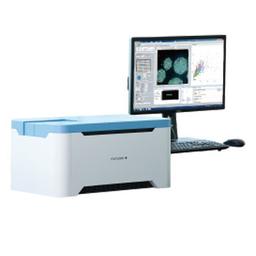
CellVoyager CQ1 Benchtop High-Content Analysis System
Compact footprint, lightweight bench-top device; no need for a darkroom.
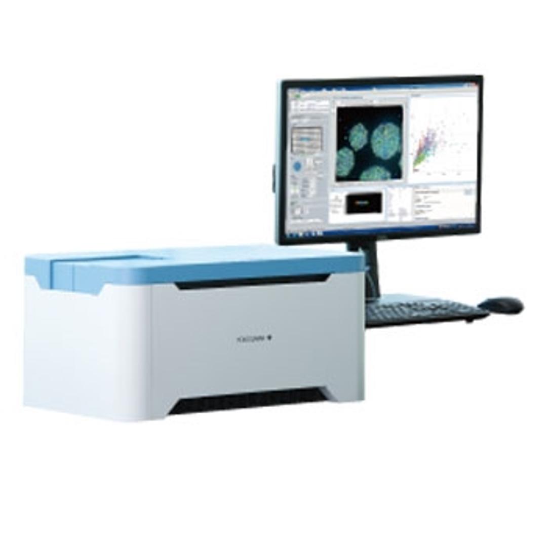
CellVoyager CQ1 Benchtop High-Content Analysis System


The supplier does not provide quotations for this product through SelectScience. You can search for similar products in our Product Directory.
Great instrument for the money!
Drug discovery
Excellent bench top confocal high content instrument with an easy to use interface. Exceptional value for the engineering and feature set.
Review Date: 30 Dec 2021 | Yokogawa Corp. of America
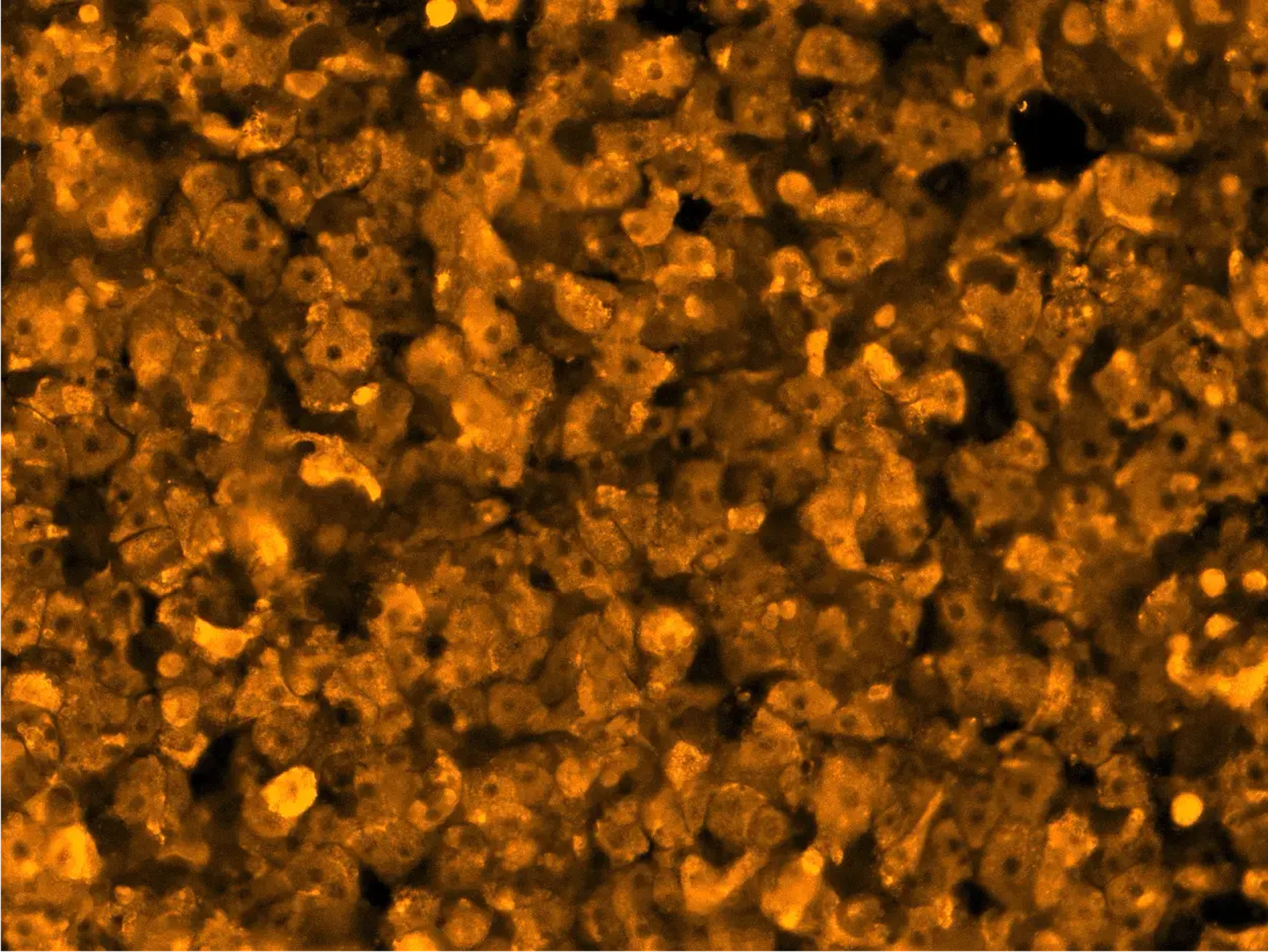
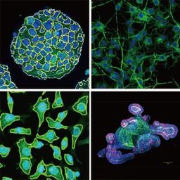
CellVoyager CQ1 enables 3D imaging and quantification of live cell clusters, such as spheroids within a 3D culture vessel, as they are, keeping the cells intact. CellVoyager CQ1 exports feature data in general formats which are readable by various third-party software for advanced data analysis. It is possible to construct a fully customized CellVoyager CQ1-based system by integrating with external systems, via robot for culture dish handling.
Enables measurement of spheroids, colonies, and tissue sections
- No need to remove cells from the culture dish, in contrast to traditional flow cytometry
- Nipkow spinning disk confocal technology allows high-speed yet gentle 3D image acquisition
- Rich feature extraction to facilitate sophisticated cellular image analysis
- Wide field of view and tiling capability enables easy imaging of large specimen
Enables analysis of time-lapse and live-cell
- High precision stage incubator and low phototoxicity of our confocal makes the analysis of time-lapse and live-cell are possible
- Max.20fps option for fast time lapse
- CQ1
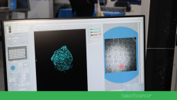
High-quality image and similar operability to a traditional flow cytometer
- Feature data and statistical graphs displayed in real-time with image acquisition
- Usable high-quality image as confocal microscope image.
- Easy to trace back to the original image from a graph spot, and make repetitive measurements
Open platform
- Connectable with external systems via handling robot
- Expandable to the integrated system as image acquisition and quantification instrument
- FCS/CSV/ICE data format readable by third-party data analysis software
- A variety of cell culture and sample dishes are applicable

Applications of high-content imaging in 3D models of disease
Generating translatable high-content imaging data from physiologically-relevant cell models, including 2D and 3D structures, is extremely valuable for drug discovery and pre-clinical research. In this webinar, James Evans, CEO of PhenoVista Biosciences presents case studies on how Yokogawa’s Benchtop CQ1 Confocal System can improve throughput and standardize processes for complex 3D cell-based phenotypic assays.
Key learning objectives:
- Strategies for designing and implementing high-content screening assays
- Approaches for deciding between 2D and 3D model systems
Who should attend?
Scientists and researchers from industry and academic backgrounds who are interested in high-content imaging and specific applications including:
- Living-cell imaging
- Organoids and spheroids
- 3D high-content imaging
- Organ on a chip
- Microfluidics
- Cell painting
- Drug screening/discovery

Biomarker detection in 3D cell models using automated immunofluorescence staining
Physiologically relevant 3D cell models are essential for drug discovery and preclinical research due to their functional and architectural similarity to solid tumors. One of the challenges faced by researchers is that many of the assays using these precious samples tend to be manual and tedious.
Using proprietary microfluidics technology, Protein Fluidics has created the Pu·MA System for automated complex 3D cell-based assays. In this webinar, application scientist Dr. Katya Nikolov will present her work on combining this novel automation technology with Yokogawa’s high-content imaging systems for biomarker detection in 3D cell models. Nikolov will demonstrate the utility of an automated immunofluorescence staining workflow followed by confocal imaging within the Pu·MA System flowchips. This automated workflow enables quantitative assessment of biomarkers which provides valuable data for further understanding disease mechanisms, preclinical drug efficacy studies, and in personalized medicine.
Attendees of this webinar will explore:
- The Pu·MA System and novel technology for automated 3D cell-based assays
- How to perform automated immunofluorescence staining for biomarkers with a “hands-off” assay workflow
- How to visualize biomarkers after the assay with high-content imaging within the flowchip
Advancing high-content imaging for biomedical discoveries
Explore how to achieve versatility, reliability and ease-of-use in your high-content cell imaging with Dr. James Evans, CEO and Founder of PhenoVista Biosciences. Dr. Evans shares how the Cell Voyager CQ1 from Yokogawa is supporting the delivery of PhenoVista's in vitro cell-based assay services, and explains how the platform excels in extracting data and providing detailed insights into subpopulation and spatial information for protein locations and disease mechanisms.
This video was filmed at SLAS2025.
Biomarker detection in 3D cell models using automated immunofluorescence staining
Watch this webinar on demand to learn how to automate your immunofluorescence staining workflows

Masterclass on the applications of high-content imaging in 3D models of disease
In this on-demand webinar, Dr. James Evans describes how he generates imaging data from physiologically relevant cell models.