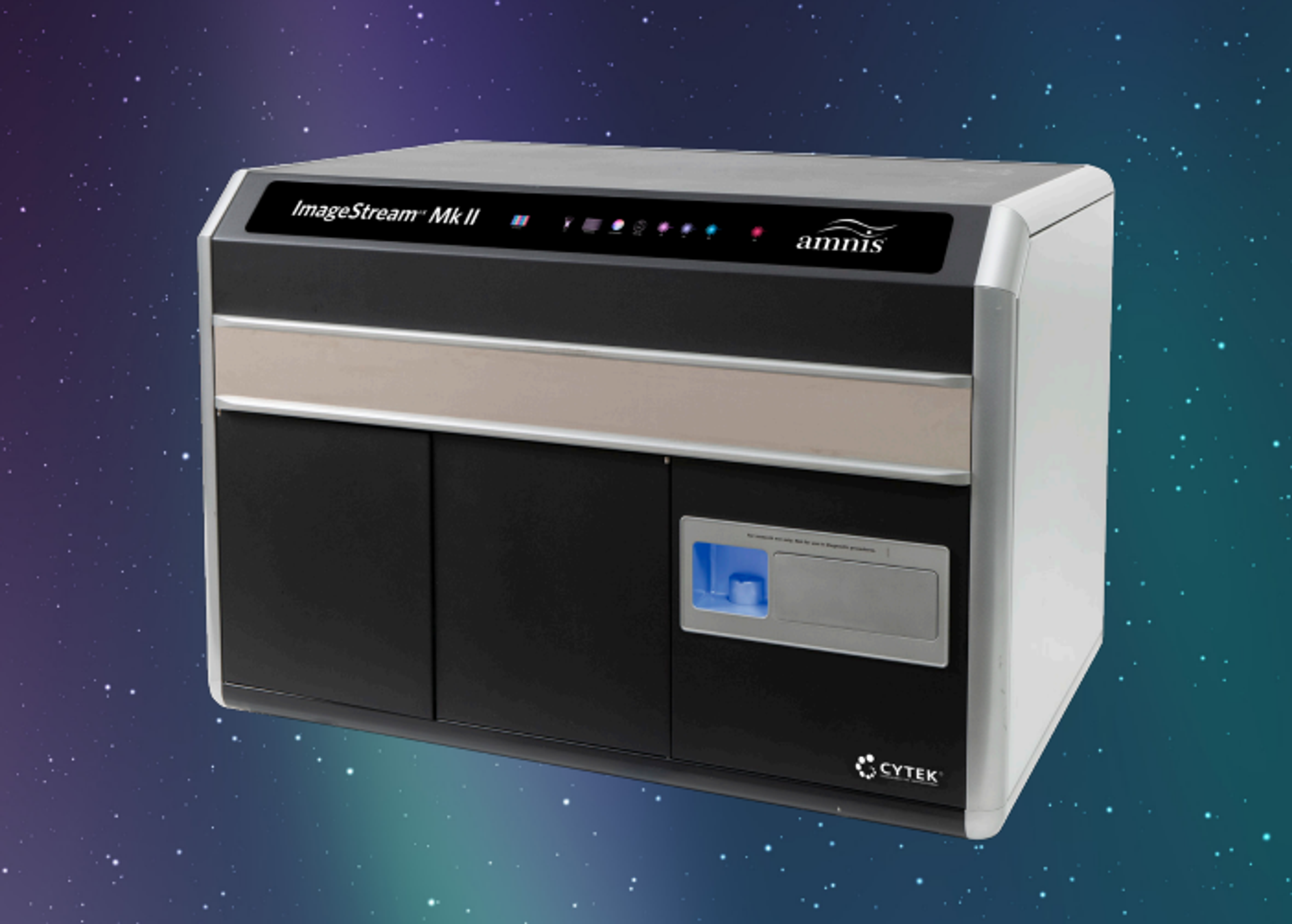
Amnis® ImageStream® XMk II
The revolutionary ImageStream ® X Mk II Imaging Flow Cytometer combines the speed, sensitivity, and phenotyping abilities of flow cytometry with the detailed imagery and functional insights of microscopy. This unique combination enables a broad range of applications that would be impossible using either technique alone.


The supplier does not provide quotations for this product through SelectScience. You can search for similar products in our Product Directory.
Excellent data quality, unique applications
Imaging Flow Cytometry
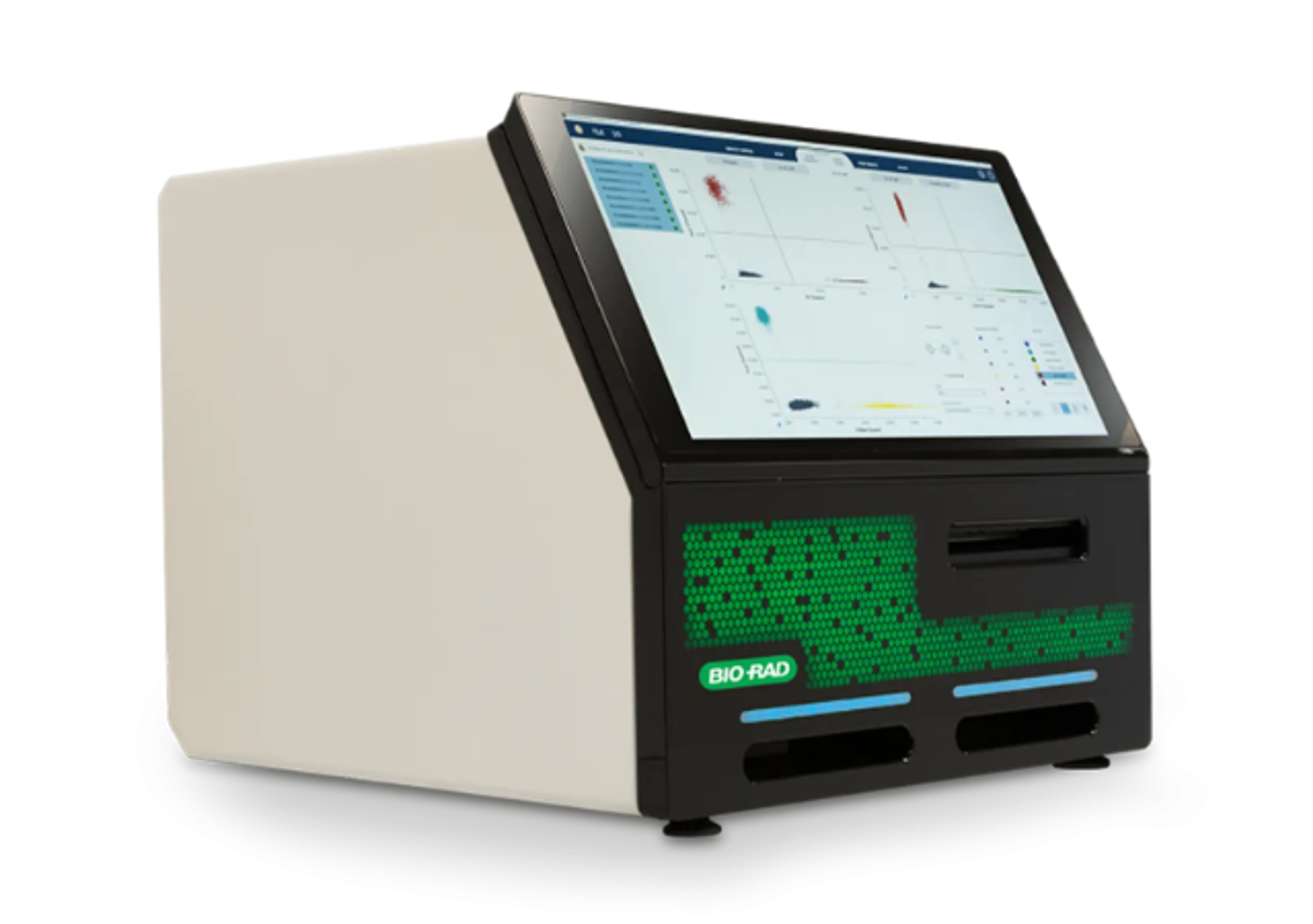
The instrument combines the information-rich imagery of microscopy with the high-throughput rapid acquisition of flow cytometry. It allows acquisition of bright-field and up 10 fluorescent images of cells in flow, up to 5000 cells / second. It has 20X, 40X and 60X magnification, the minimal pixel size is 330nm. Among the common application are co-localization, internalization, morphology, spot count and many others. The data analysis is done by a dedicated image analysis software, IDEAS. It allows robust, un-biased high throughput quantification of morphological features. I have been using it for the last 12 years, resulting in over 100 publications in numerous disciplines in biology, including immunology, cancer, cell biology, microbiology, marine biology, and many others. I can highly recommend it for all cellular applications.
Review Date: 20 Oct 2023 | Cytek Biosciences
Easy to use and versatile
Cellular biomarkers
Easy to use. It does not require much maintenance. Good resolution and versatility. However, services are difficult to obtain due to changes in service providers.
Review Date: 20 May 2022 | Cytek Biosciences
Excellent technology for high-throughput image analysis at medium resolution.
CAR T-cell synapse analysis
This is a high-throughput image analysis platform for rare events. CAR-T-cell/target cell synapses are identified with high precision and efficiency. The company provide extensive support for application advice and troubleshooting. Probably the highest-throughput image analysis system for its price.
Review Date: 29 May 2020 | Cytek Biosciences
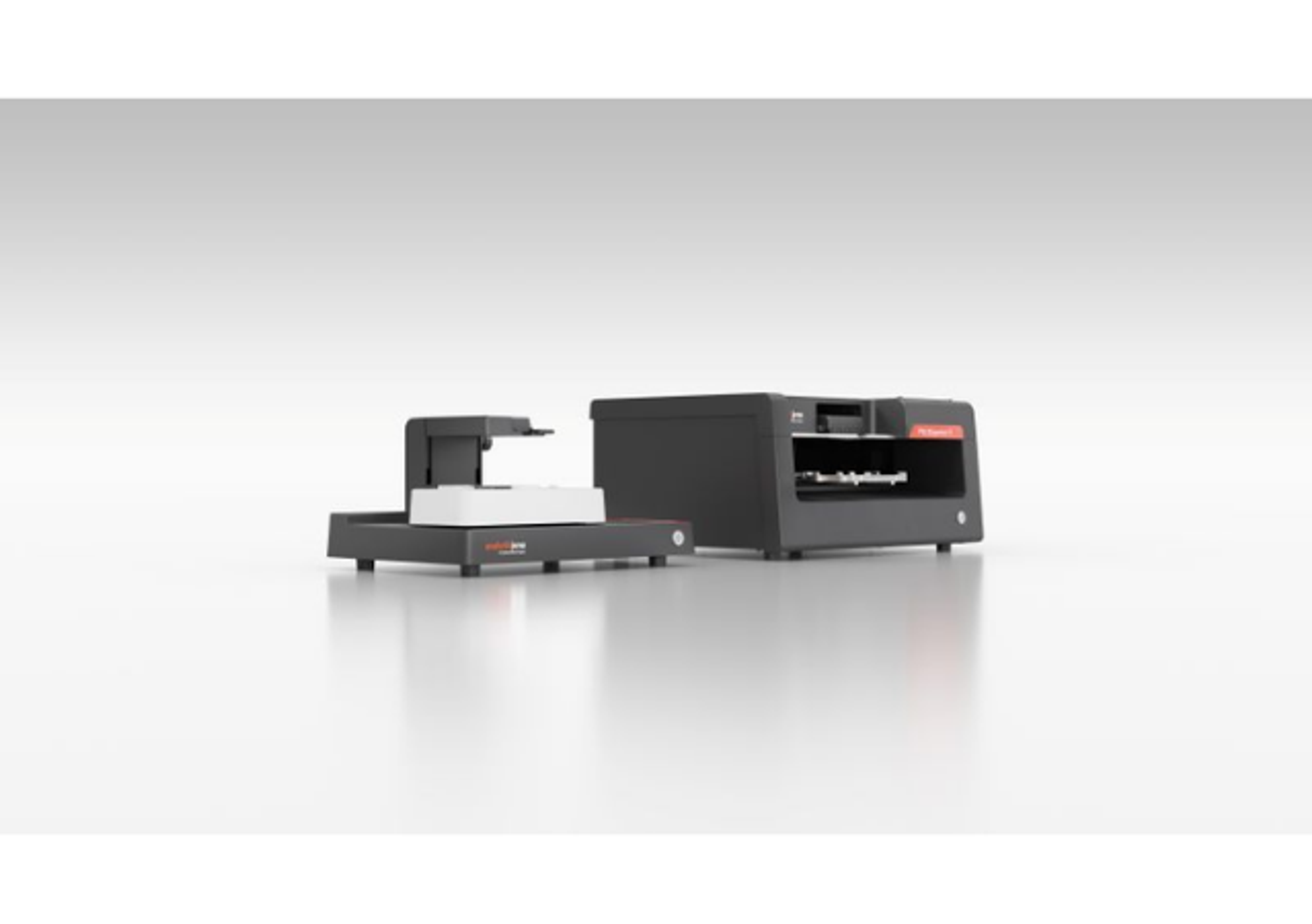
The Cytek® Amnis® ImageStream® XMk II imaging flow cytometer combines the high-throughput performance of flow cytometry with the imagery and functional insights of microscopy. This unique combination enables a broad range of applications that would be impossible using either technique alone.
The ImageStream XMk II system is a benchtop, multispectral, imaging flow cytometer designed for the rapid acquisition of millions of cells with up to 12 channels of cellular imagery. The instrument collects large numbers of digital images, performs spectral compensation, and provides high-quality images of every cell in your sample. Combined with our advanced image analysis software, where each object is measured for hundreds of parameters using not only fluorescence intensity but the fluorescence spatial location as well, the ImageStream system offers an unprecedented level of cellular information.

Detection of fluorescent nanoparticles using the Amnis CellStream flow cytometer
In this application note, Luminex describes the identification and quantification of fluorescent nanobeads (“microspheres”) on the Amnis® CellStream® Flow Cytometer.
Single cell protein secretion assays using Nanovials and the Amnis ImageStream XMk II Imaging Flow Cytometer
In this application note, Luminex describes a novel method involving a uniform hydrogel particle platform, created by Partillion Bioscience, called nanovial. Imaging flow cytometry is then used to simultaneously measure the individual cellular characteristics and secretions released from each specific cell.

Size and fluorescence calibrated imaging flow cytometry: From arbitrary to standardized units
Thursday, November 20, at 15:00 GMT | 16:00 CET | 10:00 EST | 7:00 PST
Imaging flow cytometry (IFC) has emerged as a powerful approach for single extracellular vesicle (EV) characterization, yet its broader adoption has been hindered by challenges in assay standardization and data comparability. This webinar will highlight recent methodological advances addressing these limitations. Building on earlier work that established workflows for direct detection and phenotyping of EVs in unprocessed plasma, current efforts focus on developing rigorous calibration strategies for IFC.
Recent work has introduced bead-based size calibration and fluorescence intensity standardization, enabling instrument-specific signals to be expressed in quantitative units. These approaches markedly improve reproducibility across different platforms and laboratories, thereby supporting cross-study comparability. Standardized IFC data further provide a framework for quantitative analyses in both fundamental EV research and the quality assessment of vesicle-based therapeutics.
Together, these methodological advances position IFC as a calibrated, quantitative platform for nanoparticle cytometry, with the potential to accelerate both biomarker discovery and translational applications in extracellular vesicle science.
Topics discussed in this webinar will include:
- Advances in EV assay standardization and data comparability
- Development of calibration strategies for IFC
- Introduction of bead-based standardization methodology for quantitative EV analyses
Who should attend?
- Researchers using IFC who are currently or wish to perform small particle analysis
- Anyone interested in flow cytometry applications and small particle analysis
Certificate of attendance
If you attend the live webinar, you will automatically receive a certificate of attendance, including a learning outcomes summary, for continuing education purposes.
If you view the on-demand webinar, you can request a certificate of attendance by emailing editor@selectscience.net.
For Research Use Only. Not intended for use in diagnostic procedures.
Imaging rare tumor cells in flow with Amnis systems
In this video from Luminex, Drs. Paul Wallace and Hans Minderman of the RPCCC Flow and Image Cytometry facility discuss how Amnis® systems have helped their researchers detect rare events like circulating tumor cells.
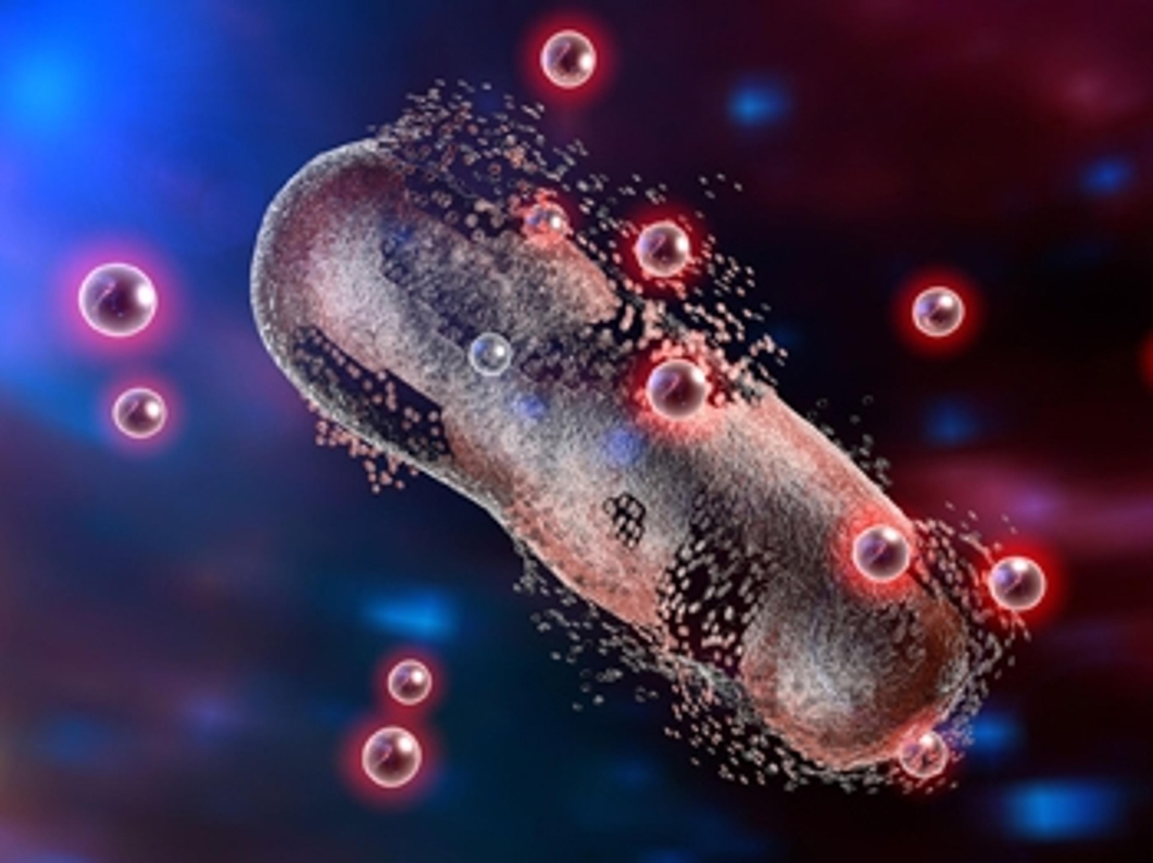
Enhanced performance of nanoparticle-mediated drug delivery systems
A Presidential award-winning laboratory has developed a new technology to address one of the most pressing challenges in nanomedicine
Using DNA repair and machine learning to improve cancer risk prediction
Scientists are harnessing machine learning to make accurate measurements of breast cancer risk from a patient’s blood cells