
Tomocube Holotomography Microscopy and Deep-Learning Network Unlock Dynamic Interaction of Immune and Cancer Cells

Rapid, label-free imaging and volumetric analysis via HT-2 microscope studies the 4D interaction dynamics of T cells and their targets in the immunological synapse for the first time

9 Apr 2019

Charlie Carter
Life Sciences Editor

Industry news

A joint team from Tomocube and the Korea Advanced Institute of Science and Technology (KAIST) has exploited the rapid, label-free imaging and volumetric analysis of cell-to-cell interactions related to immuno-oncology for the first time. Tomocube’s Holotomography (HT) microscope was exploited to study in 4D the interaction dynamics of T cells and their targets in the Immunological Synapse (IS). Using a deep-learning network to interpret the tomographic images, they tracked CD19-positive K562 cells and CD19-specific Chimeric Antigen Receptor T cells (CAR-T19 cells), hailed in immune-oncology as the next-generation, personalized, anti-cancer treatment. The results can be found at bioRxiv.

3D tomograms of the cells were acquired every 500 milliseconds to capture the refractive index (RI) and corresponding total protein density distributions before automated, real-time, three-dimensional analysis of the IS between the interacting cells was executed by the deep-learning network. This automatic and quantitative spatiotemporal analyses of IS kinetics, together with morphological and biochemical parameters related to the total protein densities of immune cells, is set to provide new perspectives for immunological studies.

According to Aubrey Lambert, Chief Marketing Officer at Tomocube, “Many research teams are focused on the study of the Immunological Synapse (IS) in their bid to understand at the nano-scale how immune cells recognize pathogens and malignant cancer cells. Although fluorescence-based techniques have proven useful in imaging this dynamically interacting junction between immune cells and their targets, photo-bleaching, photo-toxicity, and slow imaging limit their ability to address single-cell dynamic imaging successfully.

“Our powerful combination of label-free holotomography microscopy and deep learning-based automated analysis illuminates the 4D IS formation dynamics of CAR-T19 cells. However, the method is easily modified to study other areas in immunological research, ranging from TCR signalling pathways to cytotoxicity of innate immune cells. The next results, which are expected shortly, will highlight the correlative fluorescence imaging now available on the HT-2 microscope, improving the precision and accuracy of the technique and providing the chemical specificities necessary to elucidate the remaining questions about IS formation mechanisms.”

In the increasingly successful fight against cancer, CAR-T immunotherapy has hit the headlines around the world for successfully treating cancer patients, including advanced cases where other available treatments have failed. Although it is a highly complex and potentially risky treatment, CAR-T is the first therapy specifically developed for each individual patient and involves reprogramming the patient’s own immune system. CAR-T treatment takes T-cells from patients and modifies them using advanced genetic engineering to create chimeric antigen receptors (CARs), which programme the T-cells to find and destroy cancer cells when re-introduced.

The HT-2 from Tomocube is the world’s first microscope to combine both holotomography and 3D fluorescence imaging into one unit. Capable of simultaneously capturing high resolution 3D optical diffraction tomography and 3D fluorescence images, the new microscope enables long-term tracking of specific targets in live cells while minimising stress. The capability to easily deliver holotomography and fluorescence correlative analysis in 2D, 3D and 4D will enable researchers and clinicians to open new frontiers in bioscience and better understand, diagnose, and treat disease.

Register with SelectScience today for more science news straight to your inbox>>
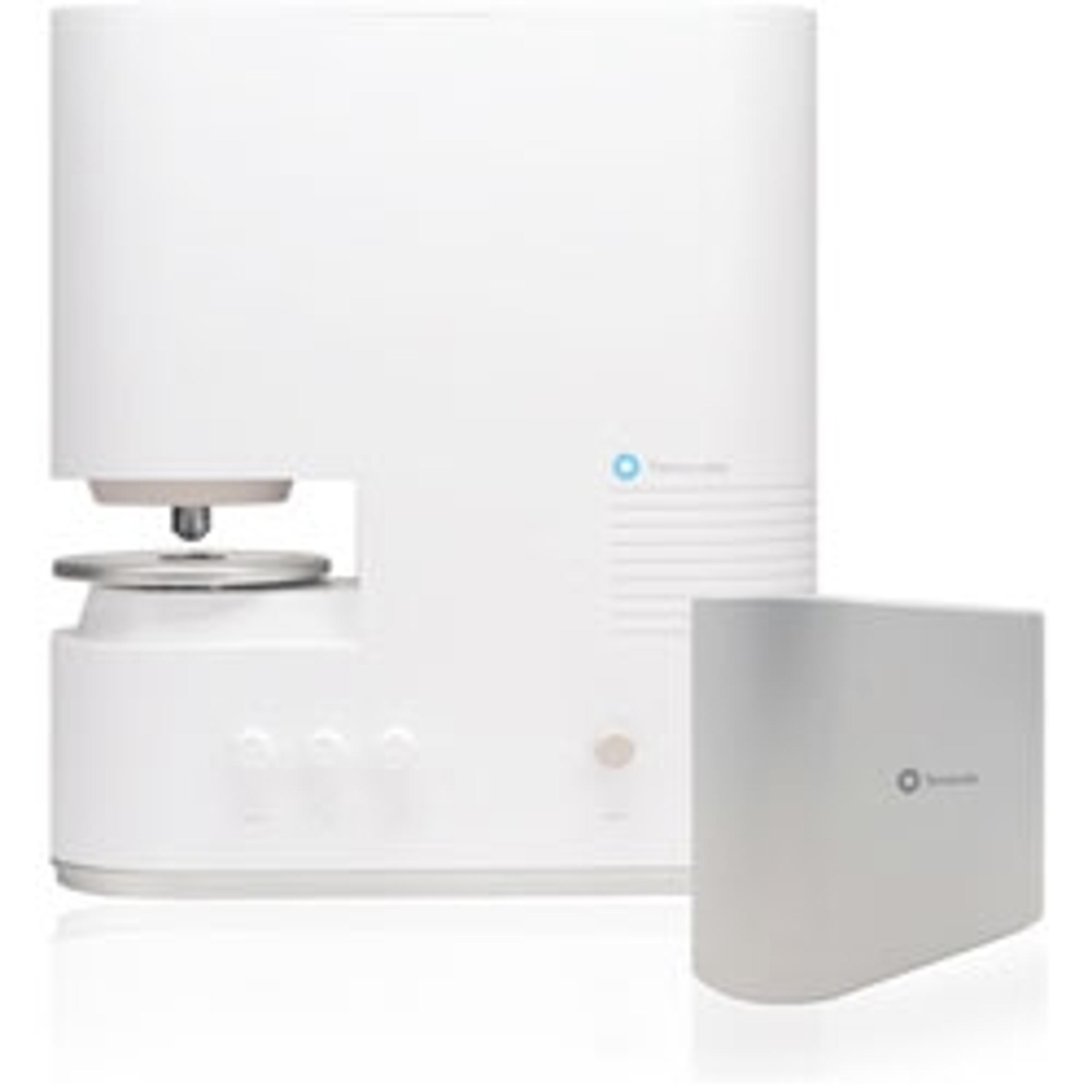
HT-2H

Tomocube

Holotomography combined with 3D fluorescence imaging

(2)

Links

Tags

ImmunologyImmunological techniques measure and characterize immune responses. Immunology kits and analysis systems often use techniques such as ELISA, radioimmunoassay (RIA) and immunodiffusion assays, Immunohistochemistry, and flow cytometry. Immunologists use equipment such as flow Cytometers, plate readers, plate washers and fluorescent microscopes.In Vivo Imaging Systems<i>In vivo</i> imaging systems, including pre-clinical imaging systems and medical imaging systems are used to non-invasively visualize and capture images of live animals and plants. Monitor the natural processes or diseases of your subjects using small-animal pre-clinical imaging systems, including single photon positron emission tomography (SPECT), positron emission tomography (PET), computed tomography (micro-CT), magnetic resonance imaging (MRI), X-ray radiography, ultrasound, fluorescence and bioluminescence imagers. Multimodal systems and software solutions are also available for correlative analysis of organ, tissue, cell, or molecular-level processes. Find the best in vivo imaging products in our peer-reviewed product directory: compare products, check customer reviews and receive pricing direct from manufacturers.Light MicroscopyLight microscopes or optical microscopes are used to visualize microscale objects under magnification, including cells, clinical specimens and materials. Lab equipment for light microscopy includes confocal microscopes, fluorescence microscopes, zoom and stereo microscopes. Microscope slides and imaging reagents are available for visualizing samples, as well as various microscope stages and incubators for large or temperature-sensitive samples. Find the best light microscopes in our peer-reviewed product directory: compare products, check customer reviews and receive pricing direct from manufacturers.ImmunofluorescenceFluorescenceThe emission of fluorescence occurs when a photon of energy is supplied to a fluorescent chemical compound by an external source, causing it to become excited. Fluorescence can be detected and measured for different purposes using microplate readers, fluorescence microscopes, fluorescence scanners, and flow cytometers.Fluorescence MicroscopyFluorescence microscopy has become an essential tool in biology, as well as in materials science. The application of many fluorochromes has made it possible to identify cells and sub-microscopic cellular components with a high degree of specificity. Using multiple fluorescence labels, different probes can simultaneously identify several target molecules.3D Imaging3D imaging technologies allow for the visualization and analysis of three-dimensional structures at high resolution. These systems are used in fields like molecular biology, material science, and medical diagnostics. 3D imaging can be applied to visualize cells, tissues, and organs, providing valuable insights into their structure and function. Browse our peer-reviewed product directory to find the best 3D imaging solutions, compare products, check reviews, and get pricing directly from manufacturers.MicroscopyMicroscopy is a technique used to observe small objects in detail, from cells to materials, using light or electron microscopes. It enables researchers to examine structures with high resolution, aiding in fields such as biology, medicine, and materials science. With advanced microscopy techniques, scientists can gain insights into cellular processes, tissue structures, and material properties. Explore the best microscopy solutions in our peer-reviewed product directory, compare products, read customer reviews, and get pricing directly from manufacturers.Cancer ResearchAlthough cancer is often referred to as a single condition, it actually consists of more than 100 different diseases. Microscopy, mass spectrometry, high throughput sequencing and flow cytometry are some of the most common techniques employed in cancer research labs.